Introduction
Heckmann et al. (Reference Heckmann, Amin and Halajian2013a) described specimens of Nephridiacanthus major (Bremser, 1811 in Westrumb, 1821) Golvan, 1962 from the central Asian and Middle Eastern long-eared hedgehog, Hemiechinus auritus (Gmelin, 1770) and the Eastern European hedgehog, Erinaceus concolor Martin (Erinaceidae, 1838) from Iran and provided histopathological observations. Heckmann et al. (Reference Heckmann, Amin and Halajian2013a) summarized the records of this acanthocephalan species from hedgehogs in Germany, Morocco, central Asia, Egypt, Bulgaria, Tadzhikistan, Lebanon, Sicily, Italy, Nigeria, Turkey and Mongolia. See Petrochenko (Reference Petrochenko1958) for references to descriptions by Bremser (Reference Bremser1811), Westrumb (Reference Westrumb1821), Porta (Reference Porta1908), Kostylev (Reference Kostylev1916, Reference Kostylev1918), Meyer (Reference Meyer1931, Reference Meyer1933) and Golvan (Reference Golvan1956) from European and Asian hedgehogs. Despite the long history of research and publications about this acanthocephalan species since its original description over 200 years ago, we could still find previously undescribed features and report chemical and molecular data for the first time. This study provides, for the first time, the molecular profile of N. major based on both small subunit ribosomal DNA (18S rDNA) and cytochrome c oxidase 1 (cox1) genes. Furthermore, its phylogenetic relationships with other members of the family Oligacanthorhynchidae and closest-related families are analysed and discussed.
Materials and methods
Collections
About 30 specimens of N. major were collected from three individuals of the hedgehog H. auratus collected in the Ferdowsi University campus in Mashhad, Razavi Khorasan province, north-eastern Iran, between 3 and 24 October 2018. Eleven mature specimens were processed for microscopical studies and four for scanning electron microscopy (SEM) studies, metal analysis and gallium (Ga) sections. Ten juvenile specimens were processed for microscopical observations but were not measured. The remaining specimens were used for molecular studies.
Methods
Freshly collected acanthocephalans were extended in water until proboscides were everted and fixed in 70% ethanol for transport to our Institute of Parasitic Diseases in Arizona, USA, for processing and further studies. Worms were punctured with a fine needle and subsequently stained in Mayer's acid carmine, destained in 4% hydrochloric acid in 70% ethanol, dehydrated in ascending concentrations of ethanol reaching 100% (24 h each) and cleared in 100% xylene then in 50% Canada balsam and 50% xylene (24 h each). Whole worms were then mounted in Canada balsam. Measurements are given in micrometres, unless otherwise noted; the range is followed by the mean values between parentheses. Width measurements represent maximum width. Trunk length does not include proboscis, neck or bursa.
Voucher specimens (whole mounts) were deposited in the University of Nebraska's State Museum's Harold W. Manter Laboratory, collection no. HWML 139975, in Lincoln, Nebraska, USA.
SEM
Specimens that had been fixed and stored in 70% ethanol were processed for SEM following standard methods (Lee, Reference Lee1992). These included critical-point drying in sample baskets and mounting on SEM sample mounts (stubs) using conductive double-sided carbon tape. Samples were coated with gold and palladium for 3 min using a Polaron #3500 sputter coater (Q150 TES, Quorum, Marly, Switzerland), establishing an approximate thickness of 20 nm. Samples were placed and observed in an FEI Helios Dual Beam Nanolab 600 (FEI, Hillsboro, Oregon, USA) scanning electron microscope with digital images obtained in the Nanolab software system (FEI, Hillsboro, Oregon, USA) and then transferred to a USB for future reference. Samples were received under low vacuum conditions using 10 KV, spot size 2, 0.7 torr using a Gaseous Secondary Electron detector.
Energy-dispersive X-ray analysis (EDXA)
Standard methods were used for preparation similar to the SEM procedure. Specimens were examined and positioned with the above SEM equipment, which was equipped with a Phoenix energy-dispersive X-ray analyser (FEI, Hillsboro, Oregon, USA). X-ray spot analysis and live scan analysis were performed at 16 Kv with a spot size of 5, and results were recorded on charts and stored with digital imaging software attached to a computer. The TEAM (Texture and Elemental Analytical Microscopy) software system (FEI, Hillsboro, Oregon, USA) was used. Data were stored in a USB for future analysis. The data included weight percent and atom percent of the detected elements following correction factors.
Ion sectioning of hooks
A dual-beam SEM with a Ga ion source (GIS) is used for the liquid ion metal source part of the process. The hooks of the acanthocephalans were centred on the SEM stage and cross sectioned using a probe current between 0.2 nA and 2.1 nA according to the rate at which the area is cut. The time of cutting is based on the nature and sensitivity of the tissue. Following the initial cut, the sample also goes through a milling process to obtain a smooth surface. The cut was then analysed with X-ray at the tip, middle and base of hooks for chemical ions with an electron beam (Tungsten) to obtain an X-ray spectrum. Results were stored with the attached imaging software, and then transferred to a USB for future use. The intensity of the GIS was variable according to the nature of the material being cut.
DNA extraction and polymerase chain reaction (PCR) amplification
Total genomic DNA was extracted from adult worms of N. major using Qiagen DNeasy blood and tissue kit (Qiagen Inc., Valencia, California, USA) according to the manufacturer's instructions. Partial nuclear 18S rDNA and partial fragments of mitochondrial cox1 gene were amplified by PCR reactions in 30 µl volumes containing 2 × red PCR premix (Ampliqon, Odense, Denmark), 20 pmol of each primer and 2 µl of isolated DNA. The partial 18S rRNA gene was amplified using the forward primer (5′-AGATTAAGCCATGCATGCGTAAG-3′) and reverse primer (5′-ACCCACCGAATCAAGAAAGAG-3′). For the cox1 gene, the primers LCO1490 (forward, 5′-GGTCAACAAATCATAAAGATATTGG-3′) and HCO2198 (reverse, 5′-TAAACTTCAGGGTGACCAAAAAATCA-3′) (1) were used. PCR conditions for 18S rDNA gene amplification comprised an initial denaturing step of 95°C for 5 min and 35 cycles followed by a denaturing step at 95°C for 30 s, an annealing step of 61°C for 30 s, 60 s of extension at 72°C and 72°C for 7 min as a final extension.
Also, the PCR cycling parameters for the cox1 gene comprised an initial denaturation at 95°C for 6 min and 35 cycles followed by a denaturing step at 95°C for 30 s, an annealing step of 55°C for 30 s, 60 s of extension at 72°C and then a final extension stage at 72°C for 6 min. The amplicons were run on a 1.5% agarose gel and visualized with UV transluminator (Vilber Lourmat, Collégien, France). Next, the amplification products were sequenced on an ABI 3730 automatic sequencer (Applied Biosystems, Foster City, California, USA) in both directions using the same PCR primers.
The obtained sequence results were manually edited and trimmed using Chromas version 2.01 (Technelysium Pty Ltd., Brisbane, Queensland, Australia) and BioEdit software version 7.0.9, Nucl. Acids. Symp. Ser. 41: 95–98. The basic local alignment search tool (BLAST; http://blast.ncbi.nlm.nih.gov/) was used to compare the sequences with available data in GenBank. The sequences of 18S rDNA and cox1 genes were submitted to GenBank database (accession numbers MN612079 for 18S rDNA and MN612080 for cox1).
Phylogenetic analysis
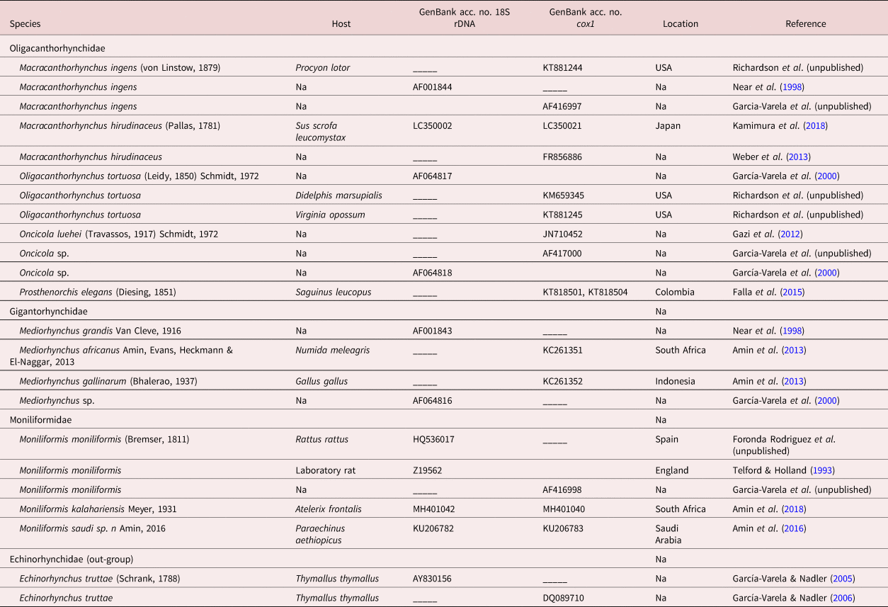
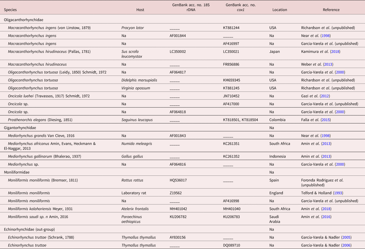
The maximum-likelihood method and Tamura three-parameter model were used for phylogenetic tree reconstruction, and genetic distances were calculated with the maximum composite likelihood model using Molecular and Evolution Genetic Analysis software version 6 (MEGA 6, Institute of Molecular Evolutionary Genetics, University Park, Pennsylvania, USA). A bootstrap value with 1000 replications was also implemented to evaluate the reliability of the tree topologies. The sequences used for the phylogenetic analysis are listed in table 1.
Acanthocephalan species represented in the phylogenetic analysis with their family, host species, GenBank accession numbers, locations and references.
Na, not available.
Results
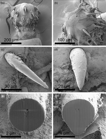
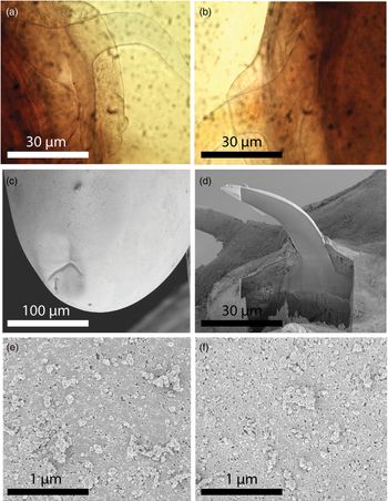
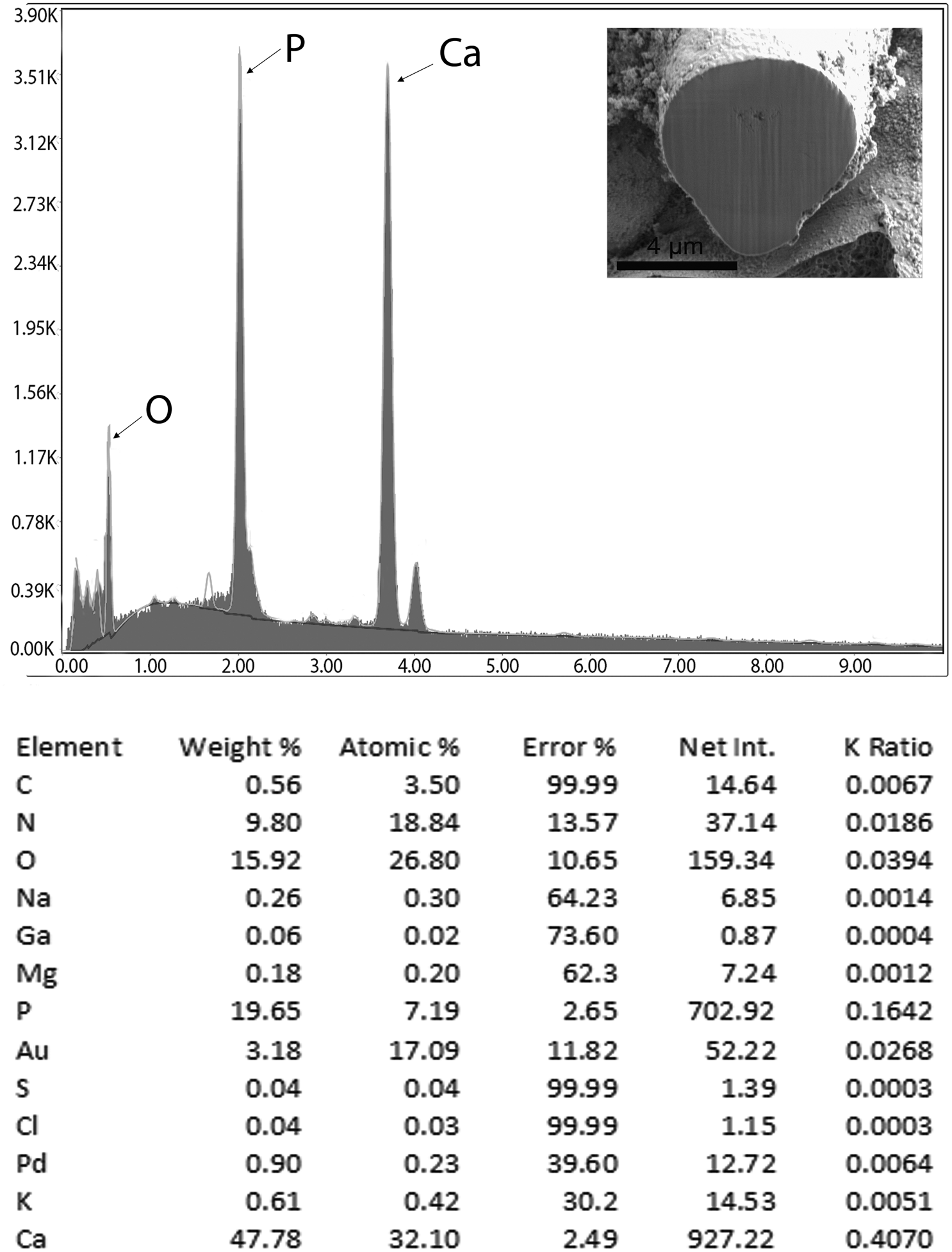
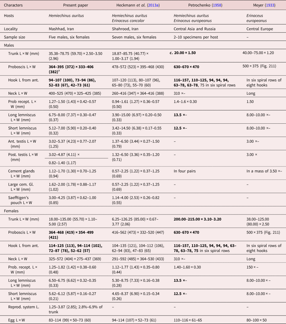
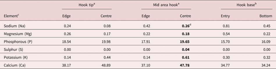
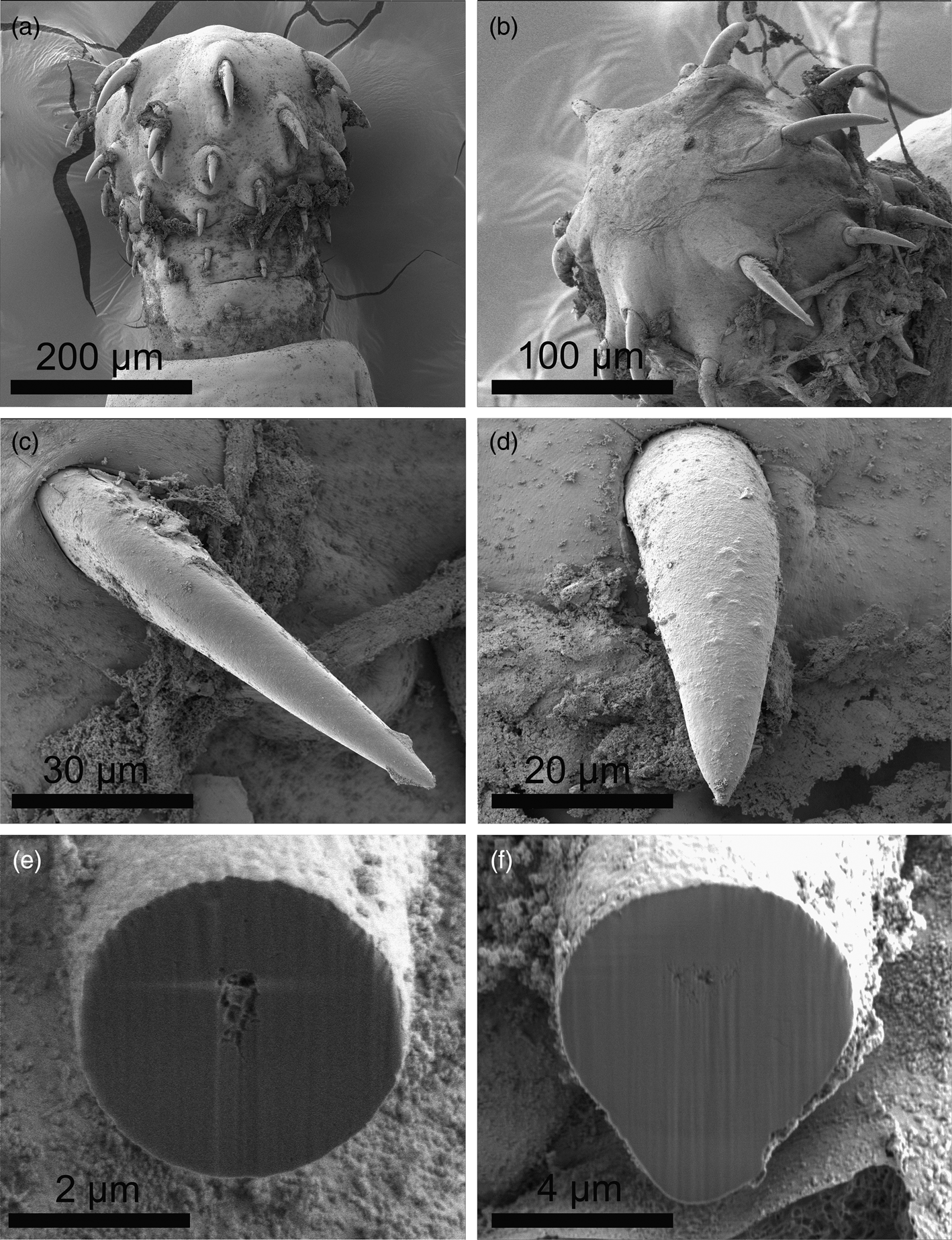
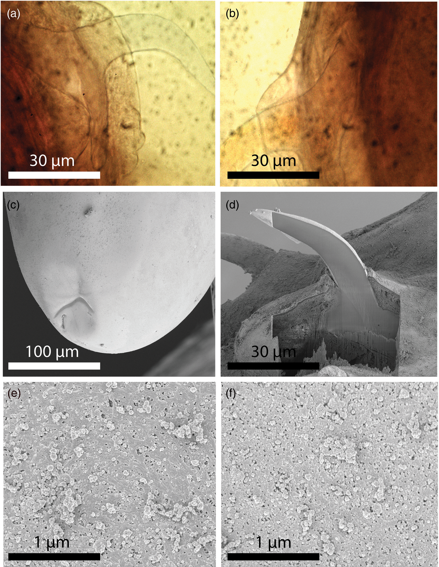
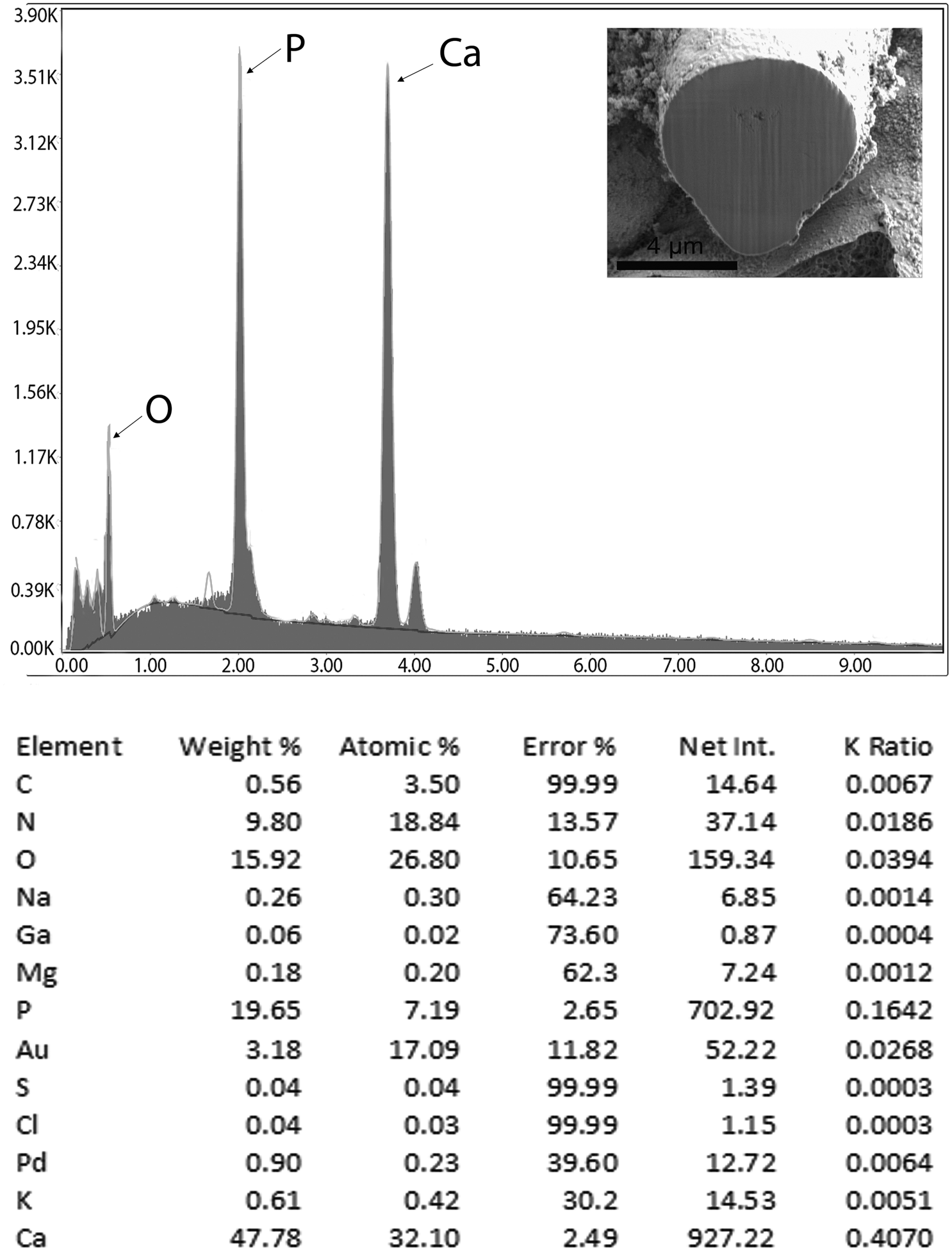
We provide a morphometric description of the present population of N. major from H. auritus in Mashhad, Iran, compared with the other population from H. auritus and E. concolor, also from Iran at Shahrood (Heckmann et al., Reference Heckmann, Amin and Halajian2013a) and from two other selected European populations – one from the west European hedgehog Erinaceus europeanus Linn, 1758 in Novocherkassk and Buzuluk, Russia, and from H. auritus in central Asia (Petrochenko, Reference Petrochenko1958); and another from E. europeanus in central Europe (Meyer, Reference Meyer1933) (table 2). Moreover, we describe additional features of N. major by SEM (figs 1 and 2), characterize the metal composition of hooks using EDXA (fig. 3 and table 3), use Ga cuts of hooks to further explain their composition and structure (figs 1e, f and 2d) and produce a molecular profile for this unusual acanthocephalan species.
SEM of specimens of Nephridiacanthus major from Hemiechinus auratus in Mashhad, Iran. (a) Lateral view of a male proboscis, showing its shape, hook arrangement and prominent neck. (b) Dorso-lateral view of another proboscis, showing anterior hooks insertions and large, rounded, slightly elevated apical organ. (c) Dorsal view of an anterior hook. (d) Face view of a shorter and plumper posterior hook. (e) Perfectly spherical gallium-cut section of a hook near its terminal end. (f) A gallium-cut section of another hook near its basal end with prominent ventral expansion. Note the solid core and thin cortical layer in both sections.
SEM and microscope images of specimens of Nephridiacanthus major from Hemiechinus auratus in Mashhad, Iran. (a) A profile of an anterior hook of a male specimen, showing the shape of the large root and angulate anterior manubrium. (b) A similar view of the second hook of the same specimen. (c) The posterior end of a female specimen, showing the sub-ventral position of the unassuming gonopore. (d) A lateral gallium-cut section of a hook, showing the same core–cortical relationship demonstrated in cross sections (fig. 1e, f). (e) Micropores of different diameters and distributions from the middle and posterior section of the trunk. (f) Micropores of different diameters and distributions from the middle and posterior section of the trunk.
Energy-dispersive X-ray spectrum of a gallium-cut mid hook of a Nephridiacanthus major specimen, showing high levels of calcium (Ca), phosphorous (P) and oxygen (O). The X-ray data are the elemental analysis of the hook centre (see bold figures in table 3). Inset: SEM of a gallium-cut hook base.
Morphometric comparisons among populations of Nephridiacanthus major from Iran and others from Europe and Russia.
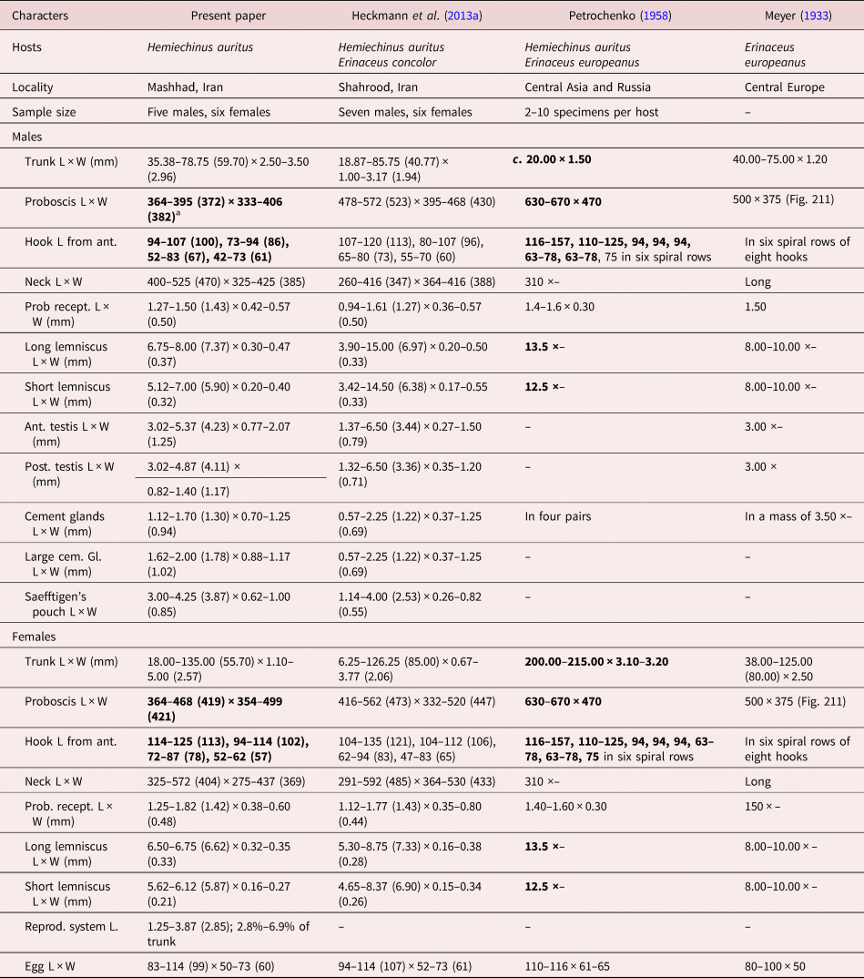
a Range (mean). Bolded numbers exhibit extreme sizes.
Notes: L, length; W, width.
Chemical composition of a gallium-cut hook of a specimen of Nephridiacanthus major.
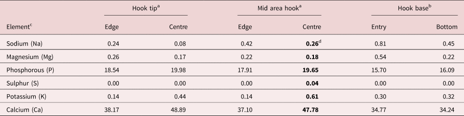
a Cross-section cut.
b Longitudinal cut.
c Common protoplasmic elements (C, N, O) and processing elements (Ga, Au, Pd) omitted from table. Listed in WT%.
d Bold weight % figures are used to generate the spectrum (Fig. 21).
New anatomical observations
The specimens studied from H. auratus in Mashhad, Iran, had similar qualitative features as the other population that we described from the same host species and from E. concolor in Shahrood, Iran, by Heckmann et al. (Reference Heckmann, Amin and Halajian2013a), and the measurements were comparable except that the sizes of the proboscis and hooks were larger in both sexes of the Shahrood specimens as well as the two European populations. Comparative measurements are provided in table 2, and new and more demonstrable morphological features are noted in figs 1 and 2. The profile of the proboscis, its shape and the comparative size and organization of its hooks, and prominent neck are shown in fig. 1a. The apical topography of the proboscis, demonstrating the slight elevation of the apical organ and the arrangement of the anterior hooks, is shown in fig. 1b. The variable shapes and sizes of anterior and posterior hooks are demonstrated in figs 1c and 1d, respectively. Hooks have a solid core and a very thin cortical layer, and are rounded at the middle (fig. 1e) but extended ventrally near their thicker base (fig. 1f). Figure 2a demonstrates the anatomy of the anterior hook of a male specimen and the massive anterior portion of its root with prominent anterior angulate manubrium. Figure 2b shows the comparable structures of the second hook of the same specimen. The sub-ventral position of the female gonopore is shown in fig. 2c, and a lateral Ga cut of a middle hook demonstrates the solid core and the thin cortical layer previously demonstrated in the cross sections (figs 1e, f).
Micropores
The variations in size and distribution of micropores in the middle and posterior sections of the trunk are demonstrated in figs 2e and 2f, respectively.
EDXA
Figure 3 and table 3 give the chemical composition of a proboscis hook at the middle centre, with calcium and phosphorous present in high concentrations, but with very low levels of sulphur and sodium.
Molecular analysis
The specimens of N. major successfully presented amplification of about 1300 bp for the partial 18S rDNA gene and about 700 bp for the partial cox1 gene. The 18S rDNA dataset (1240 nt) included ten sequences for the species of three families including Oligacanthorhynchidae, Moniliformidae and Gigantorhynchidae within the Archiacanthocephala class and the novel sequence of N. major. The cox1 dataset (597 nt) included 16 sequences from three families of class Archiacanthocephala (Oligacanthorhynchidae, Moniliformidae and Gigantorhynchidae) and the sequence of N. major.
Inter-generic differences based on a partial 18S rDNA sequence of N. major with Macracanthorhynchus hirudinaceus, Macracanthorhynchus ingens, Oligacanthorhynchus tortuosa and Oncicola sp. as other members of family Oligacanthorhynchidae was 0.2%, 0.3%, 0.7% and 0.5%, respectively. The sequence divergence based on the partial sequence of cox1 of N. major with M. ingens, M. hirudinaceus, Oncicola sp., Oncicola luehei, Prosthenorchis elegans and O. tortuosa from Oligacanthorhynchidae was 21.8–22.3%, 33.5–35.9%, 36.6%, 36.3%, 36.0–36.1% and 44.2–46.2%, respectively.
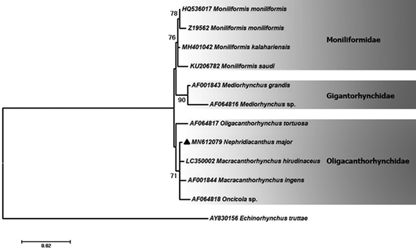
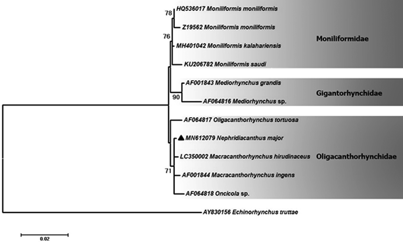
The phylogenetic tree of the 18S rDNA illustrated that the N. major sequence grouped with M. hirudinaceus (LC350002), M. ingens (AF001844) and an unidentified species of Oncicola (AF064818) in the family Oligacanthorhynchidae with strong support. The sequence of O. tortuosa (AF064817) (Oligacanthorhynchidae) is located at the basal position to the members of the clade of Oligacanthorhynchidae. Indeed, as a major sister group, families Moniliformidae and Gigantorhynchidae clustered close to the family Oligacanthorhynchidae in the tree (fig. 4).
Phylogenetic analysis of isolate of Nephridiacanthus major obtained in this study (▲) and closest-related members of class Archiacanthocephala retrieved from GenBank based on partial 18S rDNA gene. The tree was constructed using the maximum likelihood test and the Tamura three-parameter model in MEGA software version 6. Echinorhynchus truttae sequence was used as the out-group. Bootstrap values lower than 70 are omitted. The scale bar indicates number of substitutions per site.
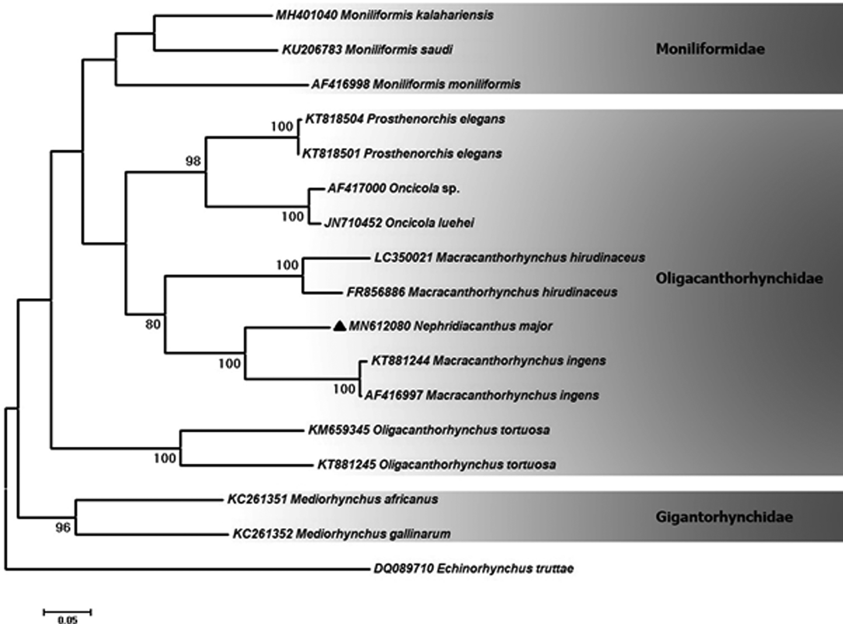
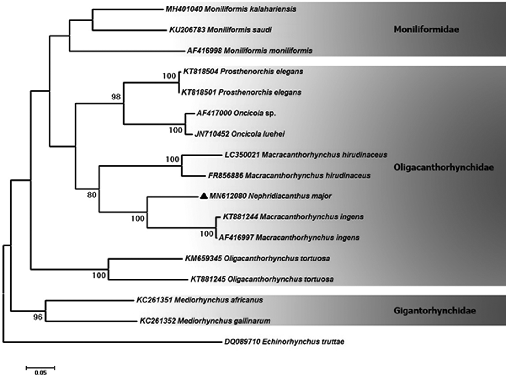
The phylogenetic reconstruction based on the partial sequence of cox1 showed that our sequence of N. major clustered with M. ingens (AF416997 and KT881244) and M. hirudinaceus isolates (FR856886 and LC350021) with high statistical support (fig. 5). This clade appeared to be a sister group consisting of sequences of an unidentified species of Oncicola (AF417000), O. luehei (JN710452) and P. elegans (KT818501 and KT818504). These two clades belong to a larger clade of the family Oligacanthorhynchidae. In addition, Moniliformis moniliformis (AF416998), Moniliformis kalahariensis (MH401040) and Moniliformis saudi sp. n. (KU206783) of the Moniliformidae family, grouped as a sister clade of the family Oligacanthorhynchidae. Additionally, O. tortuosa isolates (KT881245 and KM659345) (Oligacanthorhynchidae) located as the sister group of the major clade. Moreover, Mediorhynchus africanus (KC261351) and Mediorhynchus gallinarum (KC261352) (Gigantorhynchidae) are placed at the basal position to the members of the tree.
Phylogenetic analysis of isolate of Nephridiacanthus major obtained in this study (▲) and closest-related members of class Archiacanthocephala retrieved from GenBank based on partial cox1 gene. The tree was constructed using the maximum likelihood test and the Tamura three-parameter model in MEGA software version 6. Echinorhynchus truttae sequence was used as the out-group. Bootstrap values lower than 70 are omitted. The scale bar indicates the expected number of substitutions per site.
Discussion
Specimens of N. major are reported from only three species of hedgehogs in the Middle East, Europe and Asia, but they exhibit considerable morphological variation in size of certain taxonomically important structures that appear to be related to the geographical distribution of hosts. For instance, the trunk length of specimens from Russia and central Asia (Petrochenko, Reference Petrochenko1958) show extreme small and large values of 20.00 mm and 200.00 mm in males and females, respectively, compared to males and females from the same host species in the two collections from Iran, 18.87–85.75 mm in males and 6.25-135.00 mm in females (table 2). The lemnisci were also considerably longer in Petrochecko's (Reference Petrochenko1958) specimens than in the two Iranian populations. Similar relationships have also been observed in other acanthocephalans. For example, populations of Mediorhynchus papillosus Van Cleave, 1916 have a wide distribution in North and South America, Eastern Europe, Asia and many former Soviet republics, east to China and Taiwan. Morphometric comparisons of specimens of M. papillosus from Maryland, Colorado, Taiwan, Trans-Baikal, Lower Yenesei River, Volga, Ukraine and Bulgaria exhibited distinctive morphological variability in certain key characters, ‘with specimens from Colorado and Taiwan being at the opposite ends of the variability spectrum’ (Amin & Dailey, Reference Amin and Dailey1998). Populations of N. major may have a disjunct distribution similar to that demonstrated in M. papillosus that was attributed to ‘geographical restriction of intermediate (and adult hosts) in the apparent absence or scarcity of paratenic/reservoir hosts. Disjunct populations would promote distinct geographical diversity with consistent morphological difference from each other’ (Amin & Dailey, Reference Amin and Dailey1998, p. 199). New species would have been created from disjunct populations if it was not for their presence along the geographical west–east gradient. This proposition was not considered viable in the case of N. major.
The additional morphological features depicted in figs 1 and 2 throw additional light on the morphology of N. major that have not been presented before.
Micropores
The electron-dense micropores present throughout the epidermal surface of the trunk of N. major have not been reported previously. The micropores of N. major, like those reported from other species of the Acanthocephala, are associated with internal crypts and vary in diameter and distribution in different trunk regions corresponding with differential absorption of nutrients. We have reported micropores in a large number of acanthocephalan species (Heckmann et al., Reference Heckmann, Amin and El-Naggar2013b) and in a few more since, and demonstrated the tunnelling from the cuticular surface into the internal crypts, using transmission electron micrographs (TEM). Amin et al. (Reference Amin, Heckmann and Radwan2009) gave a summary of the structural-functional relationship of the micropores in various acanthocephalan species, including Rhadinorhynchus ornatus Van Cleave, 1918, Polymorphus minutus (Goeze, 1782) Lühe, 1911, M. moniliformis (Bremser, 1811) Travassos (1915), M. hirudinaceus (Pallas, 1781) Travassos (1916, 1917) and Sclerocollum rubrimaris Schmidt & Paperna, 1978. Wright & Lumsden (Reference Wright and Lumsden1969) and Byram & Fisher (Reference Byram and Fisher1973) reported that the peripheral canals of the micropores are continuous with canalicular crypts. These crypts appear to ‘constitute a huge increase in external surface area … implicated in nutrient up take’. Whitfield (Reference Whitfield1979) estimated a 44-fold increase at a surface density of 15 invaginations per 1 µm2 of M. moniliformis tegumental surface. The micropores and the peripheral canal connections to the canaliculi of the inner layer of the tegument of Corynosoma strumosum (Rudolphi, 1802) Lühe, 1904 from the Caspian seal Pusa caspica (Gmelin, 1788) in the Caspian Sea were demonstrated by TEM in Amin et al. (Reference Amin, Heckmann and Halajian2011).
EDXA
Results of the X-ray scans of the Ga cut hooks (dual-beam SEM) of N. major show differential composition and distribution of metals in different hook parts with the calcium and phosphorus being considerably higher at the centre of the middle of hooks where tension and strength are paramount compared to other metals (table 3). The chemical elements present in the hooks are typical for acanthocephalans (Amin & Heckmann, Reference Amin and Heckmann2017; Amin et al., Reference Amin, Heckmann and Zargar2017, Reference Amin, Heckmann and Ha2018; Ha et al., Reference Ha, Amin and Ngo2018). Note the thin outer layer (figs 1e, f and 2d) of the hook that relates to the sulphur content in the hook of N. major, which is less than what was found in other acanthocephalans (Amin & Heckmann, Reference Amin and Heckmann2017; Amin et al., Reference Amin, Heckmann and Ha2018). The hook centre in middle cuts has a completely different calcium profile than the cortical layer (table 3). X-ray scans (EDXA) provide insight into the hardened components – for example, calcium and phosphorus – of acanthocephalan hooks. The EDXA appears to be species specific, as in fingerprints, and is shown to have significant diagnostic value in acanthocephalan systematics – for example, Moniliformis cryptosaudi Amin, Heckmann, Sharifdini, Albayati, 2019 was erected based primarily on its EDXA pattern (Amin et al., Reference Amin, Heckmann and Sharifdini2019).
Molecular analysis
In recent years, DNA sequence-based methods were applied for the species identification, classification and evaluation of phylogenetic relationships among acanthocephalan species (García-Varela et al., Reference García-Varela, Cummings and Pérez-Ponce de León2002). This is the first genetic study of N. major. It resolves its phylogenetic relationships with other members of the family Oligacanthorhynchidae and also with its closest families. Inter-generic differences are noted between N. major and other members of the family Oligacanthorhynchidae based on both partial 18S rDNA and cox1 genes being 0.2–0.7% and 21.8–46.2%, respectively. These results illustrate that sequence differences between N. major and other members of the family based on the cox1 gene are higher than those for 18S rDNA. Due to the high level of divergence in the cox1 gene, it is appropriate to consider it for phylogenetic and taxonomic studies (García-Varela & de Leon, Reference García-Varela and de Leon2015). Based on the results of the cox1 analyses, the similarity of N. major with M. ingens and M. hirudinaceus was 77.7–78.2% and 64.1–66.5%, respectively. Its phylogenetic tree also confirmed the higher similarity of N. major with M. ingens, with these two genera grouped together with higher bootstrap value. Meanwhile, based on 18S rDNA, N. major illustrated more identity with M. hirudinaceus (99.8%) than with M. ingens (99.7%), which is in agreement with the 18S rDNA tree. Also, considering both genetic markers, N. major had the highest sequence differences with O. tortuosa among the members of the family Oligacanthorhynchidae. The phylogenetic analysis of the 18S rDNA sequence (fig. 1a) showed that our sequence of N. major is clustered with M. hirudinaceus (LC350002), M. ingens (AF001844), Oncicola sp. (AF064818) and O. tortuosa (AF064817), forming a clade of the family Oligacanthorhynchidae. The phylogenetic tree (fig. 4) demonstrated that the clade Oligacanthorhynchidae is well separated from those of the families Moniliformidae and Gigantorhynchidae. The phylogenetic reconstruction based on the partial sequence of cox1 illustrated that our sequence of N. major is grouped with isolates of M. ingens (AF416997 and KT881244) and M. hirudinaceus (FR856886 and LC350021) (fig. 5). This group, along with a sister clade including Oncicola sp. (AF417000), O. luehei (JN710452) and P. elegans isolates (KT818501 and KT818504), were located in a major clade of the family Oligacanthorhynchidae. Meanwhile, O. tortuosa isolates (KT881245 and KM659345) were placed separately from other members of Oligacanthorhynchidae. Due to the lack of molecular data for N. major from other parts of the world, it would be necessary to obtain specimens from other geographical regions in order to determine the genetic diversity of the species. Also, obtaining sequence data from other members of the genus Nephridiacanthus would be useful for gaining a better understanding of the phylogenetic relationships in this genus.
Acknowledgements
This project was supported by an institutional grant from the Biology Department, Brigham Young University (BYU), Provo, Utah, and the Parasitology Center, Inc., Scottsdale, Arizona. Omid Ahmadi and Yousef Sharifi, MSc students at Mashhad University of Medical Sciences, Mashhad, Iran, were instrumental in the collection of research material and recovering of specimens in field locations. We thank Madison Laurence, Bean Museum (BYU), for expert help in the preparation and organization of plates and figures, and Michael Standing, Electron Optics Laboratory (BYU), for his technical help and expertise.
Financial support
This work was supported by grants from the Departmant of Biology, Brigham Young University, Provo, Utah, and Parasitology Center, Scottsdale, Arizona.
Conflicts of interest
None.
Ethical standards
The authors assert that all procedures contributing to this work comply with the ethical standards of the relevant national and institutional guides on the care and use of laboratory animals.
Author contributions
O. Amin made the taxonomic decisions and comparisons, wrote the manuscript and made the microscopic images. M. Sharifdini made the genetic analysis and wrote the molecular text. R. Heckmann created the SEM images and the EDXA analysis. M. Zarean collected and provided specimens of N. major from trapped hosts for the study. All authors critically reviewed the manuscript.