Introduction
Churg–Strauss is a rare vasculitic syndrome first described 50 years ago comprising asthma, eosinophilic inflammation and small vessel vasculitis.Reference Keogh and Specks1 Due to the frequent occurrence of anti-neutrophilic cytoplasmic antibodies, it is usually grouped with conditions such as Wegener's granulomatosis. It can occur from adolescence to old age, but the peak incidence is in middle age.Reference Bacciu, Bacciu, Mercante, Ingengnoli, Grasselli and Vaglio2 What makes it unique compared with other primary vasculitic disorders is its allergic background. Bacciu found 43 per cent of patients had allergic rhinitis and 76 per cent suffered from nasal polyposis. One patient out of 22 patients investigated developed otitis media and two experienced a progressive hearing loss. Corticosteroid therapy associated with immunosuppressive drugs usually yielded improvement or stabilisation.Reference Bacciu, Bacciu, Mercante, Ingengnoli, Grasselli and Vaglio2 We describe a rare case of Churg–Strauss syndrome presenting to the ENT department with chronic suppurative otitis media.
Case report
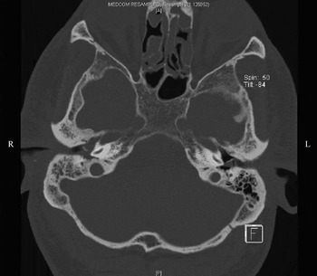
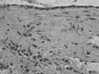
A 60-year-old man was referred to the ENT out-patient clinic with left sided sudden sensorineural hearing loss due to a suspected acoustic neuroma. His past medical history included a 20-year history of asthma and right recurrent otitis media. The patient originally presented to a regional hospital with this symptom two years earlier. He underwent a nasal polypectomy four years prior to presentation. He also suffered from diet controlled diabetes, psoriasis and thalassaemia trait. Otoscopy revealed a white discolouration behind the right tympanic membrane and a dead left ear was found on pure tone audiometry. Computed tomography (CT) showed opacification of soft tissue density in the right middle-ear cleft and mastoid cavity. There was no definite evidence of cholesteatoma as there was no bony erosion (Figure 1). However, due to the symptoms of recurrent infection the patient proceeded to surgery where he was found to have ‘rubbery’ polyps filling the middle ear. Histological examination showed pronounced chronic inflammation with scattered eosinophils (Figure 2). Anti-nuclear antibody tests, including anti-neutrophilic cytoplasmic antibodies (ANCA) were negative. A magnetic resonance imaging (MRI) scan was performed that showed a left canalicular mass measuring 7 × 3 × 3 mm. The medial border of this mass was concave and it did not enhance with gadolinium DTPA, which made the diagnosis of vestibular schwannoma very unlikely.
High-resolution axial imaging of the petrous bones with coronal reformatting. There is opacification of the mastoid air cells on the right side. The middle-ear cleft is also opacified with the opacity extending to the epitympanic recess. There is no associated bony erosion and the ossicles are intact. Minor abnormal soft tissue is also seen in the epitympanic recess on the left side, mainly superior and medial to the bony ossicles.

Middle-ear specimen showing an inflammatory infiltrate containing scattered eosinophils within the subepithelial connective tissue (H&E; × 400).

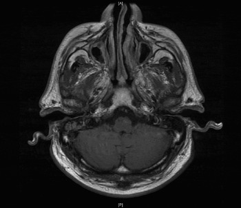
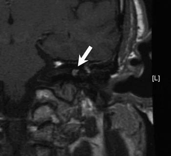
The patient was then referred to our department and he underwent a further biopsy. The anti-nuclear antibody tests were negative as before. The MRI scan was also repeated and showed enhancement of the mass in the internal auditory canal after the administration of gadolinium DTPA together with diffuse enhancement of the cochlea and vestibule on the left side (Figures 3, 4).
MRI of the head revealing some fluid retention in the mastoid air cells on both sides.

Following the injection of gadolinium DTPA there is subtle enhancement of the margins of the left auditory meatus but the neural bundle appears intact (arrow). Of significance is the diffuse enhancement of the cochlea and vestibule on the left. There is no focal parenchymal lesion demonstrated and the ventricles are of normal size. The appearances are of an inflammatory process involving the inner ear on the left.

Rheumatological assessment revealed splinter haemorrhages in both hands, but no other systemic signs. Repeated eosinophil count, inflammatory markers and ANCA were normal. The diagnosis of Churg–Strauss syndrome was based on the clinical findings of adult onset asthma, a history of nasal polyposis and marked eosinophilia noted in the histopathological sections. Moreover, the patient did not fit the criteria for other vasculitic syndromes. The diagnosis was further confirmed by the rapid and positive response to steroids and immunosuppressants.
Discussion
At present there are no clear diagnostic criteria for Churg–Strauss syndrome. The Chapel Hill Consensus defines it as an ‘eosinophil-rich and granulomatous inflammation involving the respiratory tract and necrotising vasculitis affecting small–medium sized vessels, associated with asthma and eosinophilia’.Reference Jennette, Falk, Andrassy, Bacon, Churg and Gross3 This definition aids nomenclature, but is not definite in diagnosing the condition.
Other attempts to aid the diagnosis of Churg–Strauss syndrome have been made by Lanham et al. (1984)Reference Lanham, Elkon, Pusey and Hughes4 and the American College of Rheumatology.Reference Masi, Hunder, Lie, Michel, Bloch and Arend5 The latter comprises six criteria: asthma, eosinophilia greater than 10 per cent on differential white blood cell count, mononeuropathy (including multiplex) or polyneuropathy, non-fixed pulmonary infiltrates on imaging, paranasal sinus abnormality, and biopsy containing a blood vessel with extravascular eosinophils. The presence of four or more of these six criteria yielded a sensitivity of 85 per cent and a specificity of 99.7 per cent.Reference Masi, Hunder, Lie, Michel, Bloch and Arend5 Lanham's definition includes a history of asthma, peak blood eosinophilia >1500 cells/mm3 and systemic vasculitis involving two or more extra pulmonary organs.Reference Lanham, Elkon, Pusey and Hughes4
Our patient conforms to the American definition of the disease, but not to the Chapel Hill Consensus definition or Lanham's criteria. The patient had a mononeuropathy consisting of sensorineural hearing loss, eosinophilia in the tissue from the middle ear, asthma and nasal polyposis requiring surgery four years prior to presentation.
• Churg–Strauss syndrome is a systemic vasculitis involving the respiratory system and small vessels
• Patients commonly present with adult onset asthma and allergic rhinitis
• There are no previous reports of Churg–Strauss syndrome presenting as chronic suppurative otitis media
• This report highlights the difficulty in diagnosing Churg–Strauss syndrome, but also a differential diagnosis to consider in patients with unusual presentations of chronic suppurative otitis media
Typically patients present with a history of adult onset asthma and allergic rhinitis often associated with polyposis. This phase can last for some years and is followed by a period of peripheral eosinophilia usually presenting as eosinophilic pneumonia or gastroenteritis. The final phase is characterised by polyneuropathy resulting from systemic vasculitis.Reference Bacciu, Bacciu, Mercante, Ingengnoli, Grasselli and Vaglio2 This case presented in the later stages of the disease in a very unusual way. He had an eosinophil infiltration of middle-ear mucosa, mimicking cholesteatoma on scans, but no serological eosinophilia or vasculitis as such on histology. However, he had a long history of adult onset asthma and polyposis more recently.
Considering the history of chronic suppurative middle-ear disease and the radiological findings, the other possibility in the differential diagnosis is cholesteatoma. The CT report, however, supported an inflammatory process, with mucosal thickening on the left side and no bony erosion of the ossicles or within the mastoid. The MRI was also suggestive of an inflammatory process because of the mass of soft tissue density enhanced with gadolinium DTPA. The other pathology in the differential diagnosis is Wegener's disease, but the patient did not have other vasculitic symptoms, such as renal involvement or raised inflammatory markers.
It is widely accepted that these patients respond very well to steroids and immunosuppressants as did the patient presented here. This demonstrates the importance of diagnosing this condition early to prevent hearing loss.
Conclusion
Organ involvement usually occurs in the later stages of Churg–Strauss syndrome. The otological manifestations are characterised by hearing loss, chronic thick otorrhoea and a chronic granulomatous eosinophilic infiltrate in the mastoid and middle ear. The otological pathology is steroid responsive. The aetiology of hearing loss in Churg–Strauss syndrome is not clear. In some instances, it may be a complication of chronic otitis media. This case report supports the evidence that vasculitis and ischaemic neuritis are potential pathophysiological mechanisms.
This case report illustrates the difficulties in diagnosing Churg–Strauss syndrome and reveals the importance of early medical treatment in order to prevent hearing deterioration.