Implications
In pursuit of sustainable and economically viable livestock production systems that meet consumer demands, farmers are under increasing pressure to maintain or increase fertility of their herds. Nutritional management both before and after calving has a great impact on fertility. Nutrition influences ovarian follicle development in ruminants possibly through changes in metabolic hormones and also through direct effects of nutrients on the ovary. These interactions can be manipulated to improve reproductive performance. Here, we summarize the impact of nutrients and metabolic hormones implicated in the development of ovarian follicles, oocytes and embryos in ruminants.
Introduction
In ruminants as in other mammals, alterations of energy and protein metabolism related to variations in the diet can influence reproductive functions at different levels of the gonadotropic axis. For example, in dairy cows, postpartum energy balance affects the number of follicles, their rate of growth and development, and the size of the ovulatory follicle (Lucy et al., Reference Lucy, Staples, Michel and Thatcher1991a; Boland et al., Reference Boland, Lonergan and O'Callaghan2001). In this species, negative energy balance (NEB) can also attenuate LH pulse frequency (Butler, Reference Butler2003). However, there is evidence that indicates that nutritional effects on the recruitment and growth of follicles are mediated mainly by direct metabolic signals to the ovary that vary with metabolic status (Lucy, Reference Lucy2003; Webb et al., Reference Webb, Garnsworthy, Gong and Armstrong2004). In sheep, an increase in the energy content of the diet for a few days before reproduction during the late luteal phase improves the ovulation rate (Downing and Scaramuzzi, Reference Downing and Scaramuzzi1991). Many inter-related factors are involved in the nutritional effects on fertility in ruminants. These factors are the nutrients themselves (fatty acids, glucose and amino acids) and the hormonal signals from various peripheral tissues (insulin (pancreas), IGF-I (liver), growth hormone (GH) (pituitary), thyroid hormones (T3/4) and adipokines (adipose tissue) such as leptin, adiponectin and resistin)). In the present study, we will describe the effects of these factors on the ovary (follicles and oocytes) and embryos in ruminants.
Effect of fatty acids
In this part of the review, we will detail the effects of non-esterified fatty acids (NEFAs) and also the effects of dietary supplementation with fat on ovarian function.
NEFAs
In cattle, NEB is an important risk factor for delayed postpartum cyclicity (Beam and Butler, Reference Beam and Butler1999). Follicles emerging before an NEB nadir have reduced growth and produce less oestradiol, and therefore require more time and a larger size to achieve a secretion rate of oestradiol capable of triggering the LH surge and ovulation (Beam and Butler, Reference Beam and Butler1999). It has been suggested that the harmful effects of NEB on ovarian function are exerted through decreased LH-pulsatility and reduced circulating concentrations of IGF-I, insulin and glucose (Beam and Butler, Reference Beam and Butler1999). However, metabolic alterations associated with NEB may also affect ovarian function by acting directly on the ovary. Increased lipolysis of body fat also occurs during periods of NEB, causing an elevation in the blood concentrations of NEFAs (Rukkwamsuk et al., Reference Rukkwamsuk, Geelen, Kruip and Wensing2000). For example, during periods of NEB, total serum concentrations of palmitic (C16:0), stearic (C18:0) and oleic (C18:1) acids are increased (Rukkwamsuk et al., Reference Rukkwamsuk, Geelen, Kruip and Wensing2000). Although there was no significant uptake of NEFAs by the bovine ovary in one study (Rabiee et al., Reference Rabiee, Lean, Gooden, Miller and Scaramuzzi1997), others have reported that in cattle increased concentrations of NEFAs in blood were reflected in the ovarian follicular microenvironment (Leroy et al., Reference Leroy, Vanholder, Delanghe, Opsomer, Van Soom, Bols and de Kruif2004 and Reference Leroy, Vanholder, Mateusen, Christophe, Opsomer, de Kruif, Genicot and Van Soom2005). Thus, on balance, NEFAs may affect follicular growth and fertility by acting directly on the different ovarian cells, including the oocyte within the growing follicle, and may also affect the development of the fertilized oocyte, morula and early blastocyst.
Several reports show that elevated concentrations of plasma NEFAs and in the range of concentrations frequently observed in the follicular fluid of the dominant follicle in dairy cows were associated with reduced in vitro developmental competence of the oocyte (Jorritsma et al., Reference Jorritsma, Cesar, Hermans, Kruitwagen, Vos and Kruip2004, Leroy et al., Reference Leroy, Vanholder, Mateusen, Christophe, Opsomer, de Kruif, Genicot and Van Soom2005, Aardema et al., Reference Aardema, Vos, Lolicato, Roelen, Knijn, Vaandrager, Helms and Gadella2011) and compromised the viability of the bovine granulosa cells (Vanholder et al., Reference Vanholder, Leroy, Soom, Opsomer, Maes, Coryn and de Kruif2005), but with a positive effect on the production of oestradiol by granulosa cells (Vanholder et al., Reference Vanholder, Leroy, Soom, Opsomer, Maes, Coryn and de Kruif2005). A recent study has shown that exposure of the maturing oocytes to elevated concentrations of NEFAs has a negative impact on fertility not only through a reduction in the developmental capacity of the oocyte but also through compromised quality, viability and metabolic capacity of the early embryos (Van Hoeck et al., Reference Van Hoeck, Sturmey, Bermejo-Alvarez, Rizos, Gutierrez-Adan, Leese, Bols and Leroy2011). The same laboratory suggests that these negative effects of NEFAs on the embryo could be a consequence of modified energy metabolism and particularly, mitochondrial β-oxidation of fatty acids in the oocyte (Van Hoeck et al., Reference Van Hoeck, Leroy, Arias Alvarez, Rizos, Gutierrez-Adan, Schnorbusch, Bols, Leese and Sturmey2013). A recent study has shown that bovine cumulus cells are able to protect maturing oocytes from potential harmful effects of increased local concentrations of fatty acids by intracellular storage of lipids (Aardema et al., Reference Aardema, Lolicato, van de Lest, Brouwers, Vaandrager, van Tol, Roelen, Vos, Helms and Gadella2013).
In conclusion, the current state of knowledge suggests that nutritional states that result in high-circulating concentrations of NEFAs can, and often do, compromise development of the dominant follicle and also that of the early embryo, and thus diets or metabolic states that favour high concentrations of NEFAs should be avoided during the cycle of conception and the early postconception period.
Supplementation with dietary fat
Several reviews have reported that dietary supplementation with fat influences reproduction in cows through effects on follicle growth and steroid production, oocyte maturation and embryo development (Mattos et al., Reference Mattos, Staples and Thatcher2000; Wathes et al., Reference Wathes, Abayasekara and Aitken2007; Santos et al., Reference Santos, Bilby, Thatcher, Staples and Silvestre2008; Thatcher et al., Reference Thatcher, Santos and Staples2011). The precise composition of the lipid supplement is crucial to these effects. For example, concerning the polyunsaturated fatty acids (PUFAs), the ratio of n-6 to n-3 may play an important role in reproduction in sheep and cattle (for review, Gulliver et al., Reference Gulliver, Friend, King and Clayton2012).
Effects on the follicle development
In the dairy cow, a diet enriched with long-chain fatty acids increases the number of medium follicles (6 to 9 mm) and the diameter of the preovulatory follicle (Lucy et al., Reference Lucy, Staples, Michel, Thatcher and Bolt1991b). These positive effects of long-chain fatty acids on the size of the dominant follicle have been confirmed in several studies (Mattos et al., Reference Mattos, Staples and Thatcher2000; Robinson et al., Reference Robinson, Pushpakumara, Cheng, Peters, Abayasekara and Wathes2002; Ambrose et al., Reference Ambrose, Kastelic, Corbett, Pitney, Petit, Small and Zalkovic2006; Bilby et al., Reference Bilby, Block, do Amaral, Sa Filho, Silvestre, Hansen, Staples and Thatcher2006). In addition, different effects were observed on the follicular growth between monounsaturated fatty acids (MUFA) and polyunsaturated (PUFA) fatty acids: PUFAs promoted follicular growth to a greater extent than did MUFAs (Bilby et al., Reference Bilby, Block, do Amaral, Sa Filho, Silvestre, Hansen, Staples and Thatcher2006).
Several studies have suggested that supplementation of cattle with PUFAs increased the plasma concentrations of steroid by stimulating ovarian production of steroid hormones through various mechanisms, including increased availability of lipoprotein–cholesterol (Hawkins et al., Reference Hawkins, Niswender, Oss, Moeller, Odde, Sawyer and Niswender1995; Lammoglia et al., Reference Leroy, Vanholder, Delanghe, Opsomer, Van Soom, Bols and de Kruif1996), modulation of prostaglandin synthesis (Knickerbocker et al., Reference Kolle, Sinowatz, Boie and Lincoln1986), and the direct stimulation of ovarian steroidogenesis (Staples et al., Reference Staples, Burke and Thatcher1998). In another study on cows, it was shown that diets enriched in n-6 PUFAs increased the secretion of progesterone by the granulosa cells (Wehrman et al., Reference Wehrman, Welsh and Williams1991). Although progesterone production appears in vivo to be lower with n-3 and higher with n-6, the exact mechanism through which n-3 and n-6 modulate progesterone and oestradiol is unclear (for review, Gulliver et al., Reference Gulliver, Friend, King and Clayton2012). In dairy cows, diets enriched with fatty acids (both MUFAs and PUFAs) significantly increased the concentration of cholesterol in plasma, follicular fluid and the corpus luteum (Staples et al., Reference Staples, Burke and Thatcher1998). It is well known that cholesterol is the precursor for the synthesis of progesterone by the ovarian cells. Thus, higher fertility in cattle fed diets enriched with PUFAs may be associated with increased circulating concentrations of progesterone in the luteal phases, both before and after artificial insemination. Prostaglandins are important regulators of parturition, and in ruminants they are responsible for the regression of the corpus luteum, leading to a new oestrous cycle. In cows, oestradiol had stimulatory effects on uterine secretion of prostaglandin F2α (PGF2α) (Knickerbocker et al., Reference Kolle, Sinowatz, Boie and Lincoln1986), and consequently may increase the sensitivity of the corpus luteum to PGF2α, which would promote regression of the corpus luteum. Thus, decreased plasma concentrations of oestradiol could prevent premature regression of the corpus luteum and therefore early embryonic mortality. Furthermore, the type of PUFA will also affect the synthesis of PGF2α. Indeed, dietary PUFA intake inhibits PGF2α production in bovine endometrial explants, with a more pronounced effect following n-6 rather than n-3 supplementation (for review, Wathes et al., Reference Wathes, Abayasekara and Aitken2007).
Effects on the cumulus–oocyte complex (COC) and the embryo
Compared with rodents, the oocytes and embryos of farm animals including cattle contain relatively high levels of intracellular lipids, with triglycerides being the major component. For example, the mouse oocyte contains 4 ng of triglyceride, whereas the cow oocyte contains 63 ng, and the sheep oocyte contains 89 ng (McEvoy et al., Reference McEvoy, Coull, Broadbent, Hutchinson and Speake2000; Sturmey et al., Reference Sturmey, Reis, Leese and McEvoy2009). Fatty acids are an endogenous source of energy during oocyte maturation; several lines of evidence support the idea of fatty-acid oxidation as a source of essential ATP for oocyte maturation and for early development of the embryo. For example, the triglyceride content of the bovine oocytes decreased during in vitro maturation (IVM), as well as after fertilization (Ferguson and Leese, Reference Ferguson and Leese1999). In addition, lipase activity was increased in the bovine oocytes following IVM (Cetica et al., Reference Cetica, Pintos, Dalvit and Beconi2002). In the oocytes from mice, inhibition of β-oxidation during the IVM of oocytes prevented the AMP-activated, protein kinase-mediated resumption of meiosis in the mouse (Downs et al., Reference Downs, Mosey and Klinger2009), and in cattle it impaired the developmental competence of oocytes (Dunning et al., Reference Dunning, Cashman, Russell, Thompson, Norman and Robker2010). Furthermore, Paczkowski et al. (Reference Paczkowski, Silva, Schoolcraft and Krisher2013) have shown that the oxidation of fatty acids is essential for nuclear maturation of the oocytes in mice, cattle and pigs (Paczkowski et al., Reference Paczkowski, Silva, Schoolcraft and Krisher2013).
Dietary fatty acids altered the composition of fatty acids in the cumulus cells, granulosa cells and oocytes that may also have consequences on oocyte quality (Kim et al., Reference Kim, Kinoshita, Ohnishi and Fukui2001). For example, in ewes, the number and quality of oocytes was increased when animals were fed a diet enriched in PUFAs (saponified fish oil) 74.3% v. 57%; (Zeron et al., Reference Zeron, Sklan and Arav2002). Moreover, in these animals, there was an increase in the proportion of long-chain fatty acids in the plasma and cumulus cells (Zeron et al., Reference Zeron, Sklan and Arav2002). In dairy cows, another study has shown that a diet enriched in a mixture of saturated and unsaturated fats affected oocyte development and maturation (Fouladi-Nashta et al., Reference Fouladi-Nashta, Gutierrez, Gong, Garnsworthy and Webb2007). The two fatty acids linoleic acid (LA) and linolenic acid (ALA) are both found in the plasma and follicular fluid of cows (Childs et al., Reference Childs, Hennessy, Sreenan, Wathes, Cheng, Stanton, Diskin and Kenny2008), and it has been reported that supplementation of bovine oocytes with ALA during IVM resulted in an increased maturation rate, a higher yield of blastocysts and the production of better-quality blastocysts (Marei et al., Reference Marei, Wathes and Fouladi-Nashta2009). Opposing results have been observed for supplementation with LA by the same group (Marei et al., Reference Marei, Wathes and Fouladi-Nashta2009). Indeed, they reported that treatment of bovine COCs with physiological concentrations of LA affected the molecular mechanisms that control nuclear maturation of oocytes, leading to a decreased proportion of oocytes reaching the MII stage after 24 h in culture, and the inhibition of subsequent early embryo development (Marei et al., Reference Marei, Wathes and Fouladi-Nashta2010). Further studies from this group revealed that LA induced alterations in the distribution of mitochondria and their activity, associated with increased concentrations of reactive oxygen species (Marei et al., Reference Marei, Wathes and Fouladi-Nashta2012).
Thus, the current state of knowledge suggests that PUFAs could promote the growth of follicle and could increase granulosa cell steroidogenesis in cattle. However, the ratio of n-6 to n-3 in ruminant diets is particularly important in determining the relative availability of the precursors for eicosanoid formation. The effects of PUFA on the oocyte and embryo development are unclear and need more investigations.
Effect of glucose
The published evidence suggests that the adverse effects of systemic treatment with insulin on reproductive function do not occur if the circulating concentrations of glucose are maintained at physiological levels (Downing et al., Reference Downing, Joss and Scaramuzzi1999), and concentrations of glucose that are either above or below the normal physiological range can have deleterious effects on fertility. For example, one study (Downie and Gelman, Reference Downie and Gelman1976) suggested that low-circulating glucose may be responsible for infertility in cows, and in another study pregnancy rate was higher in cows with high-circulating concentrations of glucose compared with cows with low-circulating concentrations of glucose; however, there was a trend for pregnancy rate to decline in cows with very high concentrations of circulating glucose (Pehrson et al., Reference Pehrson, Plym Forshell and Carlsson1992). Finally, Selvaraju et al. (Reference Selvaraju, Agarwal, Karche, Srivastava, Majumdar and Shanker2002) reported that insulin and glucose concentrations were higher in cows that subsequently became pregnant than in non-pregnant animals (Selvaraju et al., Reference Selvaraju, Agarwal, Karche, Srivastava, Majumdar and Shanker2002). All of this evidence suggests that fertility of cows is strongly influenced by the insulin glucose system.
Downing et al. (Reference Downing, Joss and Scaramuzzi1999) stated that there is a synergism between insulin and glucose at the ovarian level, and it is likely that the effects of short-term nutrition on ovulation rate in ewes may be mediated by direct ovarian actions of insulin and glucose (Downing et al., Reference Downing, Joss and Scaramuzzi1999). Mammals primarily rely on intracellular oxidation of glucose and fatty acids to provide the energy necessary to support most physiological processes. Although peripheral tissues may utilize both glucose and fatty acids, the ovary uses glucose as its principal source of energy (Rabiee et al., Reference Rabiee, Lean, Gooden and Miller1999; Scaramuzzi et al., Reference Scaramuzzi, Campbell, Souza and Baird2010). Glucose is available for cellular oxidation and energy production by entering the cells passively when its circulating concentrations are high and also actively at low concentrations, with the latter implying a role for insulin (Wade and Schneider, Reference Wade and Schneider1992). It seems that the effect of glucose on fertility is primarily related to its properties as a metabolic fuel.
Within the follicle, glucose can be metabolized by four different pathways: (i) by glycolysis to produce ATP and pyruvate or lactate; (ii) by the pentose phosphate pathway to provide precursors for the synthesis of purine nucleotides and NADPH for various biosynthetic pathways including antioxidant defence; (iii) by the hexosamine biosynthetic pathway to provide substrates for the glycosylation of proteins and the synthesis of the hyaluronic acid required for cumulus expansion; and (iv) by the polyol pathway producing sorbitol and fructose whose roles in follicular function remains largely unknown (for a review see Collado-Fernandez et al. (Reference Collado-Fernandez, Picton and Dumollard2012)). In cattle, the role of these four pathways for the utilization of glucose during the growth and development of follicles and their oocytes is not clearly known. Nowadays, it is believed that the follicle has well-developed systems to sense glucose and nutritional status (Webb et al., Reference Webb, Garnsworthy, Gong and Armstrong2004; Garnsworthy et al., Reference Garnsworthy, Gong, Armstrong, Newbold, Marsden, Richards, Mann, Sinclair and Webb2008; Scaramuzzi et al., Reference Scaramuzzi, Campbell, Souza and Baird2010). Different glucose transporters, including the GLUT family, are expressed in the oocyte, the somatic cells of the follicle and in the early embryo, and the expression of some of them is controlled by steroids and insulin (Purcell and Moley, Reference Purcell and Moley2009). This system allows the follicle to regulate its growth and development, mainly by altering FSH-induced effects on the synthesis of oestradiol by the granulosa cells, in accordance with the availability of glucose (Webb et al., Reference Webb, Garnsworthy, Gong and Armstrong2004; Scaramuzzi et al., Reference Scaramuzzi, Campbell, Souza and Baird2010).
The role of glucose is essential in determining the quality of the oocyte (Sutton-McDowall et al., Reference Sutton-McDowall, Gilchrist and Thompson2010) because it is its main energy source. In sheep, glucogenic treatments improve oocyte quality, evaluated by the kinetics of their in vitro development and by the production of blastocysts (Berlinguer et al., Reference Berlinguer, Gonzalez-Bulnes, Contreras-Solis, Spezzigu, Torres-Rovira, Succu, Naitana and Leoni2012). However, even if glucose is the preferred energy substrate for the cumulus cells (Sutton-McDowall et al., Reference Sutton-McDowall, Gilchrist and Thompson2010), the oocytes of most mammalian species consume little glucose, with pyruvate being the usual energy substrate (Eppig, Reference Eppig1976; Rieger and Loskutoff, Reference Rieger and Loskutoff1994). In general, oocytes take up pyruvate efficiently and have a lower capacity for glucose transport, as well as limited expression and activity of some glycolytic enzymes (reviewed by Purcell and Moley, Reference Purcell and Moley2009; Sutton-McDowall et al., Reference Sutton-McDowall, Gilchrist and Thompson2010). Thus, oocytes rely on the uptake of CC-derived oxidizable substrates, principally pyruvate for the synthesis of ATP. However, ATP and glucose can also be directly transferred to oocytes from GC/CC through gap junctions (Wang et al., Reference Wang, Chi, Schedl and Moley2012).
Effect of amino acids
In contrast with fatty acids and glucose, there are few studies reporting the effects of amino acids on ovarian functions in ruminants. Downing et al. (Reference Downing, Joss and Scaramuzzi1995) intravenously infused a mixture of branched-chain amino acids in sheep and observed an increased ovulation rate, whereas Garnsworthy et al. (Reference Garnsworthy, Gong, Armstrong, Newbold, Marsden, Richards, Mann, Sinclair and Webb2008) reported that diets with a high content of leucine increased plasma concentrations of insulin in dairy cows, but did not alter their follicular dynamics (Garnsworthy et al., Reference Garnsworthy, Gong, Armstrong, Newbold, Marsden, Richards, Mann, Sinclair and Webb2008). In dairy heifers, an association was observed between the total concentration of free amino acids in the plasma and oocyte cleavage, suggesting that dietary amino acids could mediate the oocyte quality (Rooke et al., Reference Rooke, Ainslie, Watt, Alink, McEvoy, Sinclair, Garnsworthy and Webb2009); however, Sinclair, et al. (Reference Sinclair, Sinclair and Robinson2000) also showed that in heifers excessive levels of protein in the diet negatively affected the subsequent development of oocytes in vitro (Sinclair et al., Reference Sinclair, Sinclair and Robinson2000)
In comparison with carbohydrate metabolism, and despite their important roles, there is little information about amino-acid uptake and metabolism by follicles and oocytes, especially during the earlier stages of folliculogenesis in cattle. Studies measuring amino acids in follicular fluid and the reproductive tract in mice have provided valuable information on the availability of substrates, reporting significantly higher levels of some amino acids in the follicular fluid compared with the reproductive tract (Harris et al., Reference Harris, Gopichandran, Picton, Leese and Orsi2005). In cattle, LH has been shown to increase glutamine oxidative metabolism by oocytes and COCs (Zuelke and Brackett, Reference Zuelke and Brackett1993). Furthermore, the addition of glutamine to bovine IVM media promoted the nuclear maturation of oocytes (Bilodeau-Goeseels, Reference Bilodeau-Goeseels2006), although supplementation of defined IVM media with non-essential and essential amino acids increased the levels of maternal mRNA in the oocytes and enhanced embryo development (Watson et al., Reference Watson, De Sousa, Caveney, Barcroft, Natale, Urquhart and Westhusin2000). Finally, Hemmings et al. (Reference Hemmings, Leese and Picton2012) quantified the amino-acid profiles (i.e. rates of depletion and accumulation) of bovine oocytes at MII, following IVM (Hemmings et al., Reference Hemmings, Leese and Picton2012). Glutamine, arginine and asparagine were all depleted at the highest rates, whereas alanine and glycine accumulated in the media (Hemmings et al., Reference Hemmings, Leese and Picton2012). Similar to studies carried out on embryos, oocytes with a higher potential for development after IVF and those of lesser potential had different amino-acid profiles (Hemmings et al., Reference Hemmings, Leese and Picton2012). Overall, the quality of oocytes and embryos was related to amino-acid turnover; those with the highest turnover of amino acids had the poorest quality. For example, oocytes that failed to cleave depleted more glutamine, released more alanine and had a greater depletion of total amino acids compared with oocytes that cleaved (Hemmings et al., Reference Hemmings, Leese and Picton2012). Furthermore, these data were used to predict fertilization and cleavage potential (Hemmings et al., Reference Hemmings, Leese and Picton2012). These studies provide further evidence of the importance of amino-acid metabolism to the developmental competence of oocytes.
Nutrient sensors
Metabolic hormones
The effects of acute changes in the dietary intake on ovarian activity have been correlated with changes in the circulating concentrations of metabolic hormones, including insulin, IGF-I, GH, the thyroid hormones, triiodothyronine (T3), and thyroxine (T4), ghrelin, apelin and the adipokines (leptin, adiponectin, resistin), and there is an extensive literature describing the effects of diet on the circulating concentrations of these metabolic hormones. Here, we have focused more on the action of the metabolic hormones on ovarian functions in ruminants.
Insulin (pancreas) and IGF-I (liver)
Insulin and IGF-I are produced by the pancreas and liver, respectively, and are often proposed as signalling molecules linking metabolism to fertility (Diskin et al., Reference Diskin, Mackey, Roche and Sreenan2003). They interact with the reproductive axis at the level of the central nervous system as well as in the follicle itself. Here, we only described some effects of insulin and IGF-I on the ruminant ovary.
Components of the insulin and IGF systems in the ovary
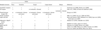
The insulin system consists of insulin itself and the insulin receptor (IR), which mediates the action of insulin. The IR has a high degree of homology with the IGF-I receptor (IGF-IR), and IGF-IR and IR hybrid receptors have been reported (Dupont and LeRoith, Reference Dupont and LeRoith2001). The IGF system is composed of several members, including IGF-I and IGF-II, two receptors (IGFR-1 and IGFR-2) and at least six binding proteins (insulin-like growth factor binding proteins: IGFBP-1, -2, -3, -4, -5 and -6) (Monget et al., Reference Monget, Fabre, Mulsant, Lecerf, Elsen, Mazerbourg, Pisselet and Monniaux2002). The essential components of both the insulin and IGF systems are all present in the follicle in ruminants (Table 1). The IGFBPs are present in biological fluids and act by inhibiting or potentiating the action of the two IGFs (IGF-I and IGF-II) in target cells (Monget et al., Reference Monget, Fabre, Mulsant, Lecerf, Elsen, Mazerbourg, Pisselet and Monniaux2002). The regulation of the bioavailability of IGF-I by IGFBPs is necessary for the co-ordinated development in vitro, of the bovine oocyte and follicle (Thomas et al., Reference Thomas, Campbell, Armstrong and Telfer2007). The receptors for insulin and IGF-I belong to the same subfamily of receptor tyrosine kinases with two extracellular α-subunits and two trans-membrane β-subunits. They have structures that are highly homologous and they induce a number of similar intracellular signalling pathways (Dupont and LeRoith, Reference Dupont and LeRoith2001). Upon activation by ligand binding, both IR and IGF-1R, which contain endogenous tyrosine kinase activity, phosphorylate several intracellular substrates such as the insulin receptor substrate (IRS) proteins (IRS-1 through -6) and Shc, leading to the activation of at least two signalling pathways, PI3K/AKT and MAPK. In ruminants, IRs and IGF-1Rs are widely distributed throughout all ovarian compartments, including the follicles (Spicer et al., Reference Spicer, Alpizar and Vernon1994; Shimizu et al., Reference Shimizu, Murayama, Sudo, Kawashima, Tetsuka and Miyamoto2008) and luteal cells (Einspanier et al., Reference Einspanier, Miyamoto, Schams, Muller and Brem1990). In bovine granulosa and theca cells, the expression of the mRNA for IR changes considerably during development from the preantral to the pre-ovulatory stage of folliculogenesis (Shimizu et al., Reference Shimizu, Murayama, Sudo, Kawashima, Tetsuka and Miyamoto2008). Similarly, the expression of IGF-1R in bovine granulosa and theca cells is increased during the final stages of follicular development and decreased at the onset of atresia (Armstrong et al., Reference Armstrong, Gutierrez, Baxter, Glazyrin, Mann, Woad, Hogg and Webb2000). Because IGF-I and/or insulin have an essential role in the final stage of follicle development, it has been suggested that abnormal concentrations of these metabolic hormones lead to follicular dysfunction, resulting in excessive atresia or the formation of follicular cysts (Braw-Tal et al., Reference Braw-Tal, Pen and Roth2009).
The expression of various metabolic factors in the ovary and embryo of ruminants

GH=growth hormone; GHS-R=growth hormone secretagogue receptor; nr=not reported in the literature; PPAR=peroxisome proliferator-activated receptors.
The effects of insulin on follicular function
Numerous studies have shown the importance of insulin as a signal mediating the effects of acute changes in nutrient intake on follicle dynamics in cattle (Webb et al., Reference Webb, Garnsworthy, Gong and Armstrong2004). The infusion of insulin into beef heifers increased the diameter of the dominant follicle (Simpson et al., Reference Simpson, Chase, Spicer, Vernon, Hammond and Rae1994). In vitro research has shown dose-dependent stimulatory effects of insulin on the proliferation and steroid synthesis of the bovine granulosa cells and theca cells; these effects can be exerted via their direct stimulatory actions on the follicle as well as by increased local responsiveness of the follicle to FSH and LH (Spicer et al., Reference Spicer, Alpizar and Vernon1994).
The effects of IGFs on follicular function
IGF-I is a hormone that modulates the maturation of the dominant follicle during the first follicular wave postpartum (Beam and Butler, Reference Beam and Butler1998), and circulating IGF-I in ovulatory cows at the first follicular wave postpartum is higher than that in anovulatory cows, regardless of parity (Beam and Butler, Reference Beam and Butler1998). As for insulin, in vitro research has shown dose-dependent stimulatory effects of IGF-I on the proliferation and steroid synthesis of bovine granulosa cells and theca cells; these effects can be exerted via their direct stimulatory actions on the follicle as well as by increased local responsiveness of the follicle to FSH and LH (Spicer and Echternkamp, Reference Spicer and Echternkamp1995). In the bovine granulosa cells, IGF-I induced the upregulation of steroidogenic and apoptotic regulatory genes via the activation of phosphatidylinositol-dependent kinase/AKT (Mani et al., Reference Mani, Fenwick, Cheng, Sharma, Singh and Wathes2010). The addition of IGF-I to the culture medium improved the in vitro development of caprine preantral follicles (Magalhaes-Padilha et al., Reference Magalhaes-Padilha, Duarte, Araujo, Saraiva, Almeida, Rodrigues, Matos, Campello, Silva, Gastal, Gastal and Figueiredo2012).
The effects of insulin and IGF on the oocyte and embryo
Both IGF-1R and IR are also present in the bovine oocytes and embryos (Nuttinck et al., Reference Nuttinck, Charpigny, Mermillod, Loosfelt, Meduri, Freret, Grimard and Heyman2004). Several studies have observed that COCs treated with IGF-I, alone or in combination with either epidermal growth factor or angiotensin II, showed increased cumulus expansion, improved rates of nuclear maturation and enhanced metabolism of pyruvate (Lorenzo et al., Reference Lorenzo, Illera, Illera and Illera1994; Stefanello et al., Reference Stefanello, Barreta, Porciuncula, Arruda, Oliveira, Oliveira and Goncalves2006). The intra-ovarian administration of IGF-I was able to improve the developmental capacity of oocytes from pre-pubertal cattle (Oropeza et al., Reference Oropeza, Wrenzycki, Herrmann, Hadeler and Niemann2004). A short in vivo exposure of oocytes to a supra-physiological IGF-I microenvironment increased the in vitro proliferation of the inner cell mass cell during the transition from morula to blastocyst (Velazquez et al., Reference Velazquez, Hadeler, Herrmann, Kues, Remy, Beckers and Niemann2012). In bovine blastocysts, insulin acted as a survival factor, blocking apoptosis, whereas IGF-I stimulated the development of blastocyst and also total cell number (Byrne et al., Reference Byrne, Southgate, Brison and Leese2002).
Collectively, these data indicate that maintaining the concentrations of IGF-I and insulin within a normal physiological range is required for proper ovarian follicular function and embryo development.
GH (anterior pituitary gland)
GH produced by the adenohypophysis exerts effects on almost every organ of the body, including the ovary, either directly after binding to specific GH receptors (GHR) or indirectly after binding to hepatic GHR, and stimulating the production of hepatic IGF-I (Webb et al., Reference Webb, Garnsworthy, Gong and Armstrong2004). Treatment of cattle with exogenous GH has significant effects on follicular development (Gong et al., Reference Gong, Bramley and Webb1993) and the function of the corpus luteum (Lucy et al., Reference Lucy, Bilby, Kirby, Yuan and Boyd1999). GH can selectively stimulate particular populations of follicles; for example, in heifers, it inhibited the development of the preovulatory follicle but stimulated the growth of the second-largest follicles (Lucy et al., Reference Lucy, Curran, Collier and Cole1994). The possibility that GH can act at ovarian sites is suggested by the detection of GH-binding activity, GHR immunoreactivity and expression of the mRNA encoding GHR in the ovarian tissue in cows (Kolle et al., Reference Kolle, Sinowatz, Boie and Lincoln1998). GH receptors have been detected in the granulosa cells, thecal cells and luteal cells (Kolle et al., Reference Kolle, Sinowatz, Boie and Lincoln1998) (Table 1), and the abundance of the mRNA encoding GHR in the bovine ovary varied in a cell-specific way during the ovarian cycle (Kolle et al., Reference Kolle, Sinowatz, Boie and Lincoln1998). However, the involvement of GH in the physiological mechanisms underlying nutritional influence on ovarian function in the cattle can be attributed to the stimulatory action of GH on the hepatic synthesis of IGF-I, and a direct action of GH on the follicle remains to be demonstrated convincingly (Webb et al., Reference Webb, Garnsworthy, Gong and Armstrong2004).
T3 and T4 (thyroid gland)
In cattle, greater nutrient intake after calving increased the concentrations of T4 in the plasma (Ciccioli et al., Reference Ciccioli, Wettemann, Spicer, Lents, White and Keisler2003), similar to the response in non-lactating cows (Delavaud et al., Reference Delavaud, Ferlay, Faulconnier, Bocquier, Kann and Chilliard2002). Follicular fluid from the bovine ovaries contains free fractions of thyroid hormones that suggests a possible role for these hormones in the regulation of follicular function. In super-ovulated Brahman cows, induced hypothyroidism improved weight gain and body score condition, increased ovarian response to FSH, and affected the ovulation, fertility and secretion of progesterone (Bernal et al., Reference Bernal, DeMoraes, Thrift, Willard and Randel1999). In cows, the physiological status of the bovine antral follicles (i.e. dominant v. subordinate) may impinge on the accumulation of T4 in the follicular fluid (Ashkar et al., Reference Ashkar, Semple, Schmidt, John, Bartlewski and King2010), and hormonally induced ovarian hyperstimulation increased the circulating levels of free T4 and the follicular fluid content of total T4 (Mutinati et al., Reference Mutinati, Desantis, Rizzo, Zizza, Ventriglia, Pantaleo and Sciorsci2010). Furthermore, the thyroid hormones (T3 and T4) directly altered in vitro steroidogenesis in the bovine granulosa and theca cells (Spicer et al., Reference Spicer, Alonso and Chamberlain2001). A recent study has shown that the supplementation of IVM media with T3 can have a beneficial effect on the kinetics of embryo development (Costa et al., Reference Costa, Cordeiro, Silva, Sastre, Santana, Sa, Sampaio, Santos, Adona, Miranda and Ohashi2013). In the ovaries of human and mice, thyroid hormone receptors (TRα-1, TRα-2 and TRβ-2) are present in the oocytes, cumulus, granulosa and luteal cells (Zhang et al., Reference Zhang, Carrillo and Darling1997) (Table 1). Thus, thyroid hormones may have direct stimulatory effects on the ovarian function and embryo development in cattle.
Ghrelin (stomach)
Ghrelin is primarily secreted from the X/A-like cells of the oxyntic gland in the stomach. The X/A-like cells constitute a distinct population of endocrine cells in the oxyntic mucosa that make up ~20% of all endocrine cells in the oxyntic gland. Ghrelin is also present in several other tissues, including the reproductive tissues of sheep and cattle (Miller et al., Reference Miller, Harrison, Brown, Doyle, Lindsay, Adam and Lea2005; Deaver et al., Reference Deaver, Hoyer, Dial, Field, Collier and Rhoads2013). The functional ghrelin receptor, GH secretagogue receptor 1 A (GHS-R1A), belongs to a large family of rhodopsin-like, G-protein coupled, 7-transmembrane domain receptors (Howard et al., Reference Howard, Feighner, Cully, Arena, Liberator, Rosenblum, Hamelin, Hreniuk, Palyha, Anderson, Paress, Diaz, Chou, Liu, McKee, Pong, Chaung, Elbrecht, Dashkevicz, Heavens, Rigby, Sirinathsinghji, Dean, Melillo, Patchett, Nargund, Griffin, DeMartino, Gupta, Schaeffer, Smith and Van der Ploeg1996). Ghrelin is an acute regulator of feed intake; circulating ghrelin concentrations increase during fasting or NEB, and exogenous administration of ghrelin stimulates feed intake in rats and cattle (Bradford and Allen, Reference Bradford and Allen2008). In these species, Ghrelin also influences energy metabolism and increases metabolic efficiency. Thus, it appears that ghrelin is a key metabolic component involved in the physiological response to energy deprivation.
Ghrelin and its receptor are present in the ovary of the adult and foetal sheep (Miller et al., Reference Miller, Harrison, Brown, Doyle, Lindsay, Adam and Lea2005) (Table 1). In the sheep ovary, ghrelin and its mRNA are present throughout the oestrous cycle with the highest levels observed during the luteal phase (Du et al., Reference Du, Xilingaowa, Cao, Wang, Li, Zhao, Siqingaowa and Cao2009). More precisely, detectable concentrations of ghrelin are found in the oocytes and thecal cells at all stages of follicular development, the granulosa cells of antral follicles and in all developmental stages of the corpora lutea (Du et al., Reference Du, Xilingaowa, Cao, Wang, Li, Zhao, Siqingaowa and Cao2009) (Table 1). In the sheep, ghrelin promotes in vitro oocyte maturation via the ERK1/2 pathway (Bai et al., Reference Bai, Zhao, Cao, Wen, Li and Meng2012) and in vitro formation of blastocysts (Wang et al., Reference Wang, Lin and Yu2013). In the Holstein heifers, the expression of ghrelin was limited to the granulosa cells in the ovarian follicles, suggesting that the follicular distribution of ghrelin is species dependent (Deaver et al., Reference Deaver, Hoyer, Dial, Field, Collier and Rhoads2013). In the dairy heifers, ghrelin and GHS-R1A and their mRNAs are also present in the ampulla and isthmus of the oviducts and the uterus (Deaver et al., Reference Deaver, Hoyer, Dial, Field, Collier and Rhoads2013).
However, despite the presence of ghrelin and its receptor in various parts of the reproductive tract, its effects, if any, in vivo on fertility in ruminants are not known.
Adipokines (adipose tissue)
The traditional view of the adipose tissue as a passive store of dormant triglycerides, which are mobilized during NEB, has been challenged by recent research that has shown that the adipose tissue also secretes a variety of hormones collectively known as the adipokines (Tersigni et al., Reference Tersigni, Di Nicuolo, D’Ippolito, Veglia, Castellucci and Di Simone2011). Leptin, adiponectin and resistin are the three best studied adipokines in relation to ovarian function in ruminants. These hormones are significant mediators of energy metabolism and they have all been implicated as mediators of nutritional influences on reproduction (Tersigni et al., Reference Tersigni, Di Nicuolo, D’Ippolito, Veglia, Castellucci and Di Simone2011).
Leptin
A positive association between nutrient intake and concentrations of leptin in the plasma has been reported in sheep (Delavaud et al., Reference Delavaud, Bocquier, Chilliard, Keisler, Gertler and Kann2000) and cattle (Delavaud et al., Reference Delavaud, Bocquier, Chilliard, Keisler, Gertler and Kann2000 and Reference Delavaud, Ferlay, Faulconnier, Bocquier, Kann and Chilliard2002). The effects of leptin can be divided into two main categories: the first is a central effect in the brain after binding to its long-form receptor (Ob Rb), and the second a peripheral effect (including the ovary). The Ob Rb is the primary mediator of the action of leptin because it is the only isoform that is capable, after ligand binding, of relaying full downstream signalling along the signal transduction pathways. There are at least six isoforms of Ob-R that are expressed in several organs including the ovary, where leptin can directly exert regulatory actions.
Leptin and its receptor are present in the follicle; the concentration of leptin in the follicular fluid is related to atresia in the small follicles (Dayi et al., Reference Dayi, Bediz, Musal, Yilmaz, Comlekci, Celiloglu and Cimrin2005) (Table 1). In the ovarian follicles, the highest coexpression of the leptin/Ob-R system was observed in the theca and in the interstitial and granulosa cells of small follicles with E2 concentration <0.5 ng/ml (Sarkar et al., Reference Sarkar, Schilffarth, Schams, Meyer and Berisha2010). The actions of leptin on ovarian and follicular function appear to be inhibitory; it inhibits in a dose-dependent manner, insulin-induced production of progesterone and oestradiol by cultured granulosa cells from small and large bovine follicles (Spicer and Francisco, Reference Spicer and Francisco1997). There is also evidence for this inhibitory effect of leptin in vivo on oestradiol in sheep during the follicular phase of the cycle (Kendall et al., Reference Kendall, Gutierrez, Scaramuzzi, Baird, Webb and Campbell2004). Similarly, leptin inhibited the insulin-induced production of progesterone and androstenedione by the thecal cells in vitro (Spicer and Francisco, Reference Spicer and Francisco1998). Leptin has also a weak inhibitory effect on gonadotropin- and/or IGF-I-induced steroidogenesis of the bovine thecal and granulosa cells (Spicer et al., Reference Spicer, Chamberlain and Francisco2000). In the bovine species, leptin and its receptor transcripts are present in the corpus luteum and their expression decrease during luteal regression (Sarkar et al., Reference Sarkar, Schilffarth, Schams, Meyer and Berisha2010). Leptin increased in vitro IGF-I-induced luteal progesterone production (Nicklin et al., Reference Nicklin, Robinson, Marsters, Campbell, Mann and Hunter2007).
Leptin enhanced bovine oocyte maturation and improved the ability of the bovine oocyte to sustain embryonic development. Indeed, the addition of leptin to the IVM medium enhanced meiotic maturation and embryo development of calf oocytes, and improved the quality of embryos derived from these oocytes (Jia et al., Reference Jia, Zhang, Wu and Tian2012). van Tol et al. (Reference van Tol, van Eerdenburg, Colenbrander and Roelen2008) confirmed that leptin enhanced meiotic maturation of bovine oocytes (van Tol et al., Reference van Tol, van Eerdenburg, Colenbrander and Roelen2008). Furthermore, they showed that this effect was cumulus cell-mediated (van Tol et al., Reference van Tol, van Eerdenburg, Colenbrander and Roelen2008), whereas Paula-Lopes et al. (Reference Paula-Lopes, Boelhauve, Habermann, Sinowatz and Wolf2007) observed that leptin enhanced both oocyte maturation and their developmental capacity by mechanisms that were both cumulus cell-independent and -dependent (Paula-Lopes et al., Reference Paula-Lopes, Boelhauve, Habermann, Sinowatz and Wolf2007). In contrast, Cordova et al. (Reference Cordova, Morato, de Frutos, Bermejo-Alvarez, Paramio, Gutierrez-Adan and Mogas2011) observed that the addition of leptin to the IVM medium used to mature oocytes from pre-pubertal cows did not increase the development potential of the oocytes (Cordova et al., Reference Cordova, Morato, de Frutos, Bermejo-Alvarez, Paramio, Gutierrez-Adan and Mogas2011).
In conclusion, it seems that elevated circulating concentrations of leptin that are a result of hunger and poor nutrition are associated with a pattern of inhibition of the ovarian function but, paradoxically, with improved oocyte quality. However, the positive effects of leptin on oocyte quality were obtained in vitro, and the relationship between the circulating concentrations of leptin and oocyte quality in vivo are not known for ruminants. In mice, leptin is essential for normal preimplantation and/or implantation processes, but it is not required for pregnancy and parturition once implantation is established (Malik et al., Reference Malik, Carter, Murray, Scaramuzzi, Wilson and Stock2001).
Adiponectin
Adiponectin is a protein produced mainly by the white adipose tissue. In humans, the level of expression of its mRNA varies depending on the location of the adipose tissue, and it is lower in the visceral adipose tissue than in the subcutaneous adipose tissue (for review, Kadowaki and Yamauchi (Reference Kadowaki and Yamauchi2005). The expression of adiponectin is higher in thin than in obese subjects (for review, Kadowaki and Yamauchi (Reference Kadowaki and Yamauchi2005). Adiponectin is involved in lipid and carbohydrate metabolism and seems to have an important role in the pathophysiology of obesity and type 2 diabetes (for review, Kadowaki and Yamauchi (Reference Kadowaki and Yamauchi2005). In healthy humans and cattle, the blood concentrations of adiponectin are of the order of 5 to 30 mg/l, which represents 0.01% of the total plasma proteins (for review, Kadowaki and Yamauchi (Reference Kadowaki and Yamauchi2005); Giesy et al. (Reference Giesy, Yoon, Currie, Kim and Boisclair2012). In humans and rodents, low concentrations of plasma adiponectin are predictive of insulin resistance and type 2 diabetes. In these species, dietary factors can modulate circulating adiponectin (Reis et al., Reference Reis, Bressan and Alfenas2010). In this latter review, it was concluded that diets rich in saturated fat reduced the concentration of adiponectin, whereas diets rich in PUFAs and supplementation with n-3 PUFAs increased both gene expression of adiponectin and its concentration in the circulation. However, an effect of dietary components on the circulating concentration of adiponectin has not yet been reported in ruminants. In the plasma from humans, mice and cattle, adiponectin circulates in a variety of high molecular weight (HMW) dimeric and trimeric forms. In dairy cows during late pregnancy, adiponectin circulated almost exclusively as HMW complexes, and this pattern was unaffected by early lactation (Giesy et al., Reference Giesy, Yoon, Currie, Kim and Boisclair2012). Decreasing plasma concentrations of adiponectin with advancing lactation are in good agreement with decreased insulin sensitivity that also occurs with advancing lactation. These are the expected adaptive responses to the increased glucose requirements of the lactating mammary gland, which preferentially diverts glucose from the adipose tissue, skeletal muscle and other tissues to the mammary gland (Mielenz et al., Reference Mielenz, Mielenz, Singh, Kopp, Heinz, Haussler and Sauerwein2013). In humans and rodents, exogenous adiponectin improved insulin sensitivity by the activation to its receptors AdipoR1 and AdipoR2 (for review, Kadowaki and Yamauchi (Reference Kadowaki and Yamauchi2005); they are receptors with seven transmembrane domains and they have an inverted topology compared with normal G-protein-coupled receptors. Once activated, AdipoR binds an adapter protein (APPL), which in turn activates adenosine monophosphate-activated kinase (AMPK) (Kadowaki and Yamauchi, Reference Kadowaki and Yamauchi2005). AMPK is a major component of the signalling pathway that regulates the metabolic effects of adiponectin (Kadowaki and Yamauchi, Reference Kadowaki and Yamauchi2005).
Some evidence suggests that adiponectin could regulate ovarian function and affect the embryo at very early stages of pregnancy during the pre-implantation period (Palin et al., Reference Palin, Bordignon and Murphy2012). In the bovine species, adiponectin and its receptors are present in different follicular cells (oocytes, theca, granulosa and cumulus cells) and luteal cells (Maillard et al., Reference Maillard, Uzbekova, Guignot, Perreau, Rame, Coyral-Castel and Dupont2010) (Table 1). Moreover, it has been shown that the physiological status of the ovary is related to the pattern of expression of adiponectin and its receptors in follicular and luteal cells from the bovine ovary (Tabandeh et al., Reference Tabandeh, Hosseini, Saeb, Kafi and Saeb2010). Indeed, the expression of adiponectin, AdipoR1 and AdipoR2 was higher in the theca and cumulus cells and the oocytes of dominant follicles compared with those of atretic follicles during both the follicular and luteal phases (Tabandeh et al., Reference Tabandeh, Golestani, Kafi, Hosseini, Saeb and Sarkoohi2012). In our laboratory, we have shown that adiponectin decreased insulin-induced steroidogenesis and increased IGF-I-induced proliferation of the bovine granulosa cells in culture possibly by a mechanism that involved the ERK1/2 MAPK pathway (Maillard et al., Reference Maillard, Uzbekova, Guignot, Perreau, Rame, Coyral-Castel and Dupont2010). Lagaly et al. (Reference Lagaly, Aad, Grado-Ahuir, Hulsey and Spicer2008), have also observed an inhibitory effect of adiponectin on bovine steroidogenesis but only in the theca cells. Concerning the oocyte and embryo, we have shown that adiponectin did not modify either oocyte maturation or embryo development in vitro in bovine species.
The available data on the physiological role of adiponectin in the follicle suggest that it has no direct actions and that its action appears to be the modulation of ovarian actions of insulin and IGF-I. However, more research is required to confirm or refute these suggestions. The role of adiponectin in vivo in the early embryonic development of cattle remains to be investigated.
Resistin
Resistin is a protein of 108 amino acids in humans and 114 amino acids in mice belonging to the family of ‘resistin-like molecules’ or ‘FIZZ’ (found in the inflammatory zone) (Schwartz and Lazar, Reference Schwartz and Lazar2011). It consists of two homodimers linked by disulphide bridges (Schwartz and Lazar, Reference Schwartz and Lazar2011). In mice, resistin is produced by adipocytes, whereas in humans it is produced by macrophages in the bone marrow and transported to adipocytes (Kaser et al., Reference Kaser, Kaser, Sandhofer, Ebenbichler, Tilg and Patsch2003; Patel et al., Reference Patel, Buckels, Kinghorn, Murdock, Holbrook, Plumpton, Macphee and Smith2003). Very little information is currently available on the mode of action of resistin. No receptor has been clearly identified, and the signalling pathway used by resistin remains obscure. Recent tudies suggest that resistin could bind to a receptor tyrosine kinase known as receptor tyrosine kinase-like orphan receptor (ROR1) in murine pre-3T3-L1 adipocytes or to the receptor known as TLR4 (Toll-like receptor 4) in the mouse hypothalamus (cited in Reverchon et al., Reference Reverchon, Maillard, Froment, Rame and Dupont2013).
In cattle, resistin has not been extensively studied. It is produced by the adipose tissue and in greater amounts in lactating compared with non-lactating cattle (Komatsu et al., Reference Komatsu, Itoh, Mikawa and Hodate2003). Resistin is also present in the bovine ovaries (Table 1). Furthermore, human recombinant resistin decreases basal but not IGF-I-induced progesterone and oestradiol production by the bovine granulosa cells (Maillard et al., Reference Maillard, Froment, Rame, Uzbekova, Elis and Dupont2011). Spicer et al. (Reference Spicer, Schreiber, Lagaly, Aad, Douthit and Grado-Ahuir2011), have also observed that resistin preferentially inhibited steroidogenesis in the granulosa cells from small follicles and inhibited proliferation of the granulosa cells from large follicles, suggesting that the ovarian response to resistin is altered during follicular development. The little data available suggest that resistin may be involved in the nutritional regulation of ovarian function and fertility in ruminants and on this basis is worthy of further investigation.
Other adipokines including chemerin and visfatin, known to regulate insulin sensitivity, have been sequenced and are present in the cattle ovaries. However, the physiological effects, if any, of these newer adipokines in ovarian function remains to be determined.
Apelin (stomach)
Apelin is a peptide isolated from extracts of the bovine stomach (Tatemoto et al., Reference Tatemoto, Hosoya, Habata, Fujii, Kakegawa, Zou, Kawamata, Fukusumi, Hinuma, Kitada, Kurokawa, Onda and Fujino1998). It has been identified as the endogenous ligand of the human orphan, APJ receptor (Tatemoto et al., Reference Tatemoto, Hosoya, Habata, Fujii, Kakegawa, Zou, Kawamata, Fukusumi, Hinuma, Kitada, Kurokawa, Onda and Fujino1998). Apelin is derived from a larger precursor, preproapelin, the cDNA for which has been cloned in humans, cattle, rats and mice (Tatemoto et al., Reference Tatemoto, Hosoya, Habata, Fujii, Kakegawa, Zou, Kawamata, Fukusumi, Hinuma, Kitada, Kurokawa, Onda and Fujino1998). Plasma apelin is upregulated in rodents and humans during obesity, there is a strong relationship between adipocyte-secreted apelin and insulin levels (Boucher et al., Reference Boucher, Masri, Daviaud, Gesta, Guigne, Mazzucotelli, Castan-Laurell, Tack, Knibiehler, Carpene, Audigier, Saulnier-Blache and Valet2005). In goats, feeding caused a slight increase in plasma apelin concentrations, and this increase was enhanced by water deprivation (Sato et al., Reference Sato, Takahashi, Kobayashi, Hagino, Roh and Katoh2012).
In the bovine ovary, the apelin/APJ system is involved in mechanisms regulating angiogenesis during follicular maturation and luteal development (Schilffarth et al., Reference Schilffarth, Antoni, Schams, Meyer and Berisha2009). Apelin and APJ are expressed in the bovine granulosa cells, and progesterone increases the expression of APJ in these cells (Shimizu et al., Reference Shimizu, Kosaka, Murayama, Tetsuka and Miyamoto2009) (Table 1). Theca cells also have both apelin and APJ and their expression is increased by exogenous LH (Shimizu et al., Reference Shimizu, Kosaka, Murayama, Tetsuka and Miyamoto2009). These latter authors suggest that the apelin/APJ system could play a role during follicle selection and dominance in cows. The apelin–APJ system is also present in the bovine corpus luteum (Shirasuna et al., Reference Shirasuna, Shimizu, Sayama, Asahi, Sasaki, Berisha, Schams and Miyamoto2008) where it could be involved in the maturation of corpus luteum and the luteolytic cascade as a regulator of intra-luteal arterioles (Shirasuna et al., Reference Shirasuna, Shimizu, Sayama, Asahi, Sasaki, Berisha, Schams and Miyamoto2008).
If similarly to the other gut hormones, such as ghrelin, apelin is nutritionally regulated, then it could be another potential regulator of nutritional influences on ovarian function in ruminants.
AMPK
Most of the metabolic hormones discussed above modulate the phosphorylation of AMPK (Tosca et al., Reference Tosca, Chabrolle and Dupont2008). AMPK is a metabolic sensor of the energy state of a cell: it has a key role in the regulation of lipid, carbohydrate and protein metabolism in the peripheral and central tissues (Tosca et al., Reference Tosca, Chabrolle and Dupont2008). It is a heterotrimeric serine/threonine kinase that is composed of a catalytic α subunit and two regulatory subunits β and γ subunits, each encoded by a different gene and for each of which, there are two or three isoforms (Hardie and Carling, Reference Hardie and Carling1997). These different isoforms allow the possible formation of 12 αβγ complexes. The balance of these complexes depends on the tissue (Hardie and Carling, Reference Hardie and Carling1997). The activity of AMPK is regulated allosterically by the binding of AMP or ATP onto the γ regulatory subunit by phosphorylation of the α subunit at threonine 172 by an AMPK kinase (either LKB1 (serine/threonine kinase 11) or CaMKKβ (calmodulin-dependent kinase kinase-β)) and its subsequent dephosphorylation by a phosphatase (protein phosphatase-1, protein phosphatase 2A or protein phosphatase 2C) (Hardie and Carling, Reference Hardie and Carling1997). The main mechanism for the activation of AMPK is a decrease in the intracellular ratio of ATP to AMP. AMPK can be activated by certain physiological conditions (exercise, stress) by the action of metabolic hormones (leptin, adiponectin, ghrelin) and by the pharmacological agents, 5-aminoimidazole-4-carboxamide-1-β-d-riboside (AICAR), metformin and the thiazolidinediones (Hardie and Carling, Reference Hardie and Carling1997). It regulates energy homeostasis maintaining a constant concentration of intracellular ATP by stimulating catabolic pathways and inhibiting anabolic pathways (Hardie and Carling, Reference Hardie and Carling1997). In the ovary, AMPK controls cellular proliferation and survival and also reproductive functions such as ovarian steroidogenesis and oocyte maturation (Dupont et al., Reference Dupont, Chabrolle, Rame, Tosca and Coyral-Castel2008).
In cattle, AMPK has been identified in different cell types of ovarian follicles (oocyte, cumulus cells, granulosa and theca) and in the corpus luteum (Tosca et al., Reference Tosca, Chabrolle, Uzbekova and Dupont2007a and Reference Tosca, Uzbekova, Chabrolle and Dupont2007b; Gallet et al., Reference Gallet, Dupont, Campbell, Monniaux, Guillaume and Scaramuzzi2011) (Table 1). In cultured bovine granulosa cells, AMPK inhibited the secretion of progesterone and/or oestradiol in response to AICAR or metformin (Tosca et al., Reference Tosca, Chabrolle, Uzbekova and Dupont2007a). This decrease can be explained by the inhibition of the steroidogenic enzymes, 3β-hydroxysteroid-dehydrogenase (3βHSD) and P450scc (p450 side-chain cleavage) and of steroidogenic acute regulatory protein (StAR) and also by inhibition of the MAPK/ERK pathway (mitogen-activated protein kinase/extracellular-regulated kinase) (Tosca et al., Reference Tosca, Chabrolle, Uzbekova and Dupont2007a). In these cells, the activation of AMPK in response to metformin also reduced cell growth and protein synthesis, MAPK ERK1/2 signalling and P90RSK phosphorylation in response to IGF-I (Tosca et al., Reference Tosca, Rame, Chabrolle, Tesseraud and Dupont2010). In vivo, we showed that the infusion of glucose (Gallet et al., Reference Gallet, Dupont, Campbell, Monniaux, Guillaume and Scaramuzzi2011) or feeding lupins (Zouaidi, unpublished data) inhibited AMPK in sheep follicles, suggesting that good nutrition or glucose can block the inhibitory actions of AMPK in the follicle.
Some studies have also established a link between AMPK and meiotic maturation of the oocyte (Downs and Chen, Reference Downs and Chen2006). In cattle, similar to the pig, pharmacological activation of AMPK blocked nuclear maturation of the oocyte (at prophase of the first meiotic division or at the germinal vesicle stage (Bilodeau-Goeseels et al., Reference Bilodeau-Goeseels, Sasseville, Guillemette and Richard2007; Tosca et al., Reference Tosca, Uzbekova, Chabrolle and Dupont2007b), whereas in mice AMPK improved the resumption of meiosis by accelerating rupture of the nuclear membrane (Bilodeau-Goeseels, Reference Bilodeau-Goeseels2011) or breakdown of the germinal vesicle (Downs and Chen, Reference Downs and Chen2006). Although there are some differences among species, AMPK appears to be important for the breakdown of the germinal vesicle during nuclear maturation of the oocyte. Recently, Pikiou et al. (Reference Pikiou, Vasilaki, Leondaritis, Vamvakopoulos and Messinis2013) suggested that activation of AMPK in response to metformin could decrease the ability of bovine oocytes to cleave following in vitro fertilization (Pikiou et al., Reference Pikiou, Vasilaki, Leondaritis, Vamvakopoulos and Messinis2013). Although the role of AMPK in nuclear maturation could be to promote early embryonic development, it is unclear whether AMPK is involved in cytoplasmic maturation of oocytes. In cows, AMPK is also found in the corpus luteum. In cultured bovine luteal cells, the activation of AMPK regulated LH-induced progesterone secretion (Hou et al., Reference Hou, Arvisais and Davis2010).
Thus, AMPK could control ovarian steroidogenesis in the granulosa cells, oocyte maturation and embryo development, and it could also be involved in the function of the corpus luteum. An improved understanding of the role of AMPK during meiosis in the oocytes will facilitate the control of this process in vitro, resulting in increased developmental competence and increased efficiency of procedures for the in vitro embryo production.
Peroxisome proliferator-activated receptors (PPARs)
PPARs are nuclear transcription factors that are classified as members of the steroid hormone receptor superfamily. To date, three related PPAR isotypes have been described: PPARα, PPARβ/δ and PPARγ. The three isotypes share a high degree of homology but differ in tissue distribution and ligand specificity (Berger and Moller, Reference Berger and Moller2002). The PPAR family (α, β/δ and γ) integrates energy regulation with lipid and glucose metabolism and affects insulin sensitivity (Kota et al., Reference Kota, Huang and Roufogalis2005). Furthermore, PPARγ expression in the adipose tissue is nutritionally regulated (Vidal-Puig et al., Reference Vidal-Puig, Jimenez-Linan, Lowell, Hamann, Hu, Spiegelman, Flier and Moller1996).
There are a variety of natural and synthetic agents that activate PPARs. Among its natural ligands, there are long-chain fatty acids, especially PUFAs, including linoleic and arachidonic acids, and some derivatives including 15-deoxy-delta 12,14-prostaglandin J2 (PGJ2), eicosapentaenoic acid and acid 9- and 13-hydroxyoctadecadienoic acid (Tontonoz et al., Reference Tontonoz, Hu and Spiegelman1995; Kliewer et al., Reference Kliewer, Sundseth, Jones, Brown, Wisely, Koble, Devchand, Wahli, Willson, Lenhard and Lehmann1997). Prostaglandins activate all members of the PPAR family, with a preferential activation of PPARβ by PGJ2 (Forman et al., Reference Forman, Chen and Evans1997). Various synthetic ligands, such as industrial plasticizers (phthalates), nonsteroidal anti-inflammatory drugs, fibrates (a class of drugs used to treat hyperlipidemia), and thiazolidinediones, activate these receptors (Forman et al., Reference Forman, Chen and Evans1997). However, there is a ligand specificity for each form of PPAR. For example, fibrates (WY-14 643, clofibrate) show a strong affinity for PPARα, but at higher concentration they can also activate PPARγ. Thiazolidinediones (troglitazone, ciglitazone, pioglitazone and rosiglitazone) activate PPARγ.
The PPARs form and function as heterodimers with retinoid-X-receptor (RXR). Once the ligand binds (e.g. long-chain fatty acids, fibrates, thiazolidinediones) to the ligand-binding domain, it produces a covalent modification of the PPAR structure (Waku et al., Reference Waku, Shiraki, Oyama, Fujimoto, Maebara, Kamiya, Jingami and Morikawa2009) activating the nuclear receptor. The activated PPAR/RXR binds to a specific DNA sequence (PPAR research response element) in the promoter region of specific target genes inducing or repressing their expression.
The three PPAR isoforms have been detected in the ovary and PPARγ is the one most extensively studied in the ovarian tissue (Table 1). This is because, in addition to the high expression level of PPARγ in the follicle of various species (for review see Froment et al., Reference Froment, Fabre, Dupont, Pisselet, Chesneau, Staels and Monget2003; Dupont et al., Reference Dupont, Chabrolle, Rame, Tosca and Coyral-Castel2008), its synthetic ligands and widely used as drugs treat polycystic ovary syndrome, one of the commonest ovarian dysfunctions causing infertility in women. In ruminants, PPARγ was detected in the ovine and bovine ovary (Sundvold et al., Reference Sundvold, Brzozowska and Lien1997; Lohrke et al., Reference Lohrke, Viergutz, Shahi, Pohland, Wollenhaupt, Goldammer, Walzel and Kanitz1998; Froment et al., Reference Froment, Fabre, Dupont, Pisselet, Chesneau, Staels and Monget2003). In the bovine species, transcripts for RXRα, RXRβ and PPARγ were detected at all stages of early pregnancy beginning from the unfertilized oocyte through to the hatched blastocyst (Mohan et al., Reference Mohan, Malayer, Geisert and Morgan2002) (Table 1). In cyclic ewes, the expression of PPARγ is mainly limited to the granulosa cells of antral follicles and corpora lutea (Froment et al., Reference Froment, Fabre, Dupont, Pisselet, Chesneau, Staels and Monget2003). In cows, the concentration of PPARγ in the corpus luteum increased after ovulation, and then decreased during regression of the corpus luteum (Lohrke et al., Reference Lohrke, Viergutz, Shahi, Pohland, Wollenhaupt, Goldammer, Walzel and Kanitz1998; Viergutz et al., Reference Viergutz, Loehrke, Poehland, Becker and Kanitz2000). Furthermore, in mice, the conditional inactivation of PPARγ in the ovary leads to reduced fertility (Cui et al., Reference Cui, Miyoshi, Claudio, Siebenlist, Gonzalez, Flaws, Wagner and Hennighausen2002). This decrease is not because of an alteration in ovarian folliculogenesis, but a drop in the number of embryos implanted and probably because of decreased progesterone secretion by the corpora lutea (Cui et al., Reference Cui, Miyoshi, Claudio, Siebenlist, Gonzalez, Flaws, Wagner and Hennighausen2002). In bovine large lutein cells, PPARγ plays a role in the arrest of the cell cycle to maintain a differentiated state (Viergutz et al., Reference Viergutz, Loehrke, Poehland, Becker and Kanitz2000). Furthermore, synthetic ligands of PPARγ increased progesterone secretion by bovine lutein cells (Lohrke et al., Reference Lohrke, Viergutz, Shahi, Pohland, Wollenhaupt, Goldammer, Walzel and Kanitz1998).
Thus, it seems that PPARγ is essential in vivo for the formation and maintenance of a functional corpus luteum capable of secretion of progesterone compatible with embryo implantation. One hypothesis is that PPARγ is involved in the beneficial effects of some PUFAs on the fertility of cattle.
Conclusions
In ruminants, fertility is a key parameter for the profitability of the farmers, and a positive nutritional status and good metabolic health are both associated with successful reproduction and help ensure high fertility. Some nutritional strategies could reduce the risk of metabolic disturbances and, consequently, not only improve herd health but also enhance fertility. The data presented in this review highlight the importance of specific nutrients (glucose, saturated and PUFAs, and certain amino acids) on ovarian function in ruminants. Nutrition influences ovarian follicular development in ruminants possibly through changes in metabolic hormones and also by the direct effects of particular nutrients on the ovary. Metabolic hormones that appear to have important established functions in these processes include insulin, leptin and the IGFs. Other metabolic hormones that may have important roles include GH, T3/4, ghrelin, apelin, and some novel adipokines produced by the adipose tissue, including adiponectin and resistin. The importance of these metabolic hormones is either debatable or unknown and they are interesting areas for future research. The adipokines, in particular, have emerged as potentially important regulatory factors in the fields of fertility and reproduction in human and possibly ruminants. These adipokines and their receptors are expressed in cattle ovary; however, further studies are necessary to determine whether their plasma concentrations are nutritionally regulated in ruminants. There is emerging evidence that dietary long-chain n-3 PUFAs can act as specific regulators of some reproductive processes. The molecular mechanisms of action of these PUFAs is still unknown in ruminants. However, PUFAs could mediate their action through the activation of PPARγ or/and AMPK by modulating plasma concentration of some adipokines such as adiponectin or resistin, as has been described in humans and rodents. AMPK appears to be a key signal regulating the amount of energy required for the growth of follicles, oocytes and embryos
Acknowledgements
This review is based on an invited presentation at the 63rd Annual Meeting of the European Association for Animal Production held in Bratislava, Slovakia, in August 2012.