INTRODUCTION AND BACKGROUND
Many types of contamination of radiocarbon analyses have been described (e.g. Olsson Reference Olsson2009 and references therein), both from field and laboratory sources. These sources include microbial contamination during prolonged or inadequate storage, especially in warm or wet environments (e.g. Wohlfarth et al. Reference Wohlfarth, Skog, Possnert and Holmquist1998). Contamination by recent rootlets that intrude or mix with samples is also well known. Pedologists (e.g. Mahaney and Boyer Reference Mahaney and Boyer1986) have long recognized that microorganisms such as bacteria and fungi along intruding roots can contaminate surrounding paleosols and produce erroneously young ages. However, there seems to be no mention in the literature describing in situ contamination by fungal hyphae that extend into the substrate, contaminating plant material not in direct contact with recent roots. This note documents and describes two cases of just such contamination as well as possible means of avoiding or overcoming it.
The first case is a 30-m-high cutbank exposure along West Inlet Creek in southern Tompkins County, NY (42°22′02′′N, 76°33′47.5′′W), dominated by lacustrine clay and silt. Based on the physical stratigraphy of the area, this proglacial lacustrine sequence was suspected to be of Erie Interstadial (Karig and Ridge Reference Karig and Ridge2015; Karig and Isacks Reference Karig and Isacks2019) rather than previously assumed Port Bruce Stadial age (e.g. Williams et al. Reference Williams, Tarr and Kindle1909; Lawson Reference Lawson1977) so an accurate radiocarbon age was of critical importance. Initial sampling of this sequence revealed that most of it contained very little if any plant material. This is probably because plant material deposited on the lake floor was generally consumed by various organisms before it could be buried and preserved. However, this site was close to the steep margin of the glacial Cayuga trough, where sediment occasionally slumped downslope from nearshore origins, transporting and rapidly burying plant material on the lake floor. Rare samples with plant material from such slump zones in this exposure gave highly variable ages with one (UCI-165567) giving a 14C age of 15,160 ± 140 yr, but another only 7060 (Beta-393533). One of these slumped zones contained relatively abundant disseminated leaves of Dryas integrifolia and other tundra plants and was chosen for more extensive sampling, but initial samples from this zone gave such young ages that surface contamination was suspected.
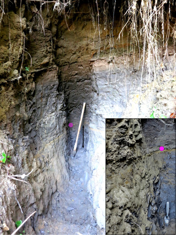
This zone was excavated to increasingly greater distances from surface in an attempt to get below the surface contamination, leading to a pit that reached a length of ~4.5 m and a depth below the surface of ~2.7 m before it collapsed (Figure 1). Strata exposed along the pit walls were jointed, with roots concentrated along these joints, almost to the final depth of pit penetration. Only Dryas leaves from the slump zone were dated, both because they were the most abundant plant species and because it was felt that they were unlikely to be recycled. It was assumed that the apparent radiocarbon ages would increase as the sampled zone increased in depth below the surface but, instead, there was a wide scatter in ages (up to 2000 yr) with no apparent increase in age with depth (Figure 2).
The pit at site 1 along the base of which the plant-bearing stratum (marked by double-headed arrow) was followed. The mattock, for scale, is about 1 m long. Inset, of the lower left section of the pit at an earlier date, shows roots along joints in the lacustrine clay. The magenta circles mark the same sand stratum.

Plot of 14C ages from samples taken from the plant bearing stratum at site 1 showing the scatter but no obvious trend in apparent ages with distance from the surface. The two samples marked with asterisks were holocelloluse extractions and the two horizontal lines mark the two alpha cellulose extractions, using Dryas leaves from several samples.

A major question was whether this age range was due to contamination by young carbon or whether it represented a scatter of real ages. If the latter, this age range would have been during the Mackinaw Interstade and the Port Huron Stade. During that interval the ice front was far to the north of the sample site (e.g. Kozlowski et al. Reference Kozlowski, Bird, Lowell, Smith, Ferenc and Graham2018) and did not subsequently readvance. However, the lacustrine sequence sampled is overlain by till and/or kame moraine (e.g. Muller and Cadwell Reference Muller and Cadwell1986; Karig and Miller Reference Karig and Miller2020), which demands that there was an ice advance after deposition of the lacustrine sequence. This led to the conclusion that the samples analyzed were contaminated with young carbon. This young carbon was suspected to be something associated with tree roots, which were observed along joints in the sediment (Figure 1).
This problem of anomalously young ages was not unique to the West Inlet Creek example. Subsequent coring of channel deposits within a lacustrine sequence beneath a surficial till in a wetland of the Owasco Inlet Valley in Tompkins County (42°32′N, 76°21′13′′W) produced “modern” ages from assorted plant leaves and spruce wood (UCI-218904, 905, 906). A 2.7-m-deep trench through this section showed that the till was jointed, with roots penetrating into the lacustrine clay beneath. Spruce wood from a channel fill in this trench gave an age of 510 ± 30 14C yr (Beta-510219). In an attempt to avoid roots, a hole was cored through the ice farther from trees. A sample of spruce wood from beneath the till there gave an age of 11,967 14C yr (AA-113007), which was still younger than was believed plausible, but indicated less contamination. The correlation of tree roots with anomalously young radiocarbon ages was even clearer in this case. Moreover, a review of our coring records indicated that the association of nearby trees with anomalously young radiocarbon ages occurred at several other sites in the region.
Based on these observations, we hypothesized that fungal contamination of the plant material, particularly by root-associated fungi, contaminated that material with young carbon, causing radiocarbon dating ages younger than expected. Fungal diversity in soil varies with seasons, depth, and soil type (Mahaney and Boyer Reference Mahaney and Boyer1986; Reitner et al. Reference Reitner, Schumann, Pedersen and Gadd2006; Santalahti et al. Reference Santalahti, Sun, Jumpponen, Pennanen and Heinonsalo2016), but in general, saprotrophic (organic matter-decaying) fungi are restricted to the upper layers of soil, and mycorrhizal fungi (mycorrhizae; obligate root symbiotic fungi) dominate deeper layers (Lindahl et al. Reference Lindahl, Ihrmark, Boberg, Trumbore, Högberg, Stenlid and Finlay2007). Mycorrhizae are tightly associated with plant roots and act as extensions of the roots, growing out into bulk soil and collecting mineral nutrients that they give to the plant in exchange for carbon in the form of photosynthesis-derived sugars (Smith and Read Reference Smith and Read2008). Mycorrhizae send out exploratory hyphae built with young carbon into bulk soil in this way (Leake et al. Reference Leake, Donnelly, Saunders, Boddy and Read2001). Although mycorrhizae must grow into or around the surface of plant roots, the exploratory hyphae of some mycorrhizae (especially ectomycorrhizae, the most common type associated with trees) can grow up to several decimeters into the surrounding bulk soil (Agerer Reference Agerer2001). These hyphae search not only for free nutrients or other roots to colonize, but in some species can actively seek and decompose organic matter in order to extract nitrogen and phosphorus from it (Bruns Reference Bruns1995; Smith and Read Reference Smith and Read2008), and when encountering leaves mycorrhizae may grow not only on their surface but penetrate into their tissue (Camenzind and Rillig Reference Camenzind and Rillig2013).
METHODS
All these samples were processed within a few days after collection. After soaking the sediment samples in a solution of sodium hexametaphosphate for a day or more to disaggregate them, they were sieved through a 177-μ screen using de-ionized water and with the usual precautions taken to avoid laboratory contamination. The lighter fraction, containing organic material, was decanted into a Petrie dish from which relevant plant material was picked under a binocular microscope. In some of the samples there were rootlets and fine filamentous aggregates, which were identified as fungal mycelia. These were noted but avoided during the picking. The plant material was then sonicated to remove as much adhering clay and any other extraneous material as possible, re-sieved and re-picked. This cleaned, selected plant material was then dried at 90°C and frozen. This scheme was tested with a sample collected for this study from a site where 41–43 ka 14C ages were earlier obtained (Karig and Miller Reference Karig and Miller2013) and obtained a 42.1 ka14C age (UCI 231804), which confirmed that recent or young carbon was not introduced during processing.
Most of the analyses in this study used the conventional ABA process, but two at the West Inlet Creek site (AA-108845 and Beta-443327) had additional holocellulose extractions (Figure 2). This removed most carbohydrate but resulted in no greater age. For this study, two alphacellulose analyses were undertaken, using Dryas leaves from several samples in the pit, both of which gave ages no older than from the less harsh analyses (Figure 2). By this time the suspected cause of the contamination was fungi having mycorrhizal relationships with tree rootlets. This suspicion was strengthened by the mycelial aggregates and by recent rootlets with associated mycorrhizal fungal threads seen in many samples.
To confirm that these threads were fungal in nature, a selection of Dryas leaves was cleared in 2.5% KOH for more than one week, until the leaves were completely pale in color, after which they were rinsed with and stored in water at 4°C. The cleared and skeletonized leaves that resulted from this process (Figure 3a) were then stained with lactophenol cotton blue, which selectively stains the chitin present in fungal cell walls.
(A) and (B) show hyphae in uncleared, teased apart leaves stained with lactophenol cotton blue. In (B), an arrow points to a hyphal septum. (C) is a skeletonized leaf after clearing. (D) shows a particularly intact hyphae, with septa indicated by arrows. (E) shows what is believed to be the best evidence that the hyphae are not just growing on top of the leaves, but penetrate into their tissue. (F) shows the thin hyphae morphotype, shown by arrows, in between the skeletonized vascular tissue. (G) shows the thick hyphae morphotype, shown with arrows, in between the skeletonized vascular tissue.

The stained Dryas leaves were placed in water on a microscope slide and teased apart with needles at 100× magnification, then covered with a cover slip and observed at 400× magnification. It was clearly evident that almost all of the sampled leaves had hyphae embedded in their tissue or attached to their surfaces, as hyphal threads were evident stretching between the leaf tissue as it was pulled apart (Figure 3A and 3B).
DISCUSSION
Microscopic examination of these leaves showed blue-stained fungal mycelia (Figure 3), which came in two general morphotypes: thinner, nonseptated, hyaline hyphae (Figure 3b), and thicker, septated, darkly pigmented hyphae (Figure 3c). The former is typical of simpler fungi in the phylum Mucoromycota and the latter is typical of higher fungi in Basidiomycota and Ascomycota; mycorrhizal fungi are found in all of these phyla (Strullu-Derrien et al. Reference Strullu-Derrien, Selosse, Kenrick and Martin2018). DNA sequencing on DNA extracted (PrepMan Ultra, Thermo Fisher) and whole genome amplified (Genomiphi V2 DNA Amplification Kit, GE) from the infected leaf tissue with fungus-specific ITS1F and ITS4 barcoding primers (White et al. Reference White, Bruns, Lee and Taylor1990; Gardes et al. Reference Gardes and Bruns1993) was unsuccessful, so taxonomic classification of fungi was not possible.
The observed fungi, presumably symbiotic mycorrhizae associated with the nearby roots but also potentially independent saprotrophic fungi that travelled down the penetration cavity created by the roots, could be a source of contaminating young carbon in the leaves. Mahaney and Boyer (Reference Mahaney and Boyer1986) reported progressively younger ages with increasing number of microbes (fungi and bacteria) present. Kilian et al. (Reference Kilian, Van der Plicht and Van Geel1995) implicated root-associated fungi as introducing older carbon contamination by directly fixing carbon dioxide derived from rising methane from much-deeper bacteria, but this is not likely, since mycorrhizae obtain the vast majority of their carbon from host-derived sugars (Bruns Reference Bruns1995). Still, Hobbie et al. (Reference Hobbie, Ouimette, Schuur, Kierstead, Trappe, Bendiksen and Ohenoja2013) demonstrate with radiocarbon dating that structural carbon in ectomycorrhiza is derived from recent photosynthesis and has recent carbon, but protein carbon can be much older for taxa with extensive exploration depths, since it can be derived from foraged organic material. Saprotrophic fungi might also introduce young carbon into Dryas leaf fossils by assimilating carbon from organic matter near the soil surface into a continuous hyphal network that penetrates the Dryas fossils deeper down. One important consideration is that the fungal hyphae observed could consist of diverse Dryas leaf endophytes that grew within leaf tissues during the life of the plant (Fisher et al. Reference Fisher, Graf, Petrini, Sutton and Wookey1995; Higgins et al. Reference Higgins, Arnold, Miadlikowska, Sarvate and Lutzoni2007; Lindsay et al. Reference Lindahl, Ihrmark, Boberg, Trumbore, Högberg, Stenlid and Finlay2007; Zhang and Yao Reference Zhang and Yao2015), which would not introduce young carbon contamination. However, the observation of fungal mycelia in the soil samples Dryas leaves were taken from seems to indicate that fungal contamination originating from the soil surrounding the Dryas leaves was possible and that at least some proportion of the hyphae observed in the Dryas leaves was not endophytic in nature.
During the preparation of this note, another area on the West Inlet Creek exposure was found that was farther from trees and which had strata with Dryas leaves. A Dryas sample from these strata had a radiocarbon age of 14,270 ± 40 14C years (UCI 231378) and also showed a small degree of hyphal contamination. This age is significantly greater than any obtained in the pit but not as great as the maximum 15,160 14C age obtained elsewhere from this exposure. These results support a correlation between fungal hyphae and sample contamination but suggests a small amount of contamination even in this sample, a problem to be addressed by further sampling.
Although our sample size is very small, there seems to be a correlation between anomalously young radiocarbon ages, trees in the vicinity of the sample site and the presence of fungal hyphae in the plant samples. Whether a sample is contaminated in this way would appear to be a function of sample depth with respect to roots and the nature of the sediment surrounding those roots. Permeable or jointed sediment would be more prone to invasion by roots and hyphae. Pits and exposures disclose the likelihood of this type contamination more readily than do core holes, where jointing is almost never revealed. The simplest method to avoid contamination by mycorrhizal fungi is to avoid sites with trees, especially when using coring methods for sampling.
If there is a possibility or probability of such contamination, either because of the sample site setting or because of questionable radiocarbon ages, testing for hyphae using the clearing and staining techniques described here might be considered. Obtaining useful radiocarbon ages from contaminated samples is presently impossible because the chitin that comprises the hyphae is apparently as or more resistant than even alphacellulose.
A possible solution is to explore methods to extract and date lignin from the sample, as little or no lignin occurs in the hyphae, but this approach presents some significant difficulties. The first stage of preparation of organic samples for radiocarbon dating involves an acid-base-acid (ABA) treatment to remove soil carbon contamination. The base-soluble fraction contains much of the easily extractable lignins from the sample but cannot be used because the soil carbon itself typically includes exogenous lignins and their breakdown products. More tightly bound lignins are extracted by bleaching with acidified sodium chlorite during cellulose preparation, but the spent bleaching solution also contains high concentrations of salt which is extremely corrosive at the high temperatures required for sample combustion to CO2; however, in principle this could be removed using dialysis or Solid Phase Extraction techniques (Khazraie et al. Reference Khazraie, Zhang, Tarasov, Gao, Price, DeMartini, Hupa and Fatehi2017; Arellano et al. Reference Arellano, Bianchi, Hutchings, Shields and Cui2018) prior to drying and combustion. An alternative would be to isolate and date the carbon content of methyl iodide derived from lignin methoxyl groups via treatment of the ABA-treated plant material with hydroiodic acid (Anhåuser et al. Reference Anhåuser, Sirocko, Markus, Esper and Keppler2014). Finally, lignin phenols have been extracted for radiocarbon dating with high performance liquid chromatography (HPLC) (Hou et al. Reference Hou, Huang, Brodsky, Alexandre, McNichol, King, Hu and Shen2010; Feng et al. Reference Feng, Benitez-Nelson, Montlucon, Prahl, McNichol, Xu and Eglinton2013), but this is chemically very challenging (A.P. McNichol, written communication, 2020).