Implications
Sedation and anaesthesia decreased the stress and probably pain responses, and especially sedation improved semen quality in electroejaculated bucks. Sedation is less risky than anaesthesia, has less practical requirements and requests of technicians to monitor animal health status. Therefore, the use of sedation could be considered for routine use when buck semen is collected by electroejaculation (EE).
Introduction
Electroejaculation is a technique that allows collecting semen in different conditions, including males untrained to artificial vagina, or collecting semen during the non-breeding season in small ruminants. However, EE provokes intense muscle contraction, struggling, vocalization and occasional recumbency all which are indicative of discomfort (Palmer, Reference Palmer2005). Also, it triggers increases in serum cortisol and total protein concentrations, glycaemia, rectal temperature (RT), heart and respiratory rates and induces changes in haematological parameters (Stafford et al., Reference Stafford, Spoorenberg, West, Vermunt, Petrie and Lawoko1996; Damián and Ungerfeld, Reference Damián and Ungerfeld2011; Abril-Sanchez et al., Reference Abril-Sanchez, Freitas-de-melo, Damián, Giriboni, Villagrá-García and Ungerfeld2017). This technique also provokes an increase in creatine kinase (CK) blood concentration (Damián and Ungerfeld, Reference Damián and Ungerfeld2011; Abril-Sanchez et al., Reference Abril-Sanchez, Freitas-de-melo, Damián, Giriboni, Villagrá-García and Ungerfeld2017), an indicator of muscle damage (Aktas et al., Reference Aktas, Auguste, Lefebvre, Toutain and Braun1993). Considering that stress is defined as ‘the biological response elicited when an individual perceives a threat to its homoeostasis’ (Moberg, Reference Moberg2000), and that all these physiological, biochemical and haematological changes are linked to a stress response in other situations, as during handling and transport in goat, sheep and pig (Grandin, Reference Grandin1997), EE is a stressful procedure.
Furthermore, EE is also painful in humans (Stafford, Reference Stafford1995). In contexts of possible pain, vocalizations may be reliable pain indicators (Prunier et al., Reference Prunier, Mounier, Le Neindre, Leterrier, Morméde, Paulmier, Prunet, Terlouw and Guatteo2013). In this sense, vocalizations are associated to electrical pulses applied during EE in rams (Damián and Ungerfeld, Reference Damián and Ungerfeld2011) and bulls (Falk et al., Reference Falk, Waldner, Cotter, Gudmundson and Barth2001; Whitlock et al., Reference Whitlock, Coffman, Coetzee and Daniel2012). Thus, EE induces vocalizations in small ruminant males, even under general anaesthesia (pampas deer: Fumagalli et al., Reference Fumagalli, Damián and Ungerfeld2015), EE has been considered as painful by several authors (Bath, Reference Bath1998; Orihuela et al., Reference Orihuela, Aguirre, Hernandez, Flores-Perez and Vazquez2009a and Reference Orihuela, Aguirre, Hernandez, Flores-Perez and Vazquez2009b). However, as it is difficult to effectively determine pain responses in animals, several pain scales have been developed. It includes visual analogue scales (VAS), numerical rating scales (NRS), simple descriptive scales (SDS) or multifactorial pain scales (Anil et al., Reference Anil, Anil and Deen2002). The VAS scale is more widely used and is considered more sensitive than NRS or SDS, because it allows intermediate values instead of only categories (Firth and Haldane, Reference Firth and Haldane1999). There are no specific studies describing the behavioural changes after EE, but it may be expected that the stressful treatment also triggers modifications in normal behaviour (Rushen, Reference Rushen2000).
The administration of anaesthesia is frequently used during EE in wild ruminants (Watson, Reference Watson1976; Beracochea et al., Reference Beracochea, Gil, Sestelo, Garde, Santiago-Moreno, Fumagalli and Ungerfeld2014). However, its application may provoke other problems, as its use has a direct risk of death (Brodbelt et al., Reference Brodbelt, Blissitt, Hammond, Neath, Young, Pfeiffer and Wood2008; Bille et al., Reference Bille, Auvigne, Libermann, Bomassi, Durieux and Rattez2012). Anaesthetised ruminants should be fasted for 12 to 24 h to prevent ruminal tympanism and regurgitation (Taylor, Reference Taylor1991; Carroll and Hartsfield, Reference Carroll and Hartsfield1996). In male ruminants it is frequently observed that untreated males attack those that are recovering from anaesthesia to establish or maintain the social rank relationships (Pelletier et al., Reference Pelletier, Hogg and Festa-Bianchet2004; Jennings, Reference Jennings2007). provoked by EE, and has been shown to increase serum cortisol Therefore, the use of general anaesthesia itself may prevent some negative effects provoked by EE, and has been shown to minimize the increase of serum cortisol concentration (Orihuela et al., Reference Orihuela, Aguirre, Hernandez, Flores-Perez and Vazquez2009a), but at the same time it involves others that may be even more important, as ruminal tympanism and regurgitation (Taylor, Reference Taylor1991; Carroll and Hartsfield, Reference Carroll and Hartsfield1996) or aggressiveness between bucks (Pelletier et al., Reference Pelletier, Hogg and Festa-Bianchet2004; Jennings, Reference Jennings2007). Furthermore, anaesthesia may also affect normal behaviour; for example, appetite decrease in sheep with anaesthesia, and the animals may remain affected during many hours (Provenza et al., Reference Provenza, Lynch and Nolan1994). An alternative to avoid the risks and concerns of anaesthetic treatments may be sedation (Brodbelt et al., Reference Brodbelt, Blissitt, Hammond, Neath, Young, Pfeiffer and Wood2008).
The aim of EE is to collect semen; thus, alternative treatments to decrease the negative responses provoked by EE should not affect semen quality. In this sense, the application of α2-adrenergic agonists stimulates the contractions of the epididymis, accessory sex glands and deferent vas in rams (Knight, Reference Knight1974). Thus, its administration facilitates the sperm ejaculation during EE in anaesthetic protocols in stallions (Mcdonnell and Love, Reference Mcdonnell and Love1991; McDonnell, Reference McDonnell2001), dogs (Dooley et al., Reference Dooley, Pineda, Hopper and Hsu1990) and cats (Dooley et al., Reference Dooley, Pineda, Hopper and Hsu1991).
Considering all this information, we hypothesized that sedation or anaesthesia decrease the negative effects of EE on bucks’ physiology and behaviour, and improve semen quality. However, as the use of sedation does not provoke death risks as anaesthesia, does not require to fast the animals, and does not provoke the problems involved in postanaesthesia recovery, the use of sedation may be preferable than the use of anaesthesia. Therefore, the aim of the present study was to compare the physiological and behavioural responses indicative of stress and possibly pain, and the semen quality in electroejaculated untreated, anaesthetised or sedated goat bucks.
Material and methods
Animals and management
All the procedures were approved by the Comisión de Ética en el Uso de Animales of the Facultad de Veterinaria (Universidad de la República, Uruguay)(CEUAFVET-344). The study was performed in October (spring) with eight healthy Gabon bucks (Capra hircus) (8 to 9 years old, 33.0±1.7 kg (mean±SEM)), initially. The eight animals were kept in an outdoor pen (17×10 m) of the Departamento de Fisiología, Facultad de Veterinaria (35°S, Montevideo, Uruguay). A veterinarian continuously supervised the conditions of the animals. This pen had free ventilation and access to artificial bedding, but did not have special bedding. All animals were fed with alfalfa hay twice/day for ad libitum intake and had free access to water. During the study, the animals were fasted for 24 h before applying each procedure to maintain the application of all methods in the same conditions. Animals were also fasted, but had free access to water during the procedures. Feed was available to the bucks from 3 h after the procedures. Activities such as lying, standing, moving (when the animal walked and/or changed the position), sleeping and/or ruminating were recorded 24 h before the procedures to monitor general health status of the bucks.
Treatments and electroejaculation procedures
Semen was collected from all the bucks using three different procedures (EE alone without pharmacological treatment as untreated bucks, EE under sedation and EE under general anaesthesia). Semen was first collected from three untreated bucks, three anaesthetised bucks, and two sedated bucks, always during the morning (0800 to 1200 h). Procedures were overcrossed in three different periods with an interval of 10 days, so one sample of semen was collected from each animal with each procedure.
The EE was performed with an electroejaculator equipped with a probe that measured 30 cm length and 1.5 cm width, with 1 cm ring electrodes (Fuhijira Industry, Tokyo, Japan) covered with duct tape in the upper side. The probe was coated with carboxymethyl cellulose gel and inserted into the rectum with the covered side positioned in the dorsal region of each buck. All bucks were stimulated with series of 10 pulses, beginning with 3 V, and increasing 1 V in each series until ejaculation. Each pulse was applied during 3 to 5 s, with resting periods of 2 to 3 s. The number of electric pulses applied was recorded.
Anaesthesia was induced with 5.0 mg/kg of 5% ketamine (phs, Pharmaservice; Norepley, Montevideo, Uruguay) plus 0.05 mg/kg of 2% xylazine (Vetcross, Portinco, Montevideo, Uruguay). Sedated animals received 0.5 mg/kg of 0.5% ketamine plus 0.05 mg/kg of 2% xylazine. The pharmacological treatments were administered by jugular vein injection. The EE began when anaesthetic and sedative drugs were acting (bucks showed muscle relaxation and block of reflex activity) (time to induce anaesthesia and sedation: 15.4±1.3 min and 15.9±1.2 min, respectively) (mean±SEM). A visual analogue scale (VAS) of sedation (Wewers and Lowe, Reference Wewers and Lowe1990) was used to confirm sedation in anaesthetised and sedated animals (mean±SEM VAS values: 70.9±9.6 mm and 49.7±9.22 mm, respectively) (see details of the VAS score used in the following subsection).
After semen collection, treatments were reversed by jugular vein injection of 0.01 mg/kg of 1% yohimbine hydrochloride (Reverze; Vetcross, Portinco, Montevideo, Uruguay). The vocalizations emitted by each buck were noted by one trained recorder during EE. Duration of the EE (only EE) and total time of the procedures (EE + anaesthesia or sedation), and the latencies to recover from anaesthesia or sedation (when bucks were completely awake, active, conscious and standing) were also recorded.
General management of the animals during the procedures
Before the procedures, the eight bucks used in the present study were maintained in the outdoor pen (17×10 m). The same operator that fed the animals and cleaned the place every day moved each animal individually to a small pen (2×2 m) separated from the other bucks by a metal grid fence. The animals were used to his handling as they had been kept at the Facultad de Veterinaria for 3 years. Also, the same operator restrained the animals before each procedure (control, anaesthesia and sedation), whereas blood was collected, and HR and RT were recorded (see the ‘Rectal temperature and heart rate’ section). Ketamine and xylacine were applied, and immediately reversed with yohimbine after ejaculation. During EE, the number of vocalizations emitted by each buck was recorded, and all the procedure was video-recorded to determine VAS scores of sedation and pain (see the ‘Visual analogue scale’ section). Immediately after EE, blood was collected again, and HR and RT were recorded again, and the animals were moved to another familiar pen where they were kept every day while the home pen was cleaned.
Bucks that were anaesthetised or sedated remained in the small pens (2×2 m) constructed into this second pen with a metal grid fences until they recovered, and control animals remained in the rest of the pen (6×6 m) altogether, maintaining visual, olfactory and auditory contact between bucks. When the bucks recovered from the anaesthesia or sedation (when they got up, and moved), they were transferred to the area of the pen where the control bucks were kept. In this pen or in the small pens, depending on the time needed for recovery, another familiar operator recorded HR and RT, and collected the blood samples. After these recordings, bucks were moved to another pen (6×6 m), also separated from the last pen by metal grid fences, where they had free water and feed. After the recordings the last buck, all the bucks went back to the initial pen. Behaviour was recorded 24 and 48 h after the procedures while animals remained in this pen.
Visual analogue scale
Animals were video-recorded during the first 30 s of EE, and also during 30 s beginning 30 min after EE. Each video recording was analysed by three blinded trained observers to provide VAS scores. The VAS score was used to assess pain and evaluate sedation. Each VAS score was calculated using a horizontal line of 100 mm where 0 indicated absence of pain and sedation, and 100 mm indicated maximum values for these (Wewers and Lowe, Reference Wewers and Lowe1990).
The pain VAS score considered the number, intensity (high or low pitched bleating) and duration of vocalizations emitted by the bucks, the contractions and movements provoked by the EE, as well as intensity and number of facial contractions, orbital tightening, abnormal ear position and abnormal nostril and philtrum shape (McLennan et al., Reference McLennan, Rebelo, Corke, Holmes, Leach and Constantino-Casas2016). The sedation VAS score considered the activity and body posture of the animals, taking into account if the animals were awake (standing up and moving), asleep, lying down, alert or standing. The values given by the three observers for all VAS scores were highly correlated (all VAS scores: Spearman’s ρ>0.8, P<0.0001), so the mean value was used for the statistical analysis.
Rectal temperature and heart rate
The RT was measured by a digital thermometer and HR was recorded counting beats during 30 s by auscultation (beats/min). Both parameters were measured before the procedures (before the administration of pharmacological treatments or immediately before EE in control bucks), and 10 and 30 min after the end of EE.
Blood samples
Blood samples were collected from all animals by jugular venepuncture before the procedures, immediately after EE and 10, 20, 30, 45, 60, 90, 120, 180 min after the end of EE, as well as 24 and 48 h later. Blood was collected with 5 ml syringes while operators restrained the animal (max duration 10 s). Then, samples were placed in tubes without anticoagulant and allowed to clot for 20 min at room temperature, and centrifuged for 20 min at 1500×g. Serum was separated and frozen at –20°C. The samples collected before the procedures, and until 120 min after EE were used to quantify serum cortisol concentration. Samples collected before the procedures, immediately after EE, and 30, 120 min, 24 and 48 h after the end of EE were used to determine serum aspartate aminotransferase (AST), CK, total protein, albumin and globulin concentrations.
Samples for haematological analysis were collected in tubes with EDTA (Eurotubo; Deltalab, Barcelona, Spain) before the procedures, and 30 and 180 min, as well as 24 and 48 h after EE. These samples were maintained at 4°C during 5-6 h until their subsequent analyses.
Cortisol concentration
Serum cortisol concentration was measured at the Laboratorio de Endocrinología y Metabolismo Animal, Facultad de Veterinaria, Montevideo, Uruguay, using a solid-phase radioimmunoanalysis kit (Cort-Ct2; Cisbio Bioassays, Parc Marcel Boiteux, France). The analytical sensitivity of the assay was 15.0 nmol/l; the intra-assay CV was 5.9%.
Blood biochemistry
The AST, CK, total protein and albumin serum concentrations were determined by colorimetry using commercial kits (Wiener Lab, Rosario, Argentina) at the Laboratorio de Análisis Clínicos, Facultad de Veterinaria, Montevideo, Uruguay. Serum globulin concentration was calculated by subtracting the serum albumin concentration from the serum total protein concentration.
Blood haematology
White blood cell (WBC) number, haemoglobin (Hgb) and leukocyte indices (relative concentration of lymphocytes (RLym), absolute concentration of lymphocytes (ALym), relative concentration of granulocytes (RGr) and absolute concentration of granulocytes (AGr)) were measured using an automated cell analyser (HumanCount 30/60TS; Human, Wiesbaden, Germany). Haematocrit (Hct) was measured manually with a microcentrifuge (Microhemato Centrifuge 2410 Model; Fanem, São Paulo, Brazil).
Behavioural recordings
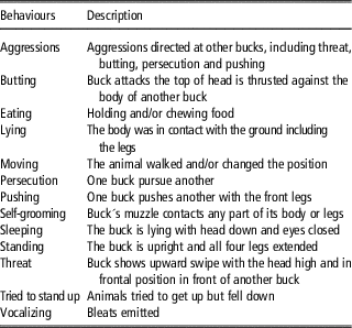
Descriptions of the behaviours recorded are included in Table 1. One blinded trained observer recorded different behaviours in each buck without disturbing them and after EE by use of scan. A period of 1 day before applying EE, beginning at 0800 h, scan recordings of each buck were performed every 10 min during 4 h (totalling 25 times). In each scan observation it was recorded if the buck was observed lying, standing, moving, sleeping and/or ruminating.
Ethogram of behaviour patterns evaluated in goat bucks before and after electroejaculation

Focal continuous recordings were performed during 10 min beginning 15 min, 3 and 24 h after the end of EE. During the focal continuous sampling performed 15 min after the end of EE, the number of times that the animal tried to stand up, moved and vocalised, as well as the frequency and duration of lying were recorded. During the focal continuous recording performed 3 h after the end of EE, the frequency, the number of vocalizations emitted, and the frequency and duration of lying were recorded. During the focal continuous sampling performed 24 h after the end of EE, the frequency of self-grooming, aggressive interactions and moving as well as the duration of eating, ruminating, lying, standing and sleeping were recorded.
Semen evaluation
The ejaculated volume was determined, and sperm concentration, sperm mass motility (scale 0 to 5), sperm vigour (0 to 5), and the percentage of motile and progressive motile spermatozoa were determined under phase-contrast microscopy as previously described Evans and Maxwell (Reference Evans and Maxwell1987). The percentage of viable sperm was determined by eosin/nigrosin staining (Campbell et al., Reference Campbell, Dott and Glover1956), and the percentage of spermatozoa with functional plasma membrane was determined with the hypo-osmotic swelling test as previously described Jeyendran et al. (Reference Jeyendran, Van der Ven, Perez-Pelaez, Crabo and Zaneveld1984). The percentage of sperm with acrosome damage (Pursel and Johnson, Reference Pursel and Johnson1974) and morphologically normal were counted in samples fixed in buffered 2% glutaraldehyde solution at 37°C, using phase-contrast microscopy. A total of 200 spermatozoa were studied in each sample for each determination. The total number of ejaculated sperm, motile and progressive motile sperm, live and normal sperm, and sperm with intact acrosome and with functional plasma membrane were calculated.
Statistical analysis
The statistical analysis was performed using the free statistical software, Statistical Analysis System (SAS) University Edition (SAS, 2016). The Shapiro–Wilk test was used to test the normal distribution of the residuals of each variable. All the recorded behaviours, the VAS score values, sperm concentration, sperm vigour, the total number of spermatozoa ejaculated and the total number of spermatozoa with acrosome damage ejaculated were not normally distributed, and thus, compared by the Friedman’s exact test. The RT, HR, cortisol concentration, all the blood biochemical and haematological parameters were compared with the mixed model of SAS. The model included the procedure (control, sedation or anaesthesia) and the time in relation to EE, as well as their interaction as main effects. The order in which each animal received each treatment (period) was included as a random effect into each treatment. A first order autoregressive covariance structure (AR1) was used to adjust the difference on data according to the differences over time. The total time required by each procedure (anaesthesia or sedation), the number of electric pulses applied, and the number of vocalizations emitted by each buck during the EE as well as the remaining sperm parameters were compared by ANOVA. The model included the procedures as main effect, and the period and the animal as random effects. Data analysed with the mixed model are presented as least square mean±SEM; other data are presented as mean±SEM. Differences were considered statistically significant if P⩽0.05 and a tendency when P>0.05 and ⩽0.1.
Results
Electroejaculation procedures
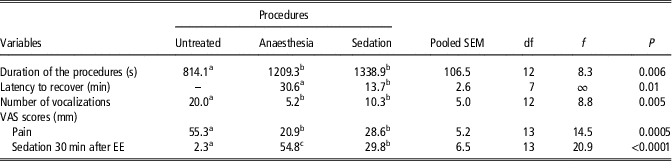
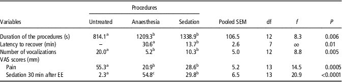
The procedure, considering the whole treatment (from the administration of the drugs until the end of the EE), was shorter when bucks were untreated than anaesthetised (P=0.02; df=12; t=−3.0) or sedated (P=0.002; df=12; t=−3.9), without differences between these (Table 2). However, there were no differences in the time needed for EE itself between the three treatments (general mean: 106±21 s). The latency needed by animals to recover was greater with anaesthesia than with sedation (P=0.01; df=7; f=infinite; Table 2).
Duration of the procedures, latency to recover, number of vocalizations and visual analogue scale (VAS) scores (data are least square means) recorded in eight electroejaculated bucks untreated (no pharmacological treatment), anaesthetised or sedated
EE=electroejaculation.
a,b,c Values within a row with different superscripts differ significantly between procedures (untreated, anaesthesia and sedation) at P<0.05.
Bucks vocalised more during the EE when untreated than when sedated (P=0.02; df=12; t=2.7) or anaesthetised (P=0.001; df=12; t=4.9; Table 2), without differences between these. The total number of electrical pulses applied was not different between the procedures (general mean: 33.7±6.0).
Visual analogue scale
The pain VAS score was greater when bucks were untreated than sedated (P=0.0002; df=13; t=5.1) or anaesthetised (P=0.002; df=13; t=4.0), without differences between these (Table 2). The sedation VAS score recorded 30 min after EE was greater when bucks were anaesthetised than sedated (P=0.009; df=13; t=3.1) or untreated (P<0.0001; df=13; t=−6.5), and was greater when bucks were sedated than untreated (P=0.005; t=−3.4) (Table 2).
Rectal temperature and heart rate
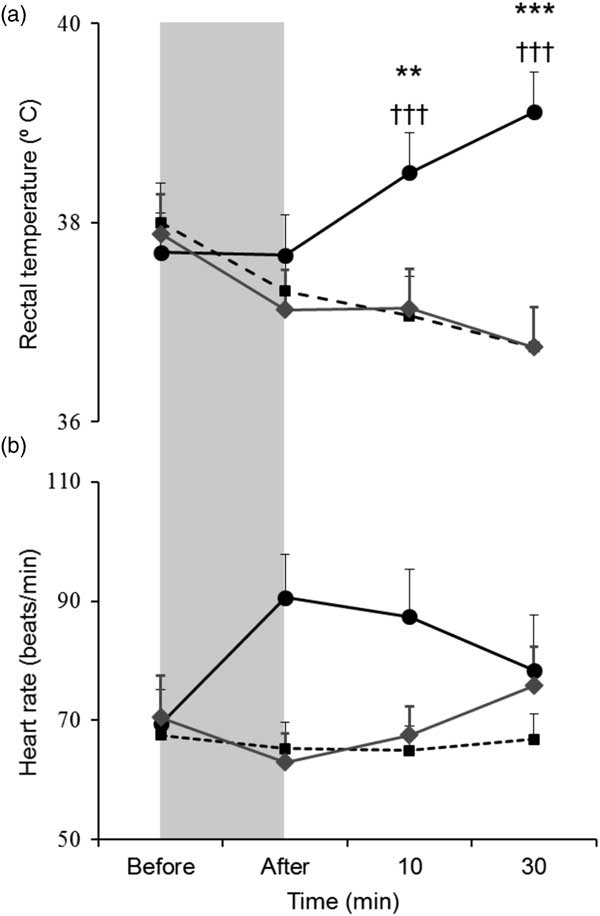
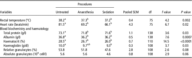
The RT was greater when bucks were untreated than anaesthetised (P=0.003; df=75; t=3.1) or sedated (P=0.002; df=75; t=3.3; Figure 1a), without differences between these (Table 3). There was an interaction between time and procedure (P<0.0001; df=75; f=20.8; Figure 1a): while RT increased 10 min (P=0.0003; df=75; t=3.7) and again 30 min (P<0.0001; df=75; t=6.3) after EE in control bucks without treatment, it did not change when bucks were anaesthetised or sedated.
(a) Rectal temperature and (b) heart rate before and after electroejaculation in untreated (–●–), anaesthetised (‐‐■‐‐) or sedated (–♦–) (least square means±SEM). Shaded area shows the period during which bucks were electroejaculated. Different symbols indicate differences between untreated and anaesthesia procedures (**P<0.001; ***P<0.0001), and between untreated and sedation procedures (†††P<0.0001) in those points.

Rectal temperature, heart rate, blood biochemistry and haematology (data are least square means) in eight electroejaculated bucks under anaesthesia, sedation or acting as untreated controls

a,b Values within a row with different superscripts differ significantly between procedures (untreated, anaesthesia and sedation) at P<0.05.
The HR was greater when bucks were untreated than sedated (P=0.03; df=75; t=2.2) or anaesthetised (P=0.008; t=2.7; Figure 1b), without differences between these (Table 3). There was no interaction between time and procedure.
Cortisol concentration
Cortisol concentration did not differ between the procedures, and there was no interaction between time and procedure. However, in general, cortisol changed over time (P=0.0006; df=200; f=3.5; Figure 2): it increased during EE (P=0.0003; df=200; t=3.7), and returned to initial values 60 min after EE (P=0.01; df=200; t=2.6).
Cortisol concentration before and after electroejaculation in untreated (–●–), anaesthetised (‐‐■‐‐) or sedated (–♦–) (least square means±SEM). Shaded area shows the period during which bucks were electroejaculated. a,b,c,d,eMean significant differences over time (P=0.0006).

Blood biochemistry
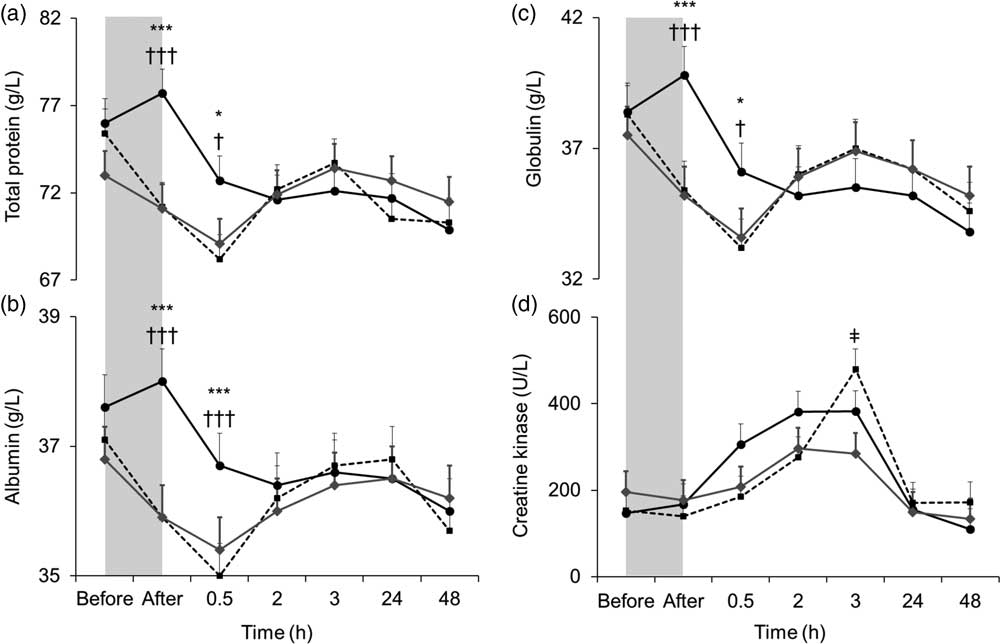
Total protein and albumin concentrations were greater when bucks were untreated than sedated (P=0.03; df=138; t=2.2 and P=0.001; df=138; t=3.3, respectively) or anaesthetised (P=0.02; df=138; t=2.5 and P=0.0008; df=138; t=3.4, respectively), without differences between these procedures (Table 3). Globulin concentration did not differ between procedures. There was an interaction between time and procedure in total protein (P=0.0003; df=138; f=3.4; Figure 3a), albumin (P=0.001; df=138; f=2.9; Figure 3b) and globulin (P=0.0002; df=138; f=3.5;Figure 3c) concentrations: all increased immediately after EE when bucks were untreated, but decreased when they were sedated or anaesthetised ( total protein: P<0.0001; df=138; t=4.8, albumin: P<0.0001; df=138; t=5.3, globulin: P<0.0001; df=138; t=5.0). A period of 30 min after EE, total protein, albumin and globulin were greater when bucks were untreated than sedated or anaesthetised (total protein: P=0.001; df=138; t=3.0, albumin: P<0.0001; df=138; t=4.2, globulin: P=0.004; df=138; t=3.0).
(a) Total protein, (b) albumin and (c) globulin and (d) creatine kinase before and after electroejaculation in untreated (–●–), anaesthetised (‐‐■‐‐) or sedated (–♦–) (least square means±SEM). Shaded area shows the period during which bucks were electroejaculated. Different symbols indicate differences between untreated and anaesthesia procedures (*P<0.05; ***P<0.0001), untreated and sedation procedures (†P<0.05; †††P<0.0001), and between anaesthesia and sedation procedures in those point (‡: P=0.02).

The CK concentration was not different between the procedures, but there was an interaction between time and procedure (P=0.02 ; df=137; f=2.1; Figure 3d): 3 h after EE CK concentrations were greater when bucks were anaesthetised than sedated (P=0.001; df=137; t=3.3), without differences with untreated bucks.
The AST concentrations did no differ between procedures, and there was not interaction between time and procedure (general mean: 83.0±5.6 U/l). In general, AST changed over time (P<0.0001; df=138; f=9.9): decreased after EE (from 83.2±5.9 U/l to 79.9±5.9 U/l; P=0.03; df=138; t=2.1), and increased 3 h (84.5±5.9 U/l; P=0.02; df=138; t=−2.4) and 24 h (88.6±5.9 U/l; P=0.0003; df=138; t=−3.7) later retuning to initial values after 48 h (85.1±5.9 U/l; P=0.002; df=138; t=3.2).
Blood haematology
Haematocrit was greater when bucks were untreated than anaesthetised (P=0.0002; df=110; t=3.9) or sedated (P<0.0001; df=110; t=5.2), without differences between the two (Table 3). There was an interaction between time and procedure (P<0.0001; df=110; f=8.0; Figure 4a): Hct increased immediately after EE when bucks were untreated, but decreased when they were anaesthetised or sedated (both: P<0.0001; df=110; t=6.3); Hct was also greater 0.5 h after EE when bucks were untreated as compared with anaesthetised or sedated (both: P<0.0001; df=110; t=5.6). In addition, Hct was greater when bucks were anaesthetised than sedated 3 h after EE (P=0.01; df=110; t=2.5).
(a) Haematocrit, (b) haemoglobin, and concentration of (c) white blood cells (WBC) and (b) absolute granulocytes (AGr) before and after electroejaculation in untreated (–●–), anaesthetised (‐‐■‐‐) or sedated (–♦–) (least square means±SEM). Shaded area shows the period during which bucks were electroejaculated. Different symbols indicate differences between untreated and anaesthesia procedures (*P<0.05; **P<0.001; ***P<0.0001), untreated and sedation procedures (†P<0.05; ††P<0.001; †††P<0.0001) and anaesthesia and sedation procedures (‡P<0.05; ‡‡‡P<0.0001) in those points. Different letters mean significant differences over time (P<0.0001).

Haemoglobin concentration was greater when bucks were untreated than sedated (P=0.007; df=108; t=2.7), without differences with anaesthetised (Table 3). There was an interaction between time and procedure (P<0.0001; df=108; f=4.8; Figure 4b): Hgb increased immediately after EE in untreated bucks, but decreased when bucks were anaesthetised or sedated (both: P<0.0001; df=108; t=4.2); Hgb was also greater 0.5 h after EE when bucks were untreated than anaesthetised (P=0.002; df=108; t=3.1) or sedated (P=0.0002; df=108; t=3.9). However, 3 h after EE Hgb was greater when bucks were anaesthetised (P=0.001; df=108; t=−3.3) or sedated (P=0.03; df=108; t=−2.2) than when they were untreated.
The WBC did not differ between the procedures, but there was an interaction between time and procedure (P=0.004; df=112; f=2.8; Figure 4c): 0.5 h after the end of EE, WBC was greater when bucks were untreated than sedated (P=0.008; df=112; t=2.7). A period of 24 h after EE it was also greater in anaesthetised than sedated (P=0.01; df=112; t=2.5) and controls bucks (P=0.002; df=112; t=−3.1), without differences between sedated and untreated bucks.
There was a tendency for differences between procedures in AGr (Table 3). There was an interaction between time and procedure (P=0.007; df=108; t=2.6; Figure 4d): 24 h after EE AGr was greater when bucks were anaesthetised than sedated (P<0.0001; df=108; t=4.2) and untreated (P=0.0002; df=108; f=−3.9) without differences between sedated and untreated bucks.
There was a tendency for differences between procedures in RGr (Table 3). There was no interaction between time and procedure. However, in general there were changes over time (P=0.002; df=108; f=4.0), RGr increased 3 h after EE (56.7±4.4%; P=0.006; df=108; t=−2.8), and returned to initial value 48 h later (47.3±4.4%; P=0.003; df=108; t=3.0).
The RLym and ALym did no differ between procedures (general means: RLym: 35.9±3.7%; ALym: 3.3±0.5×10⁹cells/l), and there was not interaction between time and procedure. However, RLym and ALym changed over time (P<0.0001; df=108; f=7.3 and P=0.003; df=108; f=3.9, respectively). The RLym decreased 3 h after EE (31.1±4.5%; P<0.0001; df=108; t=4.6), and returned to initial value 48 h later (36.9±4.5%; P=0.04; df=108; t=−2.1). The ALym decreased 0.5 h after EE (3.1±0.6 109 cells/l; P=0.004; df=108; t=−3.0).
Behavioural recordings
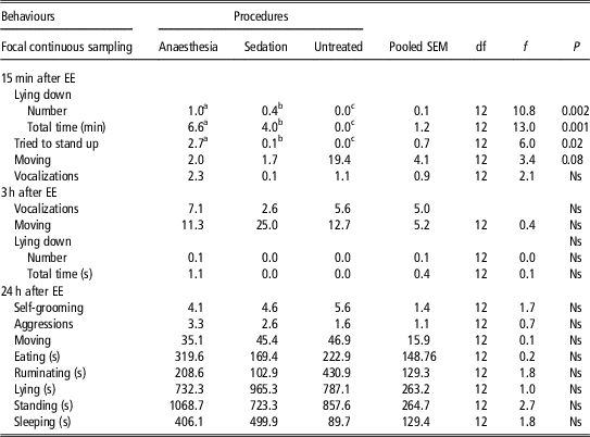
During the scan sampling performed 24 h before the EE there were no differences between the animals that later received different treatments in lying, standing, moving, sleeping or ruminating (data not shown). During the focal sampling performed 15 min after EE, the frequency (P=0.002; df=12; f=10.8) and duration (P=0.001; df=12; f=13.0) of lying were greater when bucks were anaesthetised than sedated (P=0.04; df=12; t=2.3 and P=0.01; df=12; t=2.9, respectively) or untreated (P=0.0006; df=12; t=−4.7 and P=0.0003; df=12; t=−5.1, respectively, Table 4). The frequency and length during which the bucks remained lying down were also greater when bucks were sedated than untreated (P=0.4; df=12; t=−2.3 and P=0.5; df=12; t=−2.1, respectively, Table 4).
Behaviours (least square means) observed by focal continuous recording after electroejaculation (EE) in bucks that were untreated, anaesthetised or sedated

EE=electroejaculation; Ns=no significance;
a,b,c Values within a row with different superscripts differ significantly between procedures (untreated, anaesthesia and sedation) at P<0.05.
In the same period, the number of times the bucks tried to stand up was greater when bucks were anaesthetised than sedated (P=0.02; df=12; t=2.6) or untreated (P=0.007; df=12; t=−3.3), without differences between sedated and untreated (Table 4). There was a tendency for differences between procedures in the frequency of moving (P=0.078; df=12; f=3.4; Table 4). The other recorded behaviours did not differ between procedures (Table 4).
Semen
The sperm mass motility was greater when bucks were anaesthetised (P=0.048; df=11; t=−2.2) or sedated (P=0.02; df=11; t=−2.6) than untreated, without differences between these procedures (Table 5). There were more spermatozoa with functional plasma membrane and morphologically normal spermatozoa when bucks were sedated than untreated (P=0.01; df=11; t=−3.1 and P=0.02; df=10; t=−2.8, respectively), anaesthetised bucks obtained intermediate values (Table 5). There were statistical tendencies for differences in the total number of motile spermatozoa (P=0.07; df=11; t=3.5), progressive motility (P=0.07; df=11; t=3.3) and live spermatozoa (P=0.07) (Table 5). No other sperm parameters were affected by the procedures (general means±SEM: ejaculated volume: 0.5±0.1 ml; sperm concentration: 1793.7±423.2×10⁶ sperm/ml; vigour: 3.3±0.3; motile sperm: 65.7±5.7%; progressive motile sperm: 60.8±6.2%; viable sperm: 74.8±4.1%; sperm with functional membrane: 53.3±8.2%; sperm with acrosome damage: 16.8±5%; morphologically normal sperm: 71.1±5.6%).
Parameters of sperm collected by electroejaculation from goat bucks untreated or under anaesthesia or sedation

Data are presented as least square means.
a,b Values within a row with different superscripts differ significantly between procedures (untreated, anaesthesia and sedation) at P<0.05.
Discussion
Overall, the use of sedation or anaesthesia decreased the stress responses to EE and improved the quality of the collected semen in goat bucks. In effect, when sedation or anaesthesia was applied, there were less changes in HR, which agrees with observations from Orihuela et al. (Reference Orihuela, Aguirre, Hernandez, Flores-Perez and Vazquez2009a), RT, Hct, total protein and albumin blood concentrations than when bucks did not receive pharmacological treatment. Moreover, advantages of sedation or anaesthesia were not only observed in physiological parameters, as bucks also vocalised less, and presented lower pain and vocalizations VAS scores than when they remained as control. In this study, a wide variety of physiological measurements were used; and even though not all of them were affected to the same degree, considering the responses as a whole, the application of sedation or anaesthesia decreased the negative responses as compared with untreated controls, suggesting these may be considered for routine application. It is however important to study more deeply other welfare indicators to achieve definitive conclusions.
In addition, the present study showed the use of sedation had several advantages compared with than the use of anaesthesia for goat bucks. These advantages were related to the doses and not to the drugs itself, as the same drugs were used in anaesthetic and sedation treatments. The smaller changes in Hgb and the tendencies for a lower variation in AGr and RGr when the bucks were sedated than when anaesthetised indicated that they were less affected by EE when sedated. The bucks recovered faster from sedation than from anaesthesia, lied down for a shorter time, and obtained more normal scores in the sedation VAS. It should also be considered that the used of sedation does not generate risk of death as can be observed after anaesthesia (Brodbelt et al., Reference Brodbelt, Blissitt, Hammond, Neath, Young, Pfeiffer and Wood2008). In addition, the bucks remain awake and moving while sedated, and hence, the risk of being attacked from other males is probably lower (Pelletier et al., Reference Pelletier, Hogg and Festa-Bianchet2004; Jennings, Reference Jennings2007). The recovery period was longer after anaesthesia than sedation, thus anaesthetised bucks need to be isolated, which may imply practical problems as more space is needed and more time from need to be allocated to monitor the health status. In addition, as goats are gregarious, isolation itself increases the stress response (Price and Thos, Reference Price and Thos1980; Al-Qarawi and Ali, Reference Al-Qarawi and Ali2005; Patt et al., Reference Patt, Gygax, Wechsler, Hillmann, Palme and Keil2013). Furthermore, in ruminants fasting is required under general anaesthesia to avoid tympanism and regurgitation (Seddighi and Doherty, Reference Seddighi and Doherty2016), but this can be avoided using light sedation. These risks and problems may be even greater when bucks are frequently electroejaculated, as may happen in semen production. Hence, while sedation may imply implies lower death risks, less stress, fewer practical problems, anaesthesia itself raises several constraints for its practical use associated to with EE, and the use of sedation should be recommended instead of general anaesthesia.
General anaesthesia decreased the stress and probably pain responses to EE, but triggered other negative responses, as the increase in CK and Hct that were observed 3 h after EE, and WBC and AGr observed 24 h later. The greater CK concentrations observed when bucks were anaesthetised may be a consequence of increased physical activity, including knockings, as the bucks may get up and fall down repeatedly during the recovery period (not quantified). It should also be considered that, although it was not recorded, there were aggressions from other bucks after reintroducing them their home pen. However, the muscle damage may not be so important as there were no differences between treatments in AST, an indicator of strong muscle damage (Smith et al., Reference Smith, Fry, Allen and Costa1994). The observed larger Hct 3 h after EE as well as WBC and AGr observed 24 h later when bucks were anaesthetised may have been triggered by the administration of larger doses of ketamine needed to anaesthetise the animals in agreement with previous reports in small ruminants (Ismail et al., Reference Ismail, Jawasreh and Al-Majali2010).
The increase in cortisol concentration was similar for all the procedures, as previously observed by Falk et al. (Reference Falk, Waldner, Cotter, Gudmundson and Barth2001), who did not observe differences in blood cortisol concentration after EE between anaesthetized and non-anaesthetized bulls. In contrast, Orihuela et al. (Reference Orihuela, Aguirre, Hernandez, Flores-Perez and Vazquez2009a) showed that anaesthesia decreased the blood cortisol concentrations after EE in rams that when electroejaculated for the first time. This difference may be explained by a lower response when animals used are habituated to EE, as happened in our study and in the study by Falk et al. (Reference Falk, Waldner, Cotter, Gudmundson and Barth2001). The administration of anaesthetic and sedative drugs itself, including the immobilization of the animal, general animal management and intravenous injection of drugs as well as the effect of these drugs while the animals were still conscious is probably stressful. As cortisol increases quickly (Damián and Ungerfeld, Reference Damián and Ungerfeld2011), all these effects may have masked possible differences induced by the sedation and/or anaesthesia.
In our study, the administration of anaesthetic and sedative drugs improved the semen quality. This may be a consequence of the muscle contractions induced by α2-adrenergic agonists in the epididymis, accessory sex glands and deferent vas (Knight, Reference Knight1974). Although the total number of sperm with functional plasma membrane and being morphologically normal were greater when the bucks were sedated than in the control treatment, this improvement was not observed when semen was collected under general anaesthesia. This agrees with previous observations in cats, a species in which it was reported that ketamine negatively affects sperm quality (Zambelli et al., Reference Zambelli, Cunto, Prati and Merlo2007). It has been shown that ketamine also interfere with the ejaculatory process in the chinchilla (Busso et al., Reference Busso, Ponzio, Chiaraviglio, Fiol De Cuneo and Ruiz2005). Hence, sedation seems to be useful to improve semen quality. This may be extremely important to ensure the application of this treatment in animals in which semen is frequently collected.
In conclusion, general anaesthesia or sedation decreased the stress and probably the pain response provoked by EE and improved the quality of the semen collected, being even more effective to sedate the bucks instead of anaesthetising them. Sedated bucks recovered earlier, remained less time lying and had more normal results in the sedation VAS without negative effects in any response measured.
Acknowledgements
The authors acknowledge Alice Fernández, Milton Pintos, Jesus Heras, Julio César Vázquez and Dermidio Hernández for their help during animal management and recording of data during experimental procedures. This study was supported by CSIC (Universidad de la República, Montevideo, Uruguay). S. A.-S. was the beneficiary of a research fellowship for doctoral students from Erasmus Mundus Cruz del Sur. A. T. was supported by a sabbatical grant from PASPA-DGAPA-UNAM, México.
Declaration of interest
The authors declare that there are no conflicts of interest.
Ethics statement
All procedures were approved by the comisión de Ética en el Uso de animales of the Facultad de Veterinaria (Universidad de la República, Uruguay CEUAFVET-344).
Software and data repository resources
Data are available in http://www.fvet.edu.uy/index.php/institutosfisiologia/2016-07-08-10-30-58/2016-07-08-10-32-12/fisiologia/investigacion.