Introduction
Somatic cell nuclear transfer (SCNT) in mini-pig production could facilitate various biomedical applications, such as xenotransplantation and the study of human diseases, because there are many similarities between the genomic, metabolic and cardiovascular systems of pigs and humans (Vodicka et al., Reference Vodicka, Smetana, Dvorankova, Emerick, Xu, Ourednik, Ourednik and Motlik2005; Yamanaka et al., Reference Yamanaka, Sugimura, Wakai, Shoji, Kobayashi, Sasada and Sato2007). Although many mammalian species have been cloned, the success rate of nuclear transfer is very low, especially in pigs (Im et al., Reference Im, Lai, Liu, Hao, Wax, Bonk and Prather2004) and, to our knowledge, there are only a few reports of mini-pig somatic cell nuclear transfer (Hoshino et al., Reference Hoshino, Uchida, Shimatsu, Miyake, Nagao, Minami, Yamada and Imai2005; Miyoshi et al., Reference Miyoshi, Yuki and Yoshida2005, Reference Miyoshi, Sato and Yoshida2006; Yamanaka et al., Reference Yamanaka, Sugimura, Wakai, Shoji, Kobayashi, Sasada and Sato2007).
Animal cloning by SCNT depends on several key factors: donor cell isolation and culture, oocyte collection, maturation and activation and embryo culture and transfer. The origin and extent of differentiation of donor cells can markedly influence the efficiency of nuclear transfer and the development of reconstructed embryos. It is generally accepted that relatively higher efficiencies of somatic cell cloning can be achieved using donor cells from female reproductive systems (e.g. cumulus/granulosa, oviduct and mammary gland cells), but there is little information on the use of male-specific somatic cells as donor cells (Ogura et al., Reference Ogura, Inoue and Ogonuki2000).
Phytohaemagglutinin (PHA) an N-acetylgalactosamine/galactose sugar-specific substance that has been widely used in cell biology for enhancing cell agglutination and fusion in plant protoplasts and in various types of mammalian cells, including erythrocytes, hybrid cell colonies and bone marrow mesenchymal stem cells (Du et al., Reference Du, Shen, Xu, Sung, Jeong, Lucky Nedambale, Riesen, Tian, Cheng, Lee and Yang2006), has been shown to improve embryo development when added to culture systems. Its effects on cultures of porcine parthenogenetic and cloned embryos have been investigated by various groups (e.g. Gupta et al., Reference Gupta, Uhm, Han and Lee2007a, Reference Gupta, Uhm and Leeb).
The Guangxi Bama mini-pig is a mini-pig (average adult weight: male about 60 kg, female about 50 kg) domesticated only in Guangxi. In the present paper, a modified nuclear transfer technique is described for the production of cloned embryos avoiding the use of bisbenzimide (Hoechst 33342) and exposure to ultraviolet (UV) light. The procedure involves isolation and culture of fibroblasts from the testicle of a newborn mini-pig; it produces more efficient reprogramming in the recipient cytoplast and a higher rate of blastocyst development than ear fibroblasts taken from the same newborn animal.
Materials and Methods
Unless stated otherwise, all chemicals used in this study were obtained from the Sigma Chemical Company.
Ovum recovery and in vitro maturation (IVM) of porcine oocytes
Porcine ovaries were collected from a local slaughterhouse and transported to the laboratory at 30–37°C in 0.9% (w/v) saline (0.85% NaCl, 0.02% KCl, 0.02% CaCl2·2H2O, 0.01% MgCl2·6H2O) supplemented with 75μg/ml potassium penicillin G and 50μg/ml streptomycin sulfate. Cumulus–oocyte complexes (COCs) were obtained from follicles with a diameter of 2 to 6 mm, using a 12-gauge needle connected to a 10 ml disposable syringe. The medium used for oocyte maturation was TCM-199 (Gibco) supplemented with 10% (v/v) porcine follicular fluid, 0.57 mM cysteine, 3.05 mM d-glucose, 0.91 mM Na pyruvate, 75μg/ml penicillin, 50μg/ml streptomycin, 10 ng/ml epidermal growth factor (E-4127), 10μg/ml FSH (F-2293) and 10μg/ml LH (L-5269). The COCs were washed three times with Tyrode's lactate (TL)–HEPES–PVA medium. Approximately 50–70 COCs were cultured in 400μl of maturation medium droplets at 39°C, 5% CO2 in humidified air for 42–44 h.
Cell culture and preparation of donor cells
Fibroblasts were derived from the testicle and ear of a newborn mini-pig. Testicles were cut into pieces and digested with 0.25% trypsin–EDTA (0.05%) for 12 h at 4°C and in 0.25% collagenase for 30 min. The digested cells and tissues were cultured in DMEM (Gibco, Life Technologies), supplemented with 10% fetal bovine serum (FBS; HyClone Laboratories Inc.) at 39°C, using a gas phase of 5% CO2 in air. Cells were passaged when they attained 70–80% confluence. Tissues cut from the ear were cultured directly and passaged when they grew to confluence. Testicular fibroblasts were synchronized by serum starvation in DMEM supplemented with 0.5% FBS for 1–3 days, or by contact inhibition for 2–4 days before nuclear transfer.
Cell type identification by indirect immunofluorescence
Unless otherwise stated, all steps were performed at room temperature. For antibody staining, testicular fibroblasts were cultured on sterile glass cover slips in 3.5 mm culture dishes. After 3–4 days of culture, cells were washed three times in DPBS (Gibco, Life Technologies) and then fixed with methanol and acetone (1:1) in DPBS for 15–20 min. Cells were then left in the blocking solution (1% BSA in DPBS) for 30 min before being washed three times in PBS. Fibroblasts were incubated overnight at 4°C with a mouse monoclonal antibody to vimentin (V9) or cytokeratin 1/10 (LH1) (Santa Cruz Biotechnology), both diluted 1:200. After washing with DPBS, samples were stained for 30 min with a fluorescein (FITC)-conjugated goat anti-rabbit antibody (Santa Cruz Biotechnology) in a 1:200 dilution. Finally, after washing with DPBS, samples were stained for 10 min in PBS containing 10μg/ml of PI to reveal DNA and observed under a fluorescence microscope (Nikon). For the detection of FSHR, goat polyclonal antibody FSHR (N-20) and donkey anti-goat IgG–FITC (Santa Cruz Biotechnology) were employed in a 1:200 dilution. Appropriate controls for autofluorescence and non-specific binding by the secondary antibody were included (Maguire et al., Reference Maguire, Tribley and Griswold1997; Enright et al., Reference Enright, Kubota, Yang and Tian2003, Reference Enright, Sung, Chang, Yang and Tian2005; Yang et al., Reference Yang, Yang, Beaujean, Niu, He, Xie, Tang, Wang, Zhou and Ji2007).
Nuclear transfer
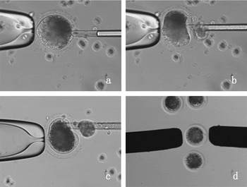
After culturing oocytes for 42–44 h, cumulus cells were removed from COCs using 0.2% (w/v) hyaluronidase in TL–HEPES–PVA medium. Oocytes were then incubated in TL–HEPES–PVA medium with 10% (v/v) FBS and 7.5 μg/ml of cytochalasin B for 15 min. Oocyte enucleation was performed using a modified two-step blind-aspirating squeezing method as shown in Fig. 1. Briefly, the first polar body and the metaphase II spindle plate with a small amount of surrounding cytoplasm were aspirated with a glass micropipette of 20–25μm outer diameter (Sutter); the zona pellucida was squeezed around the cut to express a small volume of cytoplasm and to help in ensuring the success of the enucleation process. For donor cell injection, the micropipette was introduced into the zona pellucida through the same slit made during enucleation; to facilitate close contact of membranes for subsequent fusion, the fibroblast was wedged between the zona pellucida and the cytoplast membrane (Fig. 1) (Li et al., Reference Li, Chen, Lian, Han, Zhu and Seidel2002). After manipulation, couplets were washed once and cultured in NCSU-23 with 0.4% BSA at 39°C for at least 0.5 h before fusion, using a gas phase of 5% CO2 in humidified air.
Blind-enucleation of the oocyte (a) (×200); squeezing the zona pellucida around the cut to press a little cytoplasm to further ensure the success of enucleation (b) (×200); injection of a donor cell into the enucleated oocyte (c) (×200); fusion of the donor cell and enucleated oocyte using self-made Platinum electrodes one by one (d) (×100).

Electrical activation/fusion
The couplets were washed 5–7 times at room temperature in an activation/fusion medium (0.25 M mannitol solution supplemented with 0.01% polyvinyl alcohol, 0.5 mM HEPES, 0.1 mM CaCl2·H2O and 0.1 mM MgCl2·6H2O with pH 7.2–7.4). They were activated/fused using a pair of custom-made platinum electrodes (150μm in diameter). A couplet was sandwiched between the two electrodes arranged in a straight line and attached to micromanipulators; the distance between electrodes was approximately 150μm. Three direct-current pulses of 100 v/mm for 30μs were applied to fuse the couplet, using a BTX Electro-cell Manipulator 2001 (BTX).
In vitro culture of embryos
In all experiments, except where otherwise indicated, reconstructed embryos were immediately transferred into embryo culture medium NCSU-23 containing 0.4% BSA supplemented with 7.5μg/ml cytochalasin B (CB) for 3 h (De Sousa et al., Reference De Sousa, Dobrinsky, Zhu, Archibald, Ainslie, Bosma, Bowering, Bracken, Ferrier, Fletcher, Gasparrini, Harkness, Johnston, Ritchie, Ritchie, Travers, Albertini, Dinnyes, King and Wilmut2002). They were subsequently cultured, without medium change, for 7 days at 39°C in NCSU-23 containing 0.4% BSA in groups of 10–15 in 30μl droplets under mineral oil, using a gas phase of 5% CO2 in humidified air. Embryos were examined for cleavage and blastocyst formation on day 2 and days 5–7 of culture.
Experimental design
In Experiment 1, third passage testicular fibroblasts were identified by observing intermediate filaments (vimentin, cytoceratin and FSHR) (IFs) in the cytoplast. In Experiment 2, the optimal cell cycle synchronization protocol for the treatment of testicular fibroblasts (passages 3–5) was identified by contact inhibition and serum starvation. In Experiment 3, testicular fibroblasts (passages 10–14) were introduced into enucleated oocytes to examine the ability of old cells to be morphologically remodelled after activation. Oocytes were stained with Hoechst 33342 at 0 h, 3 h, 6 h, 12 h and 7 days after checking fusion rate. The appearance of swollen nuclei or distinct pseudopronuclei in enucleated cytoplasm was considered evidence of activation. In Experiment 4, the developmental potential of reconstructed embryos in vitro was investigated, using either ear fibroblasts (passage 3–5) or two types of testicular fibroblast (passage 3–5 and 10–14). In Experiments 5 and 6, different doses of PHA were used to examine its effect on the development of parthenogenetic and reconstructed embryos.
Statistical analysis
Oocytes were randomly distributed in each experimental group; parameters examined were rates of fusion, cleavage and blastocyst formation. Data were analysed by the chi-squared test of a Statistical Analysis System 6.12 version (SAS Institute Inc.).
Results
Experiment 1
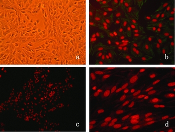
Investigating whether a new kind of somatic cell had been isolated from the male gonad involved identifying cells by indirect immunofluorescence. Results showed that testicular fibroblasts could grow and proliferate in vitro and display fibroblast cell morphology, regardless of the number of culture passages. Cell-specific marker staining confirmed that the cells were vimentin positive and cytokeratin and FSHR negative (Fig. 2).
Fixed Bama mini-pig testicle fibroblast cell (a) (×100); Bama mini-pig testicle fibroblast cell showed vimentin positive (b) (×100); Bama mini-pig testicle fibroblast cell showed cytokeratin negative (c) (×100); Bama mini-pig testicle fibroblast cell showed FSHR negative (d) (×200).

Experiment 2
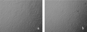
Contact inhibition and serum starvation were examined in order to determine the optimal cell cycle synchronization protocol for testicular fibroblasts. The cell fusion rate was greater using contact inhibition than serum starvation, but not significantly (66.0% vs. 58.3%, p > 0.05). The cleavage rate was similar in each group (85.1% vs. 84.1%) and although the rate of blastocyst formation was higher in the contact inhibition group (20.8% vs. 15.1%); the difference was not significant (Table 1 and Fig. 3).
In vitro development of cloned pig embryos using new-born Guangxi Bama mini-pig testicle fibroblast cells treated by inhibition and serum starvation as donors.

Note: Fused oocytes rate: no. of fused oocytes/no. of oocytes.
Cleaved embryos rate: no. of cleaved embryos/no. of fused oocytes.
Blastocysts rate: no. of blastocysts/no. of fused oocytes.
a Values in the same column are not significantly different (p > 0.05).
Contact inhibition testicle fibroblast cells (a) (×40); serum starvation testicle fibroblast cell (b) (×40).

Experiment 3
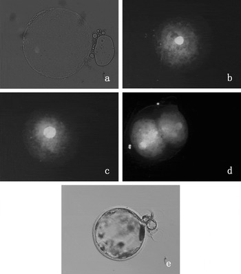
The feasibility of using high passage (old) testicular fibroblasts in nuclear transfers was examined by checking morphological remodeling regulations of the nucleus of Bama mini-pig testicle fibroblast cell in the reconstructed embryo. The nucleus was clearly visible at 3 h after activation, but with the same size as the donor nucleus in reconstructed embryos activated at 0 h. An enlarged nucleus was formed in most of the reconstructed embryos at 6 h, whilst enlarged nuclei or distinct pseudopronuclei were formed in nearly all the reconstructed embryos at 12 h and some of the reconstructed embryos even cleaved. At 7 days, some of the reconstructed embryos developed to blastocyst. In all the different stages, no premature chromosome condensation (PCC) was investigated (Fig. 4).
At 3 h after activation, the nucleus was clearly visible but with the same size as the donor nucleus in reconstructed embryos activated at 0 h (a) (×200); enlarged nucleus was formed in the most of the reconstructed embryos at 6 h after activation (b) (×200); enlarged nucleus or distinct pseudopronuclei was formed in nearly all the reconstructed embryos at 12 h after activation (c) (×200); some of the reconstructed embryos cleaved at 12 h after activation (d) (×200); the donor cell was competent to support embryo development to the blastocyst in vitro (e) (×200).

Experiment 4
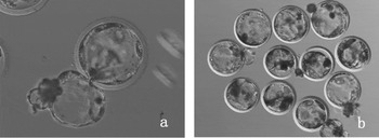
To determine the effectiveness of the testicular fibroblast as donor cells, the in vitro developmental potential of the cloned embryos was examined using either ear fibroblasts or low (3–5) and high passage (10–14) testicular fibroblasts; the lowest fusion rate followed the use of high passage fibroblasts. Similar cleavage rates were found in all three groups. Low passage testicular fibroblasts resulted in the highest rate of blastocyst production, but there was no significant difference between high passage testicular fibroblasts and ear fibroblasts (Table 2, Fig. 5).
In vitro development potential of cloned embryos using Bama mini-pig ear fibroblast cells and testicle fibroblast cell as donor cells.

Note: Fused oocytes rate: no. of fused oocytes/no. of oocytes.
Cleaved embryos rate: no. of cleaved embryos/no. of fused oocytes.
Blastocysts rate: no. of blastocysts/no. of fused oocytes.
a, bValues in the same column with different superscript significantly differ (p < 0.05).
Hatched blastocyst from nuclear transfer of testicle fibroblast cell (a) (×200); newborn Guangxi Bama mini-pig hatched blastocysts from testicle fibroblast cells (b) (×200).

Experiment 5
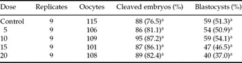
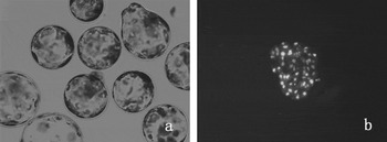
The effect of PHA on parthenogenetic development was examined by addition of different doses (5–20μg/ml) to the culture medium. Differences were not significant, although the rate of cleavage and blastocyst formation was improved at some dose levels (Table 3 and Fig. 6).
The effect of PHA on parthenogenetic embryo development.

Note: Cleaved embryos rate: no. of cleaved embryos/no. of oocytes.
Blastocysts rate: no. of blastocysts/no. of oocytes.
aValues in the same column are not significantly different (p > 0.05).
Porcine parthenogenetic blastocysts in the presence of PHA (a) (×200); nuclear staining of porcine parthenogenetic blastocyst in the presence of PHA (b) (×200).

Experiment 6
The effect of PHA on cleavage and blastocyst formation in cloned embryos reconstructed using old testicular fibroblasts was recorded, using two doses levels of the preparation in the culture medium. As shown in Table 4, development to the blastocyst stage of cloned embryos reconstructed using old donor cell was significantly higher in the presence of PHA (10μg/ml 20μg/ml) than in the control group (13.4%, 5.6% vs. 5.6%) 10μg/ml PHA is adequate to sustain cloned and parthenogenetic embryo development.
The effect of PHA on the Guangxi Bama mini-pig cloned embryos reconstructed using old donor cells.

Note: Cleaved embryos rate: no. of cleaved embryos/no. of cultured embryos.
Blastocysts rate: no. of blastocysts/no. of cultured embryos.
a, bValues in the same column with different superscript significantly differ (p < 0.05).
Discussion
The present study showed that testicular fibroblasts derived from a new-born Guangxi Bama mini-pig could be used to produce cloned blastocysts. These fibroblasts were isolated, cultured and their growth morphology investigated after identifying the cell type by staining intermediate filaments (IFs) in the cytoplasm. Different IFs are to be found in various tissues or cells: vimentin is expressed in mesenchymally derived cells and cytokeratins in epithelial cells. In adult Sertoli cells, IFs are of the vimentin type (Kopecky et al., Reference Kopecky, Semecky and Nachtigal2005). Co-expression of vimentin and cytokeratin has been demonstrated in Sertoli cells during embryonic development and under pathologic conditions in adult testes (Romeo et al., Reference Romeo, Castorina and Marcello1995). In the present study, cell-specific marker staining showed cells to be vimentin positive but cytokeratin and FSHR negative. Such results verified its fibroblast cell origin and confirmed that it was not testicular Sertoli cell, on the basis of tissue origin, method of isolation, morphology, growth characteristics and expression of IFs.
It is generally accepted that a diploid, G0/G1 stage of the cell cycle is required to initiate reprogramming after transfer of the donor nucleus into an inactivated oocyte cytoplasm. This stage is believed to ensure that the cloned embryo is normally diploid (Hashem et al., Reference Hashem, Bhandari, Kang and Lee2007). The present study attempted to improve the suitability of donor nuclei by examining methods of promoting cell cycle synchronization in testicular fibroblasts. Gibbons et al. (Reference Gibbons, Arat, Rzucidlo, Miyoshi, Waltenburg, Respess, Venable and Stice2002) reported that serum starvation of bovine and porcine cells cultured for 48 h reduced cell survival and increased DNA fragmentation. This may explain results in this work which found that the fusion rate of couplets and development to the blastocyst stage was higher using contact inhibition than using serum starvation. Testicular fibroblasts treated by contact inhibition rather than serum starvation are preferable for use in nuclear transfer.
Premature chromosome condensation (PCC) is believed to promote nuclear reprogramming and to facilitate cloning by SCNT in mammalian species. However, it is still uncertain whether PCC is necessary for the successful reprogramming of an introduced donor nucleus in an enucleated oocyte (Sung et al., Reference Sung, Shen, Jeong, Xu, Chang, Cheng, Wu, Lee, Broek, Faber, Tian, Yang and Du2007). Kawahara et al. (Reference Kawahara, Wakai, Yamanaka, Kobayashi, Sugimura, Shimizu, Matsumoto, Kim, Sasada and Sato2005) persisted that PCC is essential for subsequent development of NT embryos. In that study, they treated porcine NT embryos with caffeine to keep their MPF level high during NT. When caffeine was added to the handling medium, the donor cell was injected into the enucleated oocytes. The result showed that caffeine can increase not only the rate of chromosome condensation but also the developmental rate to the blastocyst stage of porcine NT embryos. In contrast, Sung et al. (Reference Sung, Shen, Jeong, Xu, Chang, Cheng, Wu, Lee, Broek, Faber, Tian, Yang and Du2007) reported that PCC was unnecessary for the reprogramming of a transplanted somatic genome in cattle oocyte. In their study, fused NT embryos were subjected to immediate activation (IA, simultaneous fusion and activation) and delayed activation (DA, activation applied 4 h post fusion). The morphologic changes, such as nuclear swelling, the occurrence of PCC and microtubule/aster formation, were analysed. In the IA group, the introduced nuclei gradually swelled and pronuclear-like structures formed within the oocytes, but PCC was not observed, whereas delaying embryo activation resulted in 46.5%–91.2% of NT embryos exhibiting PCC. A significantly higher rate of development to blastocysts was achieved in embryos that were immediately activated (IA, 59.1%) than in those for which activation was delayed (14.2%). Thus they concluded that IA of bovine NT embryos results in embryos with increased competence for preimplantation development. In our study, the reconstructed embryos were also subjected to IA. The results showed that enlarged nuclei were formed in the most of the reconstructed embryos at 6 h and enlarged nuclei or distinct pseudopronuclei were formed in nearly all the reconstructed embryos at 12 h. However, PCC was also not observed and some of the cloned embryos developed to blastocysts. It is concluded that high passage testicular fibroblasts could be successfully reprogrammed, indicating their potential to support cloned embryo development.
To determine the effectiveness of testicular fibroblasts as donor cells, in vitro development of cloned embryos after transfer of testicular fibroblasts of low and high passages and ear fibroblasts from the same piglet was examined. Results showed that both low and high passage testicular fibroblasts had higher capacities development than ear fibroblasts to support cloned embryo. Donor cells from the reproductive system have been used in other reports on nuclear transfers. In nuclear transfer studies in mice, Wakayama et al. (Reference Wakayama, Perry, Zuccotti, Johnson and Yanagimachi1998) reported the higher rates of blastocyst production in embryos reconstructed with cumulus cells than with Sertoli or neural cells. The present study showed a 20.8% rate of blastocyst production with testicular fibroblasts from a newborn piglet; such fibroblasts showed greater potential to support development than ear cells. High passage testicular fibroblasts were significantly less able to support cloned embryo development in the present work and an attempt was made to improve the blastocyst production rate by adding PHA to the culture system. Recent papers by Gupta (Reference Gupta, Uhm, Han and Lee2007a, Reference Gupta, Uhm and Leeb) reported that parthenogenetic embryo development to the blastocyst stage in the presence of PHA (15μg/ml) was significantly higher than in control groups: as with parthenogenetic embryos, the development of cloned embryos was also significantly improved. In our study, parthenogenetic embryo development to the blastocyst stage was higher in the presence of PHA (10μg/ml) than in the control group, but the difference was not significant. However, with cloned embryos reconstructed using old donor cells development to the blastocyst stage was significantly higher in the presence of PHA (10μg/ml) than in the control group and 20μg/ml group (13.4% vs. 5.6% and 5.6). The influence of PHA on parthenogenetic and cloned embryos was dose dependent and 10μg/ml PHA is sufficient to sustain cloned and parthenogenetic embryos development.
Acknowledgements
This research was funded by the grants from the National Natural Science Foundation of China (No. 30660127 and 30860039); Guangxi Program of Scientific Research and Technology Development (0815011–6–3); Guangxi Science Foundation of China (No. 0640002); the Open Foundation of Guangxi Key Laboratory of Subtropical Bioresource Conservation and Utilization (SB0701). We are very grateful to Emeritus Professor Ian Gordon at University College Dublin and Professor Qing-Yuan Sun at the Chinese Academy of Science for their critical advice.