Introduction
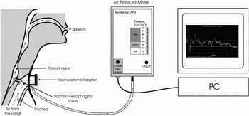
Surgical voice restoration by creation of a tracheoesophageal fistula (TOF) following laryngectomy was first described by Singer and Blom,Reference Singer and Blom1 and has become the preferred method since its widespread introduction in the 1980s. This method of voice production diverts air from the trachea through a one-way valve into a neopharynx, shown diagrammatically in Figure 1. Compared with normal laryngeal voicing (during which high pressure air used for speech is contained in the trachea and not the oesophagus), patients with a TOF may generate a wide range of air pressures in their neopharynx during speech, and these air pressures can in turn be transmitted throughout the whole oesophagus.
Diagram showing how high pressure air from the trachea is diverted into the upper oesophagus.

Previous studies of oesophageal function have identified cases of upper oesophageal disorder following simple laryngectomy. However, to date no study has reported changes in oesophageal body motility or lower oesophageal sphincter function following TOF.
This study assessed the symptomatic and functional changes in the body and lower part of the oesophagus, which may be due to the pressures applied to the oesophagus and neopharynx following creation of a TOF.
Methods
Following invitation, 16 patients who had undergone TOF reconstruction following laryngectomy volunteered and consented to participate in a study of oesophageal function. Ethical approval was obtained from the Sunderland research ethics committee.
Each patient underwent the following: completion of an 11-point dyspepsia symptom questionnaire; five-channel oesophageal manometry; videofluoroscopy and barium swallow; and tracheal (neopharyngeal) air pressure measurements.
Dyspepsia symptom questionnaire and manometry
Prior to attending for oesophageal manometry, each patient was provided with written and verbal information about the procedure. Written consent was obtained from all patients. Patients then completed an 11-point dyspepsia symptom questionnaire, which scored oesophageal symptoms by both frequency and severity (Appendix 1).
Each patient fasted for four hours prior to the manometry procedure. A five-channel manometric oesophageal probe (Model CTO 5e; Gaeltec, Dunvegan, Scotland) with sensors 5 cm apart was introduced through the nose to 60 cm. This was withdrawn at centimetre intervals until the distal sensor was positioned in the lower oesophageal sphincter (LOS) (reference point 0 cm). Proximal sensors were thus positioned in the lower (5 cm), mid- (10 and 15 cm) and upper oesophagus (20 cm; the latter includes the neoglottis).
A series of four ‘dry’ swallows were recorded, followed by four controlled wet swallows of 5 ml of water. Once these recordings had been made, the probe was withdrawn further to locate the neoglottis, as identified by a rise in pressure in the proximal pressure channel. A number of dry swallows were performed in this position to record neoglottic function during swallowing. Following collection of all data, the manometric probe was removed and the patient discharged.
Each study was analysed by the clinical physiologist and results recorded for: LOS pressure, position and function; respiratory reversal point; and oesophageal body contraction characteristics and amplitudes.
Any observed abnormalities were also noted, such as absent LOS relaxation or disordered oesophageal motility.
Videofluoroscopy and barium swallow
Patients underwent video-recorded fluoroscopic examination of pharyngoesophageal function during swallowing and speech production via the TOF. Recordings were obtained of the entire pharynx and oesophagus during speech using a long ‘/a:/’ vowel and counting from one to 20.
Examinations were performed with standard fluoroscopic equipment in the erect position, in lateral and lateral-oblique orientations. A standard metallic marker (a two pence coin, 26 mm diameter) was placed on the skin of the left anterolateral chest wall to calibrate subsequent measurements. Swallowing was assessed using a high density barium suspension and a semi-solid (chewed) marshmallow bolus. Additional barium suspension was taken as required to facilitate swallowing.
The clinician performing the videofluoroscopic examinations was blind to the findings of other investigations and, as far as possible, to the date of surgery or other interventions. Video tapes were reviewed by the same person who assessed pharyngoesophageal diameter and motility during swallowing and phonation. Recordings were analysed further by transfer to digital versatile disc; in this medium, the video recording could be assessed frame by frame, and measurements made by software using the two pence coin as a reference. Estimates were recorded for the maximum diameter of the neopharynx below the neoglottis (to represent the new upper oesophagus) and for the lower oesophageal maximum diameter.
Tracheal and pharyngeal air pressure measurements
All patients underwent tracheal manometry using the Sunderland air pressure meter (winner of the National Health Service innovation award 2005; manufactured by Canford Audio PLC, Washington, UK) on the same day they attended for oesophageal manometric assessment. A 10-second spontaneous speech sample was recorded on a personal computer. The equipment is shown in Figure 2. Free speech, rather than reading aloud, was considered to be more representative of everyday tracheal air pressure levels. Mean pressure levels recorded during speech were calculated by spreadsheet analysis.
Typical arrangement of equipment, enabling measurement of tracheal air pressure and data recording on a personal computer (PC). The air pressure meter is connected via a tube to the patient's stoma housing.

Results
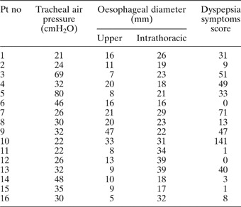
The majority of patients were male (87.5 per cent) and all but one patient had smoked. The time since surgery ranged from one to 16 years. The mean age of the patients at the time of study was 64 years and the range was 58 to 81 years. See Table 1 for results to the questionnaire and other investigations.
Patient results for questionnaire and investigations

Pt no = patient identification number
Dyspepsia symptom questionnaire
A symptom severity score was used to indicate how troublesome the individual found each symptom. Based on a score of one to 10, symptoms were rated as follows: one to three, mild; four to six, moderate; and seven or more, severe.
The following symptoms and severity scores were noted. Eleven patients (69 per cent) reported high dysphagia to solids. Six patients reported belching (three mild, two moderate and one severe). Five patients reported high heartburn (two mild, two moderate and one severe). Four patients reported bloating (one mild, one moderate and two severe). Three patients reported a feeling of early satiety (one mild and two moderate).
Three patients were taking dyspepsia medication, and 50 per cent of the study group reported strong or significant predictors of gastroesophageal reflux disease, according to the system of Dennison et al. (unpublished data) (i.e. heartburn, regurgitation, belching and bloating).
To obtain numerical data from the questionnaire, each patient's answer was scored by summing the product of the frequency and severity for each experienced symptom. These data were then compared with other clinical data.
Manometry
Manometric studies showed abnormal lower oesophageal sphincter (LOS) function in six cases, usually demonstrating poor relaxation or coordination on swallowing. Six patients had low LOS pressure (<6 mmHg) and two showed no relaxation on swallowing. One LOS examination was not possible due to a hiatus hernia and a Schatzki ring.
Oesophageal body motor function was abnormal in nine patients. Simultaneous contractions were observed in four of these patients. Five studies recorded low amplitude contractions (normal median, 70 mmHg; range, 30–160 mmHg), and one patient had completely disordered propagation throughout the oesophagus. Six studies showed oesophageal body dysmotility with incomplete, abnormal or absent contraction propagation.
Barium swallow and videofluoroscopy
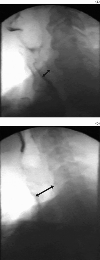
The upper oesophageal mean diameter during speech was 16 mm (range, 5 to 47 mm) (Figure 3). Oesophageal dysmotility was observed in seven patients, six of whom had dysmotility demonstrated on manometric study. Two studies showed a dilated intrathoracic oesophagus, defined as a diameter greater than 35 mm.
Two videofluoroscopy images showing a dilated upper esophagus. (a) An upper oesophagus of 16 mm diameter, which was the mean value found in our study. (b) An oesophageal diameter of 33 mm.

Tracheal and pharyngeal air pressure measurements
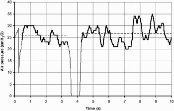
Data recorded from the air pressure meter were stored on a microcomputer and analysed using a spreadsheet. A graph showing a typical set of tracheal air pressure data recorded during speech is shown as Figure 4. The range of pressures in this recording varied from peaks of 35 cmH2O to troughs of 0 cmH2O when the patient inhaled. The parts of the graph in bold indicate the pressure levels when the patient was speaking. Using the spreadsheet, the mean pressure during speech was calculated for each patient; in Figure 4, the mean pressure for this particular patient is indicated by dotted lines. The mean tracheal pressure during speech for all patients was 36 cmH2O (standard deviation = 17) and the range was 21 to 80 cmH2O.
Graph showing tracheal air pressure data recorded during speech (bold trace). The dotted trace indicates the mean pressure during speech. S = seconds

Correlation between clinical measurements
To investigate the relationship between our clinical data sets, scatter diagrams were plotted and are shown in Figure 5. These scatter diagrams compare: (1) air pressure vs oesophageal diameter; (2) air pressure vs dyspepsia symptoms score; (3) upper oesophageal diameter vs dyspepsia symptoms score; and (4) time elapsed since surgery vs dyspepsia symptoms score.
Scatter plots of various study parameters. Pearson correlation coefficients and confidence intervals (CI) were as follows: (a) r = −0.31, CI = −0.61 to 0.37; (b) r = −0.08, CI = −0.55 to 0.43; (c) r = 0.55, CI = 0.08 to 0.82; and (d) r = −0.16, CI = −0.61 to 0.37. Yrs = years

There were no observable or statistically significant relationships between our data sets. The Pearson correlation coefficient and the confidence limits for each set of data are given for each Figure 5 graph; there were low correlation coefficients with large confidence limits. Only one of the correlation coefficients was above 0.5; however, this result is unreliable because of the limited number of patients and the large range in the confidence limits and outliers in our data.
Discussion
To the best of our knowledge, no previously reported study has examined TOF speakers' oesophageal function and dyspeptic symptoms. While Artazkoz del Toro and Lopez MartinezReference Artazkoz del Toro and Lopez Martinez2 have compared manometry and videofluoroscopy of the pharyngoesophageal segment in 29 such patients, most such studies have considered only dysphagia after laryngectomy, and have not assessed any other symptoms of oesophageal function. A study by Ward et al. Reference Ward, Bishop, Frisby and Stevens3 reported only that 58 per cent of their laryngectomy patients managed a normal diet by three years post-surgery; they did not state whether the remaining 42 per cent had high, mid- or low dysphagia. Roed-Petersen et al. Reference Roed-Petersen, Jorgensen and Larsen4 conducted manometric studies on 17 oesophageal speakers, but did not report symptoms. We believe that the current study is the first to report total oesophageal function, using both subjective and objective measurements, in a select group of patients following TOF formation.
Few studies have investigated oesophageal body and lower sphincter manometry in tracheoesophageal speakers. Dantas et al. Reference Dantas, Aguiar-Ricz, Gielow, Filho and Mamede5 studied proximal oesophageal contractions in laryngectomees, some with oesophageal speech and others without. They concluded that the amplitude and duration of proximal oesophageal contractions were reduced after surgery. Another study by the same groupReference Dantas, Aguiar-Ricz, de Oliveira, Mello-Filho and Mamede6 measured intra-oesophageal pressures during oesophageal speech and found a higher pressure in the upper oesophagus in those patients with oesophageal speech compared with those without. The authors proposed that laryngectomy may alter oesophageal motility and LOS function in some patients, due to damage to the innervation of the pharynx and oesophagus during laryngectomy, i.e. injury of the branches of the vagus nerve.Reference Dantas, Aguiar-Ricz, Oliveira, Mello-Filho and Mamede7 These authors did not consider the possibility that oesophageal speech could cause abnormalities of oesophageal function.
Choi et al. Reference Choi, Hong, Kim, Yoon, Nam and Son8 used manometry to investigate oesophageal motility after total laryngectomy, but did not comment on the method of voice rehabilitation. They concluded that the nature of the surgery and the altered anatomy affected the upper oesophageal sphincter and the upper oesophageal motility, but not the function of the lower oesophageal sphincter. Hanks et al. Reference Hanks, Fisher, Meyers, Christian, Postlethwait and Jones9 studied oesophageal motility before and after laryngectomy and found significant changes in the upper oesophageal sphincter, but no changes in the proximal or distal oesophagus. The method of voice production was again not stated. The authors found no dramatic alterations in motility after post-operative radiation. Dworkin et al. Reference Dworkin, Meleca, Zacharek, Stachler, Pasha and Abkarian10 compared swallowing and speech function in 10 total laryngectomy patients (TOF speakers) with that of 10 patients who had undergone partial laryngectomy. They found that none of the total laryngectomy patients reported swallowing difficulties, and concluded that further investigations were thus unnecessary in this group of patients.
In their responses to our dyspeptic symptom questionnaire, all but two patients reported suffering from one or more upper gastrointestinal (GI) symptoms. Over 30 per cent of our patients group would appear to suffer from gastroesophageal reflux disease, based on the reported incidence of heartburn and regurgitation. These symptoms are strong predictors of gastroesophageal reflux disease and have been shown to correlate well with oesophageal pH and manometry investigations. This is a higher incidence than one would expect for a normal population of similar age. At the age of 60 years, 10 per cent of the population would be expected to suffer gastroesophageal reflux disease.Reference Wilkinson11 Belching and bloating were common in our patients, but should be expected considering the pathophysiology associated with tracheoesophageal voicing. Other symptoms such as chest pain, nausea and vomiting were less common.
A high proportion of patients also had disordered oesophageal function, demonstrated by both manometric and radiological methods, even when no mid- or low dysphagia was reported. Videofluoroscopy demonstrated distension of the oesophagus to a variable extent in TOF patients, due to accumulation of air below the neoglottis. Van As-Brooks et al. Reference van As-Brooks, Hilgers, Koopmans-van Beinum and Pols12 found that higher tone in the neoglottis resulted in greater distension of the oesophagus. This air is either expelled through the neoglottis or swallowed, or both.
• This was a comprehensive study of oesophageal function in 16 laryngectomee patients who used tracheoesophageal speech as their main means of communication
• Fifty-six per cent of patients had disordered oesophageal function as determined by manometry and/or videofluoroscopy; 50 per cent reported symptoms of disordered oesophageal function and 69 per cent reported high dysphagia
• Increased tone of the pharyngoesophageal segment may predispose to oesophageal dysmotility. Correction of pharyngoesophageal segment hypertonicity may prevent further deterioration of oesophageal function
• An increase in oesophageal symptoms in such patients merits investigation of oesophageal function, either by manometry or videofluoroscopy. There appears to be good correlation between these two methods of investigation
Tracheoesophageal speakers can generate a wide range of air pressures in their trachea–pharynx, and one may postulate that those patients producing a high pressure may be more at risk of altered oesophageal function. The amplitude of these pressures will depend upon various factors, including the effort the patient exerts, the pulmonary function (e.g. lung capacity and maximum flow rate), the resistance to air flow through the valve and the neoglottis tonicity. Some patients attempt to compensate for what they regard as a low volume voice by increasing the pressure in their trachea–pharynx. This may enable a small gain in volume, but may also apply excessive pressure to the tissue of the neopharynx. The optimum tracheal pressure for TOF speakers is recommended by BlomReference Blom13 as 30–35 cm H2O. This level is thought to be a good compromise between the voice volume and the durability of the tracheostoma housing seal. However, it may be that, over time, such an unusually high pressure applied to the neopharynx could cause dilatation of the oesophagus, with resultant oesophageal dysfunction.
In the majority of our cases, the increased intrathoracic pressure generated by tracheoesophageal voice production may protect the mid- and lower oesophagus from dilatation. The cervical oesophagus, i.e. the neopharynx below the neoglottis, is not protected in this way and could be prone to permanent dilatation. However, only two of our patients had an abnormal degree of upper oesophageal dilatation (>30 mm diameter), even though their tracheal air pressure was not elevated. Interestingly, those patients with a raised air pressure measurement did not have an abnormal degree of oesophageal dilatation but did demonstrate abnormal oesophageal function.
Those patients who had been using fistula voice for longer periods appeared to be no more vulnerable to oesophageal symptoms or abnormal investigation findings than those using fistula voice for a shorter time. However, those patients using fistula voice for only a few years (one to five) had a higher incidence of heartburn.
In our group of patients, 81 per cent underwent pre- or post-operative radiotherapy. It is therefore possible that factors other than surgery and TOF speech could have caused oesophageal dysfunction. However, Hanks et al. Reference Hanks, Fisher, Meyers, Christian, Postlethwait and Jones9 found no dramatic alterations in motility after post-operative radiation following laryngectomy. Gaze et al. Reference Gaze, Wilson, Gilmour, McDougall and Maran14 found that laryngeal irradiation had no effect on upper or lower sphincter function, but they did demonstrate an increase in wave duration and a reduction in wave velocity in the pharyngoesophageal segment. However, only one of their 19 patients complained of dysphagia, and dyspeptic symptoms were not reported.
Conclusions
On the basis of this study, we conclude that there is no contraindication to proceeding with TOF voicing even in patients with a history of oesophageal dysfunction. Regular post-operative assessment of dyspeptic symptoms may alert the clinician to a change in oesophageal function and prompt further investigation, either radiological or manometric.
Attention should be paid to the neoglottal segment; if the pressure is raised or there is videofluoroscopic evidence of hypertonicity, chemical myotomy could be performed using botulinum toxin, particularly if there is evidence of oesophageal dilatation. In addition, the lower oesophageal sphincter should be examined and advice sought from an upper GI specialist.
We acknowledge that this study involved a small number of subjects. This is not surprising as many of our patients were elderly and reluctant to undergo invasive investigations. The study was time-consuming for both patients and investigators. As with any retrospective study, our patients may have self-selected, coming forward because they were suffering symptoms.
Acknowledgements
The authors are grateful to the City Hospitals Sunderland Research and Development Strategy Board and Research and Development Scientific Review Committee for the purchase of the five-channel manometry equipment. Statistical advice was also gratefully received from Professor Tony Hildreth, Trust Medical Statistician.
Appendix 1. Oesophageal questionnaire
All patients were questioned regarding the frequency and severity of their epigastric and oesophageal symptoms.
Frequency scores were as follows:
6 = Constant pain/symptom
5 = Very frequently throughout the day
4 = At some point every day/night
3 = Majority of week
2 = At least once per week
1 = Less frequently
Severity scores were as follows:
0 = no symptoms/pain
1–3 = mild symptoms/pain
4–6 = moderate symptoms/pain
≥7 = severe symptoms/pain
The oesophageal symptoms score was derived from the product of the frequency and severity scores.
The patients were questioned on the following symptoms:
Stomach pain/discomfort
Chest pain/pain behind breastbone
Heartburn during the day/while upright
Heartburn during the night/while supine
Regurgitation
Excessive belching
Early satiety
Vomiting
Nausea
Pain upon swallowing*
Difficulty swallowing*
Bloating/fullness after eating
*The patient was also asked in what position they experienced pain or difficulty and whether it occurred with solids, liquids or both.