Introduction
The increase of electricity consumption has led to the need of finding new sources for lighting with a power consumption as low as possible. Knowing that light-emitting diodes (LEDs) have a long life correlated with low-energy consumption, there is a need to manufacture devices based on semiconductor chips that can replace conventional lighting system.[Reference Chiang, Tsai and Hon1] In this context, the LEDs technology has experienced a real boom for generating white light. Currently there are two main methods for emission of white light using semiconductor devices. The first devices with white light emission are based on the use of individual colored LEDs, the RGB (red–green–blue) system. The RGB devices are widely used in video displays, rather than in a lighting system. Starting with the development of the blue LED based on indium gallium nitride (2014-Nobel Prize in Physics) has opened the door to the possibility of replacing conventional lighting systems (incandescent bulb, fluorescent, etc.) with a low-energy devices. Using a system for converting light emitted from a blue or UV chip by one or more corresponding phosphors represents the most promising way of emission of white light, but still does not solve all the problems so as to can replace conventional lighting systems as a whole.[Reference Akasaki, Amano, Nakamura and Nakamura2, Reference Ryu3]
Development of materials with luminescent properties experienced a real expansion since 1998 when it was demonstrated their capacity of converting light for white LEDs manufacturing. This luminescent material known in the semiconductor lighting area as phosphors that can emit yellow, red, blue, green light when stimulated by absorption of energy (photons).[Reference Haaheim and DenBaars4, Reference Jang, Choi, Wu, Lim and Yoo5] The yellow ceramic phosphors with garnet structure and general formula A3B2C3O12 doped with rare earth have become extremely popular and finding their applicability in areas as the light sources (from fluorescent lamps to optical filters and white LED), display devices (i.e., cathode-ray tubes), detector systems (i.e., x-ray screens), paint, biologic application, and so on. The broad range of application is based on thermal and chemical stability at elevated temperature, able to withstand corrosive environments, radiation resistance, interesting optical and mechanical properties, non-toxic biologic material, etc.[Reference Tsukamoto and Isobe6–Reference Guo, Ma, Zhao, Qiu, Liu, Sang, Claverie and Liu10] The first white LEDs are based on InGaN blue LEDs and yellow yttrium aluminum garnet doped with cerium (YAG:Ce) as phosphor. YAG:Ce is currently the most widely used phosphors, with superior optical properties, but seems to be cold and suffer from the lack of a red component. In the last years, special attention is given to the development of the nanostructured complex oxide phosphors that can emit light in an efficient and controlled way.[Reference Chiang, Tsai and Hon1, Reference Jang, Choi, Wu, Lim and Yoo5, Reference Lin, Meijerink and Liu11] Terbium aluminum garnet (TAG, Tb3Al5O12) appears as an alternative to YAG:Ce, having the advantage of an emission spectrum slightly shifted to the red range, but TAG:Ce is very hard to obtain.[Reference Kim, Chung and Ryu12–Reference Tsai, Liu and Chung14]
For both types of ceramic phosphors, the synthesis routes play a determining role in establishing the properties and performances of these materials. Each processing step of yellow phosphors manufacturing can influence the structural and morphological properties of these (i.e., increase particles size, secondary reaction products) that can lead to an alteration of the chemical, electrical, optical, mechanical, and thermal properties, and thus limited the ability to the optoelectronic applications used to generate white light (i.e., light scattering, low light efficiency, etc.). Whichever method is used for the synthesis of TAG ceramics, the processing steps are more difficult to be controlled compared with YAG. In both cases of the materials studied, some problems appear in terms of purity, composition, structure, morphology, etc. which limits their applicability or have lower production yield.[Reference Guo, Ma, Zhao, Qiu, Liu, Sang, Claverie and Liu10, Reference Kim, Kim, Hong, Lee and Kang15] The top–down (solid-state, laser ablation) methods are the most common technologies to obtain phosphors for optoelectronic application. Despite the solid-state methods being the only mass production at industrial level for synthesis of yellow phosphors, it is hard to control the shape, size, and distribution of the particles. For solid-state process synthesis of yellow phosphor, it is necessary that oxides or nitrides (that have the refractory nature), temperatures of about 1600–1900 °C, and longtime reactions are required. Solid-state reaction is the most used method for TAG:Ce synthesis for applications in white light emission devices. The literature shows a number of wet chemical routes for getting the YAG phosphor, but only few publications report the formation of TAG phosphor by bottom–up processes. In practice, the TAG:Ce has a low applicability because of technological limitations for its synthesis and the higher cost price than YAG:Ce.[Reference Haaheim and DenBaars4, Reference Jang, Choi, Wu, Lim and Yoo5, Reference Kim, Chung and Ryu12, Reference Tsai, Liu and Chung14, Reference Nazarov16–Reference Cho, Huh, Park and Do20] As a result of the many advantages [i.e., a better process control, lower processing temperatures (below 1200 °C), etc.], the bottom–up methods [sol–gel, (co)precipitation, spray pyrolysis, hydro-/glyco-/solvothermal processes] are intensively studied in order to improve the optical properties.[Reference Chiang, Tsai and Hon1, Reference Kim, Kim, Hong, Lee and Kang15, Reference Kim, Zakharov and Chashchin21–Reference Yixuan, Li and Junguo28]
To obtain nanoscale ceramic phosphors at relatively low temperature, we propose a modified sol–gel method to synthesize phosphors like YAG and TAG. To form a phosphor with a garnet structure with applicability in white light-emitting devices, Ce3+ was adopted to substitute the Y3+ and Tb3+, respectively. The garnet phase was analyzed by energy-dispersive x-ray spectroscopy (EDX), Fourier transform infrared (FTIR) spectrometry, and x-ray diffraction (XRD) method, the morphology was observed by scanning electron microscopy (SEM), and luminescence properties were studied with a photoluminescence (PL) spectrometer.
Experimental
Materials
The following raw materials were used to synthesize the yellow YAG:Ce and TAG:Ce phosphors: yttrium oxide (Y2O3), terbium oxide (Tb4O7), aluminum nitrate [Al(NO3)3·9H2O], cerium nitrate [Ce(NO3)3·6H2O], dimethyl sulfoxide [DMSO, (CH3)2SO], acetylacetone (AcAc, CH3COCH2COCH3), cetrimonium bromide [CTAB, (C16H33)N(CH3)3Br], nitric acid (HNO3), ammonia solutions (NH4OH) (from Sigma Aldrich, all analytical grade), and deionized water (ADI).
Yellow phosphors (YAG:Ce and TAG:Ce) nanopowders preparation
The YAG:Ce and TAG:Ce powders were prepared by a modified sol–gel method and subsequently annealed at 1100 °C. In the present paper, we proposed the similar methods of manufacturing both the yellow phosphors.
Hydrolysis and condensation
The sol–gel process for YAG:Ce and TAG:Ce manufacturing was realized in acid catalyst, using HNO3 both as the reaction medium and as a dispersion medium. Yellow phosphors synthesis was started by dissolving the oxides in hot nitric acid solution. For a quantitative dissolution, the reagents are maintained at 80 °C. After 30 min. 100 mL of deionized water was added while stirring. At the same time, the other salts were dissolved in deionized water and heated to 80 °C. A solution from AcAc in DMSO was also prepared in a volumetric ratio of 3 : 1. In two glasses, in the stoichiometric report: (1) Y, Al, and Ce nitrates—for YAG:Ce, and (2) Tb, Al, and Ce nitrates—for TAG:Ce were added. The premixed solution AcAc–DMSO was added to the obtained solutions, and stirring was continued for another 10 min at 80 °C, under the reflux condition. In the present study, AcAc act as chelating agents and was added in excess. The molar ratio of metal ions to the AcAc used is 1.5 higher than the theoretical one.
In the process, we used stoichiometric inorganic amounts for synthesizing the phosphors with general formula: (Y0.93Ce0.07)3Al5O12, YAG:Ce and (Tb0.93Ce0.07)3Al5O12 or TAG:Ce.
We use CTAB in a ratio of ADI:CTAB 10 : 1. CTAB is a cationic surfactant having a determinant role in the size and shape control as a result of the anchoring capacity or selectivity to various crystal facets.
The sol–gel process was continued under vigorous stirring at 80 °C and reflux condition for another 5 h to produce the final sol. The pH value of the solution was adjusted with HNO3 or NH4OH. To prevent the precipitation, we worked at pH 4. In order to produce the gel, we removed the reflux and permitted a slow evaporation followed by a rest period to allow bulk polymerization.
Thermal treatment
Thermal treatment is one of the most important steps in the yellow phosphor manufacturing process and the largest consumer in terms of time. First, the gel transforms into a macroporous powder by thermal treatment at 150–200 °C. Second, the powder was presintered at maximum 400 °C until it transforms into a brownish powder. The presintering steps are characterised by nitride (beginning at 200 °C) and organic decomposition. The use of DMSO can prevent cracks and avoid pore formation during drying the gel, having a favorable influence on the shape and size of the final particles. Finally the temperature was raised until 1100 °C, and the end product was two yellow-emitting phosphors, YAG:Ce and TAG:Ce. Thermal treatment has followed the protocol: (i) 0–150 °C with 5 °C/min and maintained for 2 h, (ii) 150–200 °C with 2 °C/min and maintained for 1 h, (iii) 200–400° with 7 °C/min and maintained for 3 h, (iv) 400–900° with 5 °C/min and maintained for 5 h, and (v) 850–1100° with 5 °C/min and maintained for 12 h, in air atmosphere.
Characterization of YAG:Ce and TAG:Ce powder
The crystallinity and phase identification of YAG:Ce and TAG:Ce powders were carried out by Rigaku SmartLab x-ray Diffraction System using Cu Kα radiation in the 2θ range from 20° to 70°. Scherrer's equation was used to calculate the average crystallite size. FTIR spectrometry (Bruker Optics, Tensor 27, Ettlingen, Germany) and KBr disk technique was used to study the bond configuration of YAG:Ce and TAG:Ce phosphors. The spectra were taken in the wavenumber 4000–400 cm−1 by averaging 64 scans and with a resolution of 4 cm−1. Our research uses EDX for the phosphors elemental composition analysis. EDX spectra were collected with a TESCAN having an EDAX facility, operated at 10 kV. Microstructure of the YAG:Ce and TAG:Ce phosphors samples were studied by field emission SEM. The SEM images for the powder samples were taken with an FEI Nova NanoSEM 630 system. Before the examination, the yellow phosphors were dispersed into ethanol, ultrasonication for 30 min, dip-cast onto the surface of silicon wafers and coated with a thin gold layer. The PL spectra of the YAG:Ce and TAG:Ce phosphors were analyzed by fluorescence spectrometer at room temperature using a FLS920 Edinburgh Instrument spectrometer equipped with a 450 W Xenon lamp as an excitation source, and a red sensitive photodetector, in the range of 250–800 nm. The excitation wavelength was at 470 nm.
Results and discussion
Phase analysis of the sample
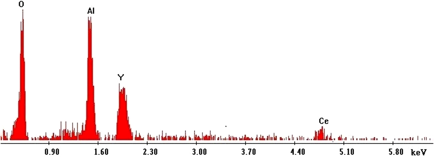
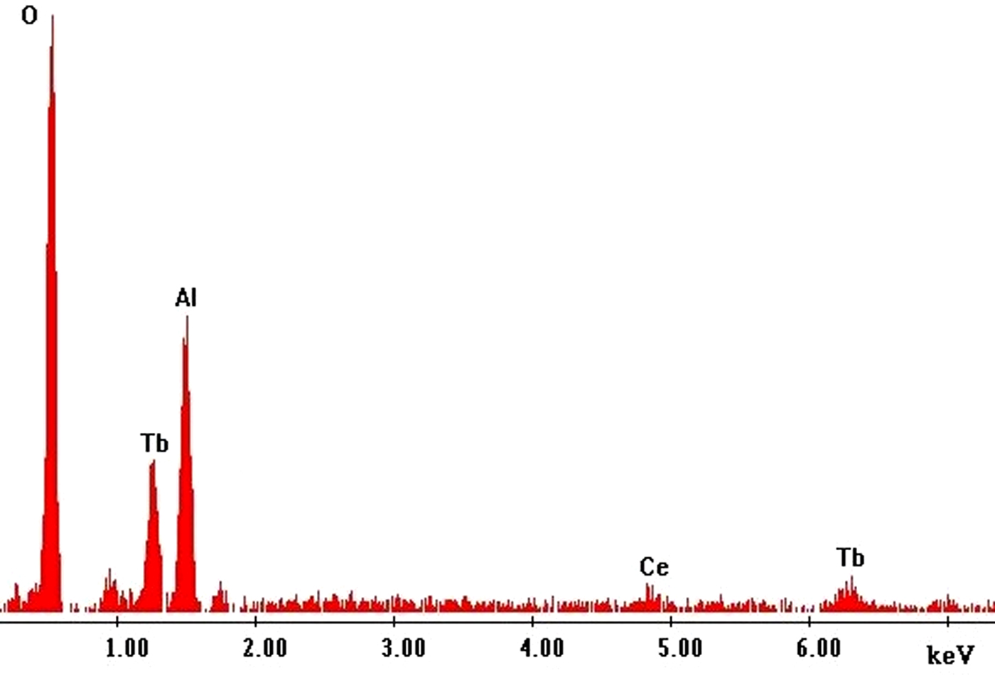
A qualitative elemental analysis of YAG:Ce and TAG:Ce phosphor powders annealed at 1100 °C was made using EDX. The EDX spectra are presented in Figs. 1 (for YAG:Ce) and 2 (for TAG:Ce) in order to evaluate the composition at the atomic level. For both phosphors, the presence of O(K) peak at 0.53 keV and the Al(K) peak at 1.49 keV was determined. Also, the presence of dopant Ce(L) in the samples was signaled for both phosphors at about 4.8 keV. In the TAG:Ce EDX spectra, at 0.9 keV a peak may be observed that suggests an atom of Ce(M). For YAG, the Y(L) peak was found at 1.92 keV, and for TAG, two peaks were found that may be attributed to Tb(M) at 1.24 keV and Tb(L) at 6.27 keV. In the case of phosphor based on yttrium, the observed peaks suggesting the formation of the YAG:Ce and the atomic percentage correspond to theoretical Y2,78Ce0,20Al5,00O12,02 formula, which indicates good crystallinity and very close to that of the target materials. Also, for the yellow phosphor based on terbium, the observed peaks suggesting the formation of the TAG:Ce, correspond to theoretical Tb2,83Ce0,17Al4,83O12,17 formula, but the atomic percentage of aluminum was slightly lower than theoretical. A possible explanation is based on the existence of the TAP (TbAlO3 with a perovskite structure) and/or TAM (Tb4Al2O9 with a monoclinic structure) metastable phases. The purity of the phosphors was confirmed by the absence of other peaks.
Energy-dispersive x-ray (EDX) spectrum showing the main elemental peaks for YAG:Ce phosphor powder after annealing at 1100 °C (quantitative analysis).

Energy-dispersive x-ray (EDX) spectrum showing the main elemental peaks for TAG:Ce phosphor powder after annealing at 1100 °C (quantitative analysis).

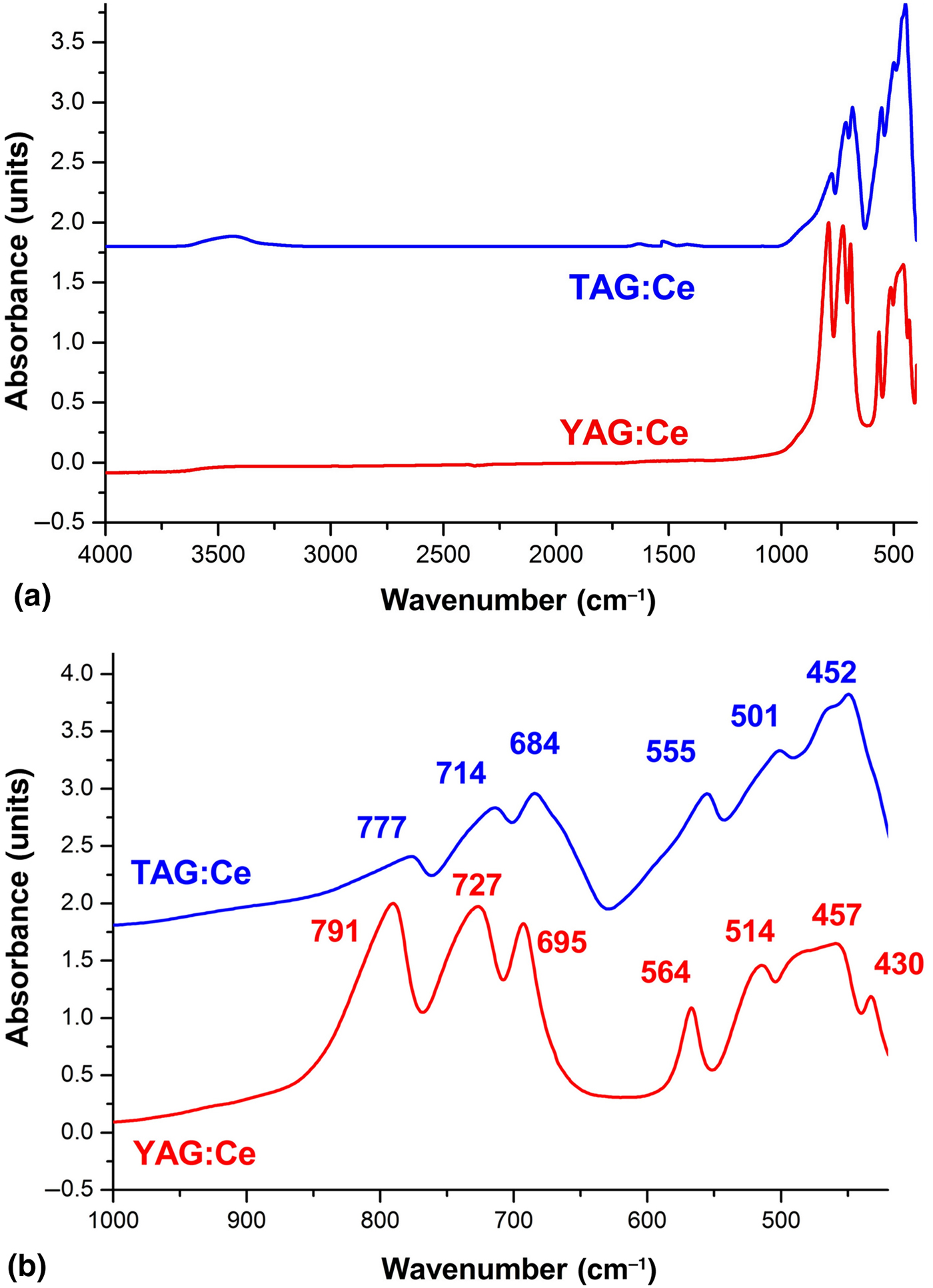
FTIR spectra from Fig. 3 are used to determine the structural configuration of the YAG and TAG yellow-emitting phosphors. By studying the peaks we observed: (i) 4000–800 cm−1 peaks that can be attributed to the water absorption during processing of samples for characterization by FTIR spectrometry; (ii) there is no maximum absorption that would indicate the presence of organic bonds or impurities; (iii) transition from the amorphous to crystalline phase confirmed by the presence of peaks below 800 cm−1 that may be assigned to the metal–oxygen bonds. Similar structure of the two phosphors is confirmed by the similarities between the two FTIR spectra, the slight spectral shift is attributed to the differences between the ray ions of Y and Tb, which leads to different lengths of bonds Y–O–Al and Tb–O–Al, respectively. The FTIR spectrum of YAG:Ce sample was characterized by absorption bands centered at 727 and 564 cm−1 that can be assigned to Y–O vibrations, while Tb–O bonds in the sample TAG:Ce can be seen about 714 and 555 cm−1. The peaks that can be assigned to Al–O bond are observed at 791, 695, and 430 cm−1 for YAG:Ce and 777, 684, and 501 cm−1 for TAG:Ce. Presence of the Ce dopant in phosphor molecules was confirmed by spectral bands centered at 514 and 457 cm−1 for YAG:Ce and 452 cm−1 for TAG:Ce, peaks that can be assigned to Ce–O bond. All the FTIR data are in agreement with our previous work and were found to be consistent with the TAG:Ce and YAG:Ce phase crystallization process observed by XRD.[Reference Schiopu, Matei, Dinescu, Danila and Cernica29, Reference Tucureanu, Matei, Mihalache, Danila, Popescu and Bita30]
Fourier transform infrared (FTIR) spectra of the (red) YAG:Ce and (blue) TAG:Ce phosphor powder after annealing at 1100 °C (a) FTIR spectra on throughout the MIR range, 4000–400 cm−1 and (b) peak indexing on spectral range 1000–400 cm−1.

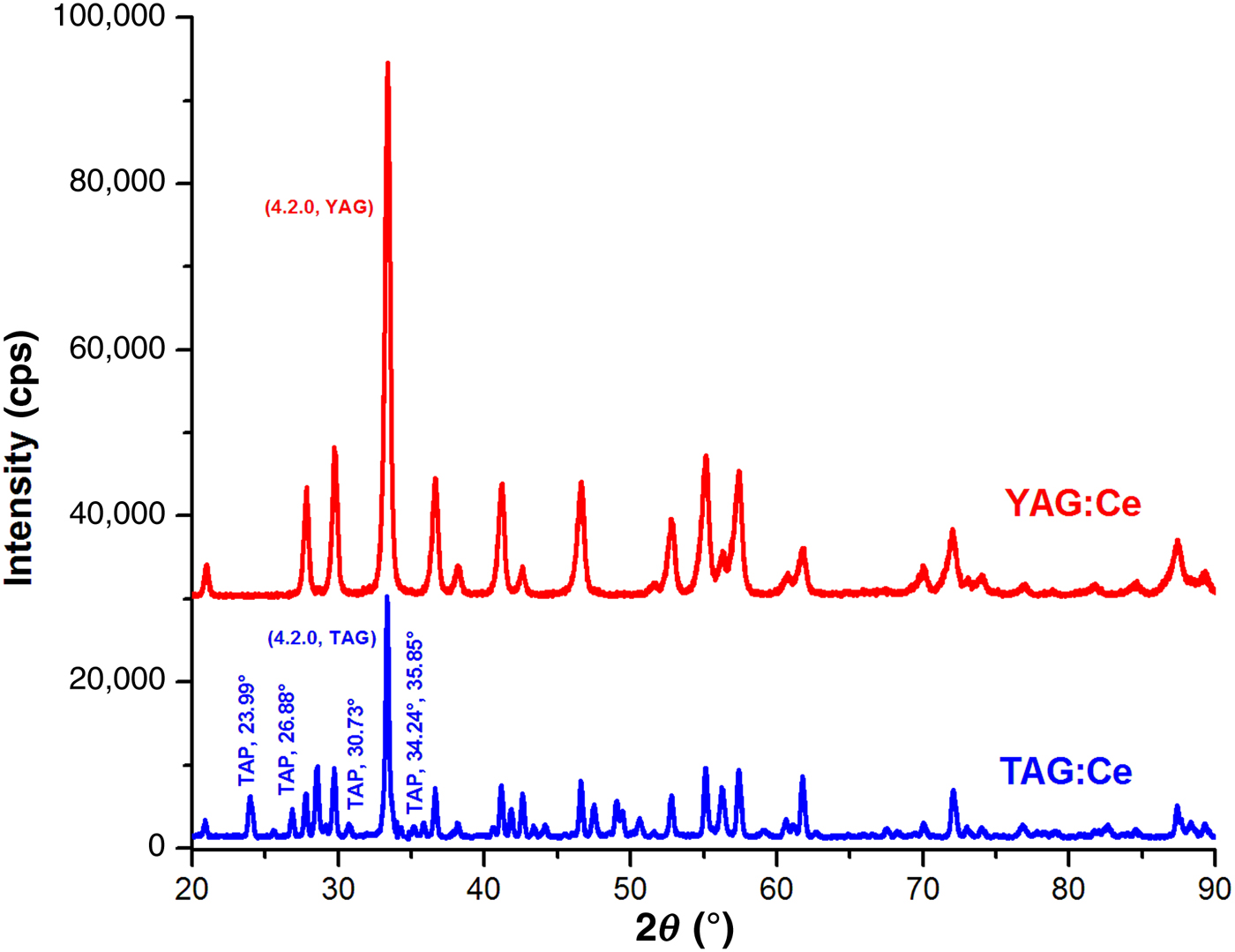
In Fig. 4, we present XRD pattern of YAG: Ce and TAG:Ce powders after sintering at 1100 °C. By indexing the lines in agreement with JCPDS—file no. 01-082-0575 and using Scherrer's equation [sin2θ = λ 2(h 2 + k 2 + l 2)/4a2] we obtained information on the crystal structure, purity, lattice parameter, and crystallite size. For both yellow phosphors, the transition from amorphous structure to the crystalline phases and formation of the YAG and TAG garnet structure was confirmed by the sharp peaks and the possibility to index the Miller indices of the all lines according to the database [at about 2θ (hkl) = 20.9° (220), 27.8° (321), 29.7° (400), 36.6° (422), 38.1° (431), 41.2° (521), 42.6° (440), 46.6° (532), 52.8° (631), 55.1° (640), 56.2° (552), 60.7° (651), 61.7° (800), 70° (840), 72° (842), 76.9° (932), 82.6° (10.1.1), 87.4° (864), 89.3° (10.4.2)]. The main peak corresponds to the garnet crystalline plane with Miller indices of (420) and may be found at 2θ = 33.47° for YAG:Ce sample and at 2θ = 33.35° for TAG:Ce sample. The diffraction peaks of TAG:Ce are shifted slightly to smaller 2θ angles, this can be attributed to the large size of terbium atoms. In case of YAG:Ce, the phase purity was found to be excellent, but for TAG:Ce, we observed some minor lines that suggest the presence of some intermediated terbium aluminum perovskite structure (TbAlO3, TAP at 2θ = 23.99°, 26.88°, 30.73°). The presence of terbium aluminum monoclinic structure (Tb4Al2O9, TAM) was not observed. For TAG:Ce sample, the XRD data confirm deviation from theoretical formula, observed by EDX analysis, but as can be seen from the PL analysis, the presence of TAP phase appears to be in minor amounts, so it does not affect the optical properties of the material.
X-ray diffraction pattern of a (red) YAG:Ce and (blue) TAG:Ce phosphor powder after annealing at 1100 °C.

The absence of specific peaks of cerium oxide indicates that cerium ions are well activated in the matrix of the garnet phosphors. In the YAG:Ce and TAG:Ce phosphors, the Ce3+ ion substitutes a Y3+ or Tb3+ to modify the lattice parameters, but does not disturb the crystalline structure. Due to the difference in the ionic radii [Y3+ (1.019 A°), Tb3+ (1.04 A°)] and to the cerium ions, the lattice parameters are modified and the garnet cell parameters of our samples have found to be 12.0273 A° for YAG:Ce and 12.0509 A° for TAG:Ce, and the strain was found to be 0.006% and 0.08%, respectively. These XRD diagrams confirm the formation of the garnet structure, polycrystalline, having a unit cell with eight Y3Al5O12 or Tb3Al5O12 molecular with bcc structure and Ia-3d (230) space group. In the garnet structure, unit cells are interconnected octahedrons [centered on Al that occupy the 16(a) site], tetrahedrons [centered on Al that occupy the 24(d) site], and dodecahedrons [centered on Y/Tb that occupies the 24(c) site], with oxygen at the corners [occupy the 96(h) sites, each O is linked to two Y/Tb inside the AlOx] one Alocta. and one Altetr.. An AlO6(octa.) is connected with six AlO4(tetr.) and an AlO4 is connected with four AlO6 by sharing the corners.[Reference Tucureanu, Matei and Avram7, Reference Wang, Han, Lang, Tu and Peng8, Reference Nazaraov13] At the peak corresponding to the (420) reflection, we calculated the average size of the crystallites with Scherrer's equation, obtaining 37 nm for YAG:Ce and 48 nm for TAG:Ce, with both samples assumed to have spherical shape.
Morphology characterization
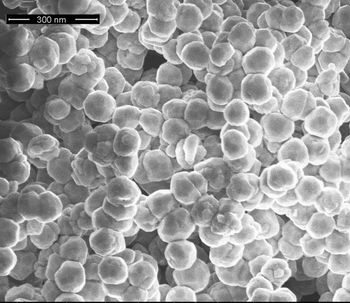
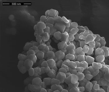
In general, the optical properties of luminescent materials are strongly influenced by morphology and the microstructure of particles. To obtain a white light emission with high quality, it is desired to synthesize spherical particles with nanometric dimensions and low agglomeration tendency, so as to limit the scattering effect, to high quantum efficiency, to avoid the agglomeration in the packaging step, and avoiding damage to other properties that could affect the quality of the white light emitted.[Reference Tucureanu, Matei and Avram7, Reference Nazaraov13] Using SEM, we studied shape, size, defects, and agglomeration tendency. In Figs. 5 and 6, we present the SEM micrograph for YAG:Ce and TAG:Ce powder, which are of relevance for the phosphor nanoparticles characterization. The form of the YAG:Ce particles proves to be spherical with smooth surfaces and uniform distribution, while for TAG particles, the distribution is uneven, some particles have approximately spherical shape while others being irregular. However, the particles of TAG:Ce have better shape and more uniform distribution than in a classic solid-state process for TAG:Ce powder synthesis. For the same manufacturing conditions, the TAG:Ce particles seem to be slightly bigger than YAG:Ce particles. We observed that the two yellow phosphor powders consist of crystallites with sizes ranging from the 40 nm to about 80 nm, with different degrees of agglomeration tendency. In both samples, we did not notice the presence of surface defects, which would indicate the presence of the dopant at the surface or in the crystal lattice.
SEM micrograph of the YAG:Ce phosphor powder after annealing at 1100 °C.

SEM micrograph of the TAG:Ce phosphor powder after annealing at 1100 °C.

Luminescence properties
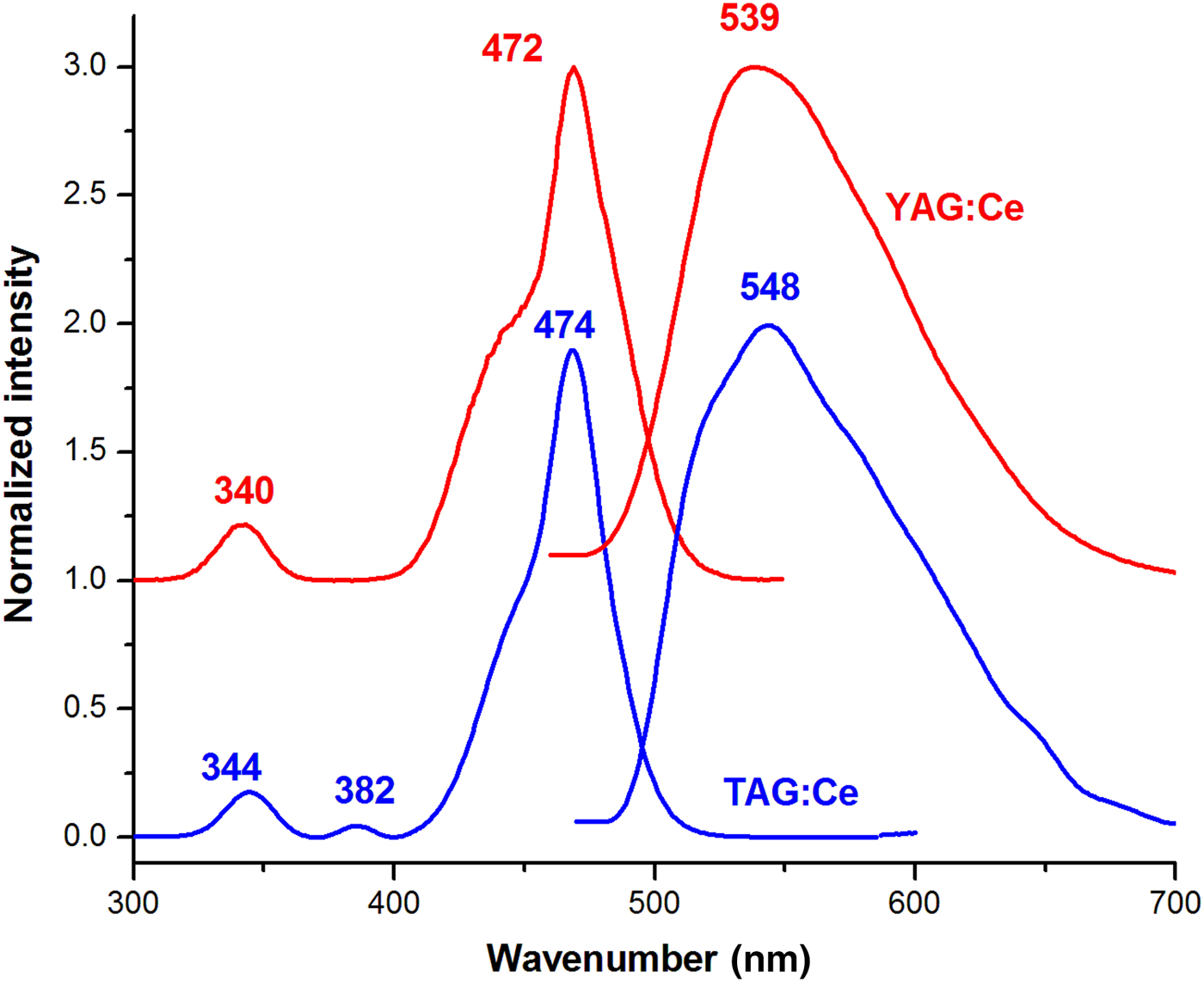
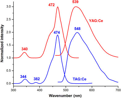
Garnet phosphors are luminescent materials most used for the development of LEDs with white light emission. By excitation with light emitted by a blue LED chip, the garnets phosphor will emit a broad yellow band that by combined with the remaining blue light leads to the generation of the light that the human eye perceives as white light. Commercial blue LED chips emit at 450–470 nm; thus, in this chapter, we study the optical properties of both phosphors by excitation at 470 nm. In Fig. 7, the normalized PL excitation and emission spectra of YAG:Ce and TAG:Ce yellow-emitting phosphor are presented, spectra that show the phase transition from amorphous to crystalline of both phosphors. The YAG garnet phosphor is an insulator material, but by adding lanthanide ions, new acceptor–donor levels and electron transitions between these energy levels appear. In our YAG and TAG samples, the use of Ce3+ as doping element involves light absorption due to transitions 4f (4f 1 configuration, split in two 2F 5/2 and 2F 7/2 levels) → 5d (4f 05d 1 configuration, split in two 2D 3/2 and 2D 5/2) what is seen in the PL excitation spectrum by two bands centered at about 340 and 470 nm. In addition, for TAG:Ce phosphor, a band centered at 382 nm is observed, that is due to the allowed transitions 4f 8 → 4f 8 in Tb3+.[Reference Wang, Han, Lang, Tu and Peng8, Reference Berezovskaya, Zadneprovski, Poletaev, Doroshenko, Efryushina, Zubar and Dotsenko17] The band located at about 472 and 474 nm (recorded at λ em = 620 nm) confirms the possibility of using blue emitting chips with emission at 460–470 nm. The yellow emission of garnets is due to 5d → 4f transition of the Ce3+, which absorbs blue excitation light and is sensible to process condition (i.e., Ce4+ → Ce3+ transition are influenced by sintering temperature) crystal field, crystallographic environment. In our PL emission spectra, a broad yellow band is observed, having the maximum at 539 nm for YAG:Ce and 548 nm for TAG:Ce, shifted in spectrum to the red by comparison with similar garnet phosphors obtained by other sol–gel methods. In the case of the TAG:Ce, a little shift to higher wavelengths is observed, which might be due to reduced local symmetry determined by the difference in ionic radius. YAG:Ce and TAG:Ce yellow emitting phosphors showed high PL quantum yield efficiency (QY) of above 46% and 42%, respectively, upon 470 nm excitation, which is a superior value compared with the quantum efficiency of YAG:Ce phosphor presented in literature for the other sol–gel methods. In comparison with the commercial Phosphor Tech QMK58/F-U1 (phosphor obtained by solid-state reaction, under a reducing atmosphere at a temperature of 1500 °C), our phosphors have lower QY.[Reference Tucureanu, Matei and Avram7, Reference Nazaraov13, Reference Tucureanu, Matei, Mihalache, Danila, Popescu and Bita30] Less QY in the case of TAG:Ce could be due to the presence of TAP in the crystal structure and to the irregularities in the morphologic structure of phosphor.
Normalization PL excitation spectra (left) and emission spectra (right—observed after excited at 470 nm) of (red) YAG:Ce phosphor and (blue) TAG:Ce phosphor.

Conclusions
The YAG:Ce and TAG:Ce powders were prepared by a modified sol–gel method. Starting from oxides and nitrides, the sol–gel process for YAG:Ce and TAG:Ce manufacturing was realized in acid catalyst, and AcAc was used as chelating. The thermal treatment started with a presintered step at 400 °C and subsequently annealed at 1100 °C. The use of DMSO in the process can improve the morphology of the particles. Using EDX analysis, we determined the presence of the main components and the composition was calculated and corresponds to Y2,78Ce0,20Al5,00O12,02 formula and, respectively, Tb2,83Ce0,17Al4,83O12,17 formula. The TAG:Ce deviation from the theoretical formula can be explained by the emergence of TAP phase with TAG-desired garnet phase. FTIR spectra confirm the transition from the amorphous to crystalline phase and the absence of impurities. A similar structure of the two phosphors is confirmed by the similarities between the two FTIR spectra and the presence of peaks below 800 cm−1 that may be assigned to the metal–oxygen bonds. Also, the presence of the Ce dopant in phosphor molecules is confirmed by FTIR spectroscopy analysis. In the XRD diagrams, all lines are indexed and assigned to cubic garnet structure for both yellow phosphors. In the case of YAG:Ce, the phase purity was found to be excellent, but for TAG:Ce, we observed the presence of TAP intermediated phase, but does not affect the optical properties of the material. The parameters have found to be 12.0273 A°, a 0.006% strain and crystallites of 37 nm for YAG:Ce, and 12.0509 A°, a 0.008% strain and crystallites of 48 nm for YAG:Ce, respectively. The SEM micrograph shows that using the same manufacturing condition, the TAG:Ce particles seem to be bigger than YAG:Ce particles, having crystals with sizes ranging from the 40 nm to about 80 nm, with different degrees of agglomeration tendencies. The PL excitation and emission spectra of YAG:Ce and TAG:Ce confirms the yellow emission of phosphors by the broad band centered at 539 nm for YAG:Ce and 548 nm for TAG:Ce. PL spectra also confirm the phosphors ability to be used with blue chips that emit at 470 nm for white light generated with a QY of above 46% for YAG:Ce and 42% for TAG:Ce. By comparing the properties of the two YAG:Ce and TAG:Ce phosphors, we can say that our modified sol–gel method is a viable alternative to the already known techniques for obtaining the yellow phosphors.
Acknowledgments
This work was supported by a grant of the Ministry of National Education and Scientific Research, RDI Program for Space Technology and Advanced Research—STAR, project number 639/2017. This work was also supported by UEFISCDI in the Partnership Framework: PN-III-P2-2.1-PTE-2016-0145 (Project No. 46PTE/2016), PN-II-PT-PCCA-2013-4-0366 (Project No. 208/2014) and by the National Basic Funding Programme TEHNOSPEC (Project No. PN1632/2016). The authors would like to acknowledge to researchers: Mihai Danila for technical support in samples characterization and Carmen Iorga, Tamara Dobre, Elena Nistor, and Marioara Paznicu for cooperation.