Case report
Cardiac involvement is the major cause of morbidity and mortality in hypereosinophilic syndrome, but sequential changes from thrombotic to fibrotic stages are rarely reported.
A 12-year-old boy with no significant past medical history presented with fatigue, fever, progressive dyspnoea, and loss of weight since 3 weeks. He immediately required inotropic support and non-invasive ventilation. His chest X-ray revealed bilateral infiltrates. Laboratory investigations demonstrated marked hyperleucocytosis (118×109/L) with 71% eosinophils. Extensive workup excluded all causes of secondary hypereosinophilia.
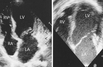
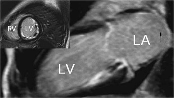
Transthoracic echocardiography showed moderate mitral regurgitation due to thickening of the posterior mitral leaflet and embedding of the valvar apparatus to a thick plaque adhering to the ventricular wall (Fig 1); ventricular function was preserved. Cardiac MRI confirmed early necrotic stage endomyocardial involvement associated with mural thrombi (Fig 2).
On the left, initial echocardiography shows thrombotic mass in the left ventricular posterior wall and mitral valve thickening (arrows). On the right, preoperative transoesophageal echocardiography shows the mass, with embedding of the posterior leaflet apparatus, resulting in severe mitral regurgitation (arrows). LA=left atrium; LV=left ventricle; RA=right atrium; RV=right ventricle.

Cardiac MRI at the time of diagnosis shows in the long-axis view typical subendocardial enhancement in the subvalvular region. Inset, short-axis, midventricular, two-chamber view shows a thin area of delayed enhancement distributed along the endocardium of the left ventricle (LV). LA=left atrium; RV=right ventricle.

The patient was commenced on steroids and responded clinically with respiratory improvement, but no change in eosinophil counts occurred. He was also anticoagulated with warfarin to prevent cardioembolism. As marked hypereosinophilia persisted and cardiac disease progressed, multiple second-line agents were administrated sequentially but were overall inefficacious: hydroxyurea, vincristine, methylprednisolone (intravenous pulses), interferon a-2b, nilotinib, mepolizumab, and lenalidomide had no persistent effect on the eosinophil count, whereas imatinib mesylate was interrupted because of hepatotoxicity.
After 4 months, severe mitral regurgitation due to endomyocardial fibrosis with invasion of the posterior leaflet and chordae tendinae (Fig 1) led to uncontrollable heart failure despite maximal medical management. Finally, eosinophil level was controlled with fludarabine. The patient underwent urgent heart surgery with Carpentier–Edwards porcine bioprosthesis replacement and endomyocardectomy under extracorporeal circulation. A bioprosthesis was preferred because mechanical valves have a high incidence of recurrent obstructive thrombosis. Pathological examination of the tissue removed during endomyocardectomy showed marked fibrous thickening of the endocardium with some areas of eosinophil infiltration within the fibrous tissue and immediate subendocardium. A few weeks later, the patient underwent unrelated cord blood haematopoietic stem cell transplantation without serious complications. Hypereosinophilia relapsed 2.5 years later and he died 3 years later of septic shock, while being treated for secondary B cell acute lymphoblastic leukaemia. Frequent follow-up visits from the time of surgery to his death included electrocardiography – sometimes Holter recording – and echocardiography. Recurrence of Loeffler endocarditis, occurrence of rhythm or conduction disorders, and development of ventricular dysfunction or restrictive cardiomyopathy were never observed or suspected. An autopsy was not performed.
Discussion
Hypereosinophilic syndrome is currently diagnosed in case of idiopathic hypereosinophilia and organ damage or dysfunction due to this haematological disorder. It is very rare in paediatrics.Reference Keivanidou, Giannopoulos, Papageorgiou, Hatzipantelis, Pana and Athanasiadou 1 It often results in eosinophil infiltration of cardiac tissues, and then is responsible for necrosis of the myocardium, damage to the endothelium with formation of intracavitary thrombi and systemic embolisation, and finally endomyocardial fibrosis resulting in restrictive cardiomyopathy and progressive valvar dysfunction.Reference Keivanidou, Giannopoulos, Papageorgiou, Hatzipantelis, Pana and Athanasiadou 1 , Reference Ogbogu, Rosing and Mc Donald 2 In this case, subvalvar damage aggravated the mitral valve regurgitation. The experience with valve replacement in such patients is very limited due to the rarity of the disease.Reference Kleinfeldt, Nienaber and Kische 3 Despite anticoagulation, mechanical valves may have a higher incidence of thrombosis, so that bioprosthetic valves are preferred even if it may require more frequent replacement.Reference Kleinfeldt, Nienaber and Kische 3
Acknowledgement
None.
Financial Support
This research received no specific grant from any funding agency, commercial, or not-for-profit sectors.
Conflicts of Interest
None.
Ethical Standards
The authors assert that all procedures contributing to this study comply with the ethical standards of the relevant national guidelines on human experimentation and with the Helsinki Declaration of 1975, as revised in 2008.