Introduction
Bipolar disorder (BD) is a psychiatric illness affecting 1% to 2% of the adult population.Reference Merikangas, Akiskal and Angst 1 The etiology and pathogenesis of BD are complex and not well understood. So far, a significant amount of effort placed in studies aiming at clarifying the biological basis of BD, including the role of oxidative stress, inflammation, DNA methylation, and microRNAs.Reference Camkurt, Karababa and Erdal 2 –Reference Kuratomi, Iwamoto and Bundo 5 Similarly, a large number of structural and functional neuroimaging studies have investigated putative alterations in BD patients.Reference Strakowski, DelBello and Adler 6
Diffusion-weighted imaging (DWI) is a specific neuroimaging approach based on water diffusion in tissues.Reference Taylor and Bushell 7 This technique is used to identify white matter tracts in the brain and their integrity.Reference Catani and De Schotten 8 Fractional anisotropy (FA) is a value used to describe the diffusion process and ranges from zero to one; zero stands for unrestricted diffusion. One stands for entirely restricted diffusion on one direction.Reference Le Bihan, Mangin and Poupon 9 Fractional anisotropy is considered an indirect indicator of white matter myelination, diameter of the axons, and density of fibers.Reference De Erausquin and Alba-Ferrara 10 , Reference Cohen-Adad, Descoteaux and Rossignol 11
The term endophenotype is mainly used in the field of psychiatry. Endophenotypes are applied in categorizing, describing, and differentiating psychiatric disorders from healthy controls or unaffected relatives. It also has implications of detecting under risk populations.Reference Gottesman 12 There are specific criteria in psychiatric research to describe a biological marker as an endophenotype, such as being heritable, segregated with illness, co-segregated with illness within the same family, measured reliably, more common within affected families and, finally, being independent of the stage of the illness.Reference Beauchaine 13
Given the noninvasive nature of imaging techniques, the identification of neuroimaging endophenotypes is of high interest. Various brain regions have been proposed to be altered in BD. Vederine et al.Reference Vederine, Wessa and Leboyer 14 performed a meta-analysis of whole-brain diffusion tensor imaging (DTI) studies in BD and found decreased FA in the parahippocampal gyrus and the subgenual cingulate cortex. A systematic review and voxel-based meta-analysis of DTI studies in BD pointed out three clusters of decreased FA; one cluster in the right posterior temporoparietal and two clusters in the left cingulate.Reference Nortje, Stein and Radua 15 A more recent meta-analysis by Yang et al.Reference Yang, Li and Hu 16 using tract-based spatial statistics showed significant FA reductions in the genu and body of the corpus callosum. Finally, mega and meta-analysis of 3033 individuals focusing on white matter abnormalities in BD revealed the corpus callosum and cingulum regions with the largest effect size. They also found that lithium treatment was associated with higher FA in multiple regions of interests (ROIs).Reference Favre, Pauling and Stout 17
Given the accumulating past evidence, decreased FA seems promising as a potential endophenotype for BD.Reference Hajek, Carrey and Alda 18 Several studies investigated this possible association by comparing healthy controls with BD patients and their relatives and described decreased FA in different brain regions as possible endophenotypes for BD. The regions reported before are the right temporal lobe, the corpus callosum, the fornix, the anterior and posterior thalamic radiata, the cingulum, the right middle frontal gyrus, and the right superior temporal gyrus.Reference McDonald, Bullmore and Sham 19 –Reference Mahon, Burdick and Ikuta 22 In addition to the regional abnormalities, widespread white matter abnormalities have been found throughout the brain regarding individuals at high genetic risk for bipolar disorder.Reference Sprooten, Sussmann and Clugston 23 , Reference Chaddock, Barker and Marshall 24
As mentioned above, endophenotypes have the potential to categorize and differentiate psychiatric disorders and to determine under risk populations. Here in the present study, we compared the white matter integrity of three groups with the ROI approach: BD patients, youth at high risk for BD, and healthy controls (HC). We hypothesized that both BD offspring and BD patients would have decreased white matter integrity represented by decreased FA compared to HC. Moreover, the offspring group would demonstrate a level between BD and HC, allowing FA’s characterization as a possible endophenotype for BD.
Methods
Subjects
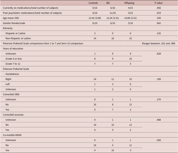
Study approval was obtained from the local institutional review board at the University of North Carolina at Chapel Hill. Written informed assent and consent were obtained from all subjects and parents/guardians. Study participants included 15 unaffected (nonbipolar) children of patients with BD, 16 pediatric patients with BD, and 16 healthy controls matched for gender, age, pubertal status, ethnicity, years of education, and handedness (Table 1).
Clinical and Demographic Characteristics of the Groups

This table demonstrates clinical characteristics of groups in terms of age, gender, current and past psychiatric medication status, ethnicity, handedness, years of education, pubertal status, comorbid oppositional and defiant disorder, enuresis, attention deficit, and hyperactivity disorder. P value determined as .05.
Abbreviations: ADHD, attention deficit hyperactivity disorder; ODD, oppositional and defiant disorder; SD, standard deviation.
Patients were diagnosed with BD type I, type II, or BD not otherwise specified (BD NOS) according to the Diagnostic and Statistical Manual of Mental Disorders, fourth edition (DSM-IV) criteria. 25 The exclusion criteria included substance abuse within the past 6 months, as well as general medical problems. BD offspring were characterized by the presence of BD in at least one biological parent according to the Structured Clinical Interview for DSM disorders. Axis I disorder (other than mood disorders or psychotic disorders) were not considered exclusion criteria for offspring. For healthy controls, the inclusion criteria were the absence of a history of any psychiatric disorders or neurologic disorders or mood disorders in first degree relatives. Exclusion criteria for both patients and healthy controls included head injury with loss of consciousness, presence of metallic objects in the body, family history of hereditary neurological disorders, and pregnancy.
All participants completed a semi-structured interview, the Schedule for Affective Disorders and Schizophrenia for School-Age Children-Present and Lifetime Version (K-SADS-PL).Reference Kaufman, Birmaher and Brent 26 The K-SADS-PL is the most traditionally used questionnaire to evaluate Axis I disorders in children and adolescents. Parents and children interviewed separately.
No subjects in any group met criteria for conduct disorder, binge eating disorder, or substance use or dependence (alcohol, cannabis, cocaine, and other substances). Of note, comorbidity with attention deficit hyperactivity disorder (ADHD) was present in 10 out of the 15 patients (66.7%).
Magnetic resonance imaging (MRI) protocol
DWI scans were acquired using a 3.0T Siemens Allegra scanner using a spine echo-planar imaging protocol with the following acquisition parameters. Echo time = 79 ms, repetition time = 9200 ms, slice thickness = 2 mm, voxel size = 2 mm, image matrix = 104 × 128 with 21 diffusion encoding directions, b-value = 1000 s/mm2 and one nondiffusion weighted volume. At the same time, T1-weighted scans were acquired using a three-dimensional magnetization prepared rapid gradient echo (3DMPRAGE) protocol with the following parameters. Repetition time (TR) = 1750 ms, echo time (TE) = 4.38 ms, flip angle = 8°, slice thickness = 1 mm, matrix size = 256 × 208, and voxel size = 1 mm.
Image pre-processing
All scans were visually inspected to rule out gross artifacts. DWI scans were pre-processed using the FMRIB software package (FSL) version 5.0.7Reference Smith 27 in the following steps: (a) Correction for “eddy currents” using the “eddy_correct” routine. (b) Removal of the skull and nonbrain tissue using FSL’s brain extraction tool (BET). (c) Calculation and fitting of a diffusion tensor at every voxel using the “dtifit” routine. (d) Estimation of FA, axial diffusivity (AD), and radial diffusivity (RD) volumes for each subject to facilitate atlas-based regional measurement extraction.
T1 weighted scans were automatically segmented using the FreeSurfer software library version 5.3.0 (http://surfer.nmr.mgh.harvardu.edu).Reference Fischl 28 Brain white matter (WM) tissue was parcellated using an automated technique utilizing morphometric landmarks and gray matter/WM boundary information, as explained in detail elsewhere.Reference Salat, Lee and Van der Kouwe 29 This process resulted in 68 WM parcellations, that have been previously used in multiple neuroimaging studies, including pediatric ones.Reference Salat, Lee and Van der Kouwe 29 –Reference Østby, Tamnes and Fjell 32
Statistical analysis
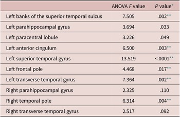
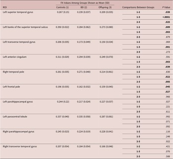
A previous study from our group has identified a number of atlas-based regions that can be used to differentiate pediatric BD patients’ brains from those of healthy controls, using a multivariate support vector machine learning approach.Reference Mwangi, Wu and Bauer 31 Using these published results, we selected the 10 regions (see Table 2) most relevant to distinguishing patients from healthy controls and included them in our analysis. Regions found to be statistically different were then included in a matched three-group comparison across BD offspring, BD patients, and healthy controls. This secondary analysis was done using ANOVA, and Tukey test was used for post-hoc analysis of ROIs, and each comparison between groups represented in Table 3. ANOVA test was used to compare parametric continuous variables across groups, while the Kruskal–Wallis test was used for nonparametric continuous variables. Chi-Square test was used to compare categorical variables.
Comparison of FA Values Across Groups with ANOVA

ANOVA test result for regions of interests (ROIs) investigated.
Abbreviation: FA, fractional anisotropy.
* P value determined as 0.05.
** ROIs showed significant differences between groups.
Results
Comparison of current medication status and history of psychiatric medications is shown in Table 1. The control group consisted of drug naïve subjects. 37.5% (6/16) of the BD patients and 26.6% (4/15) of the offspring group were currently on psychotropic medications, P-value: .456. 68.7% (11/16) of the bipolar patients and 33.3% (5/15) of the offspring group had a history of using psychotropics, P-value: .019.
When we compared FA between BD patients, offspring group and healthy controls, we found significantly decreased FA in 6 of the 10 areas analyzed (Figure 1), LSTG (F = 13.519 and P < .0001), LBSTS (F = 7.505 and P = .002), LTTG (F = 7.364 and P = .002), LAC (F = 6.500 and P = .003), RTP (F = 6.314 and P = .004), and LFP (F = 4.468 and P = 0.017). Overall, mean FA values among groups were consistently in the following order: controls > BD > offspring, except for two ROIs; left and right parahippocampal gyrus.
Fractional anisotropy (FA) values in significant brain regions.

We further performed a post-hoc analysis to determine the origin of the significant findings. Post-hoc analysis of the ANOVA results shown in Table 3 for each comparison. Out of these six significant ROIs, three showed higher FA only in the control group compared to BD patients and offspring group: LBSTS (control vs BD P-value: .009, control vs offspring P-value: .003), LFP (control vs BD P-value: .045, control vs offspring group .027), LTTG (control vs BD group P-value: .069, control vs offspring group P-value: .001). On the other hand, LSTG showed significance with all comparisons (control vs BD P-value .039, control vs offspring P-value < .0001, BD vs offspring .025). LAC showed significantly increased FA values in controls and BD group compared to offspring group (control vs offspring P-value: .003, BD vs offspring P-value: .039, control vs BD P-value: .591). In addition, RTP showed similar statistical significance as LAC (control vs offspring P-value: .022, BD vs offspring P-value: .005, control vs BD P-value: .830.)
Fractional Anisotropy Values in Different Brain Regions Among Bipolar Patients, Nonbipolar Offspring, and healthy controls

Post Hoc analysis of ROIs between groups.
Comparisons between groups performed as following: 1-2 Controls vs BD, 1-3 Controls vs Offspring, and 2-3 BD vs Offspring.
Abbreviations: FA, fractional anisotropy; BD, bipolar disorder patients; SD, standard deviation.
Discussion
Our results indicated significant differences across groups with regards to FA values in six ROIs. In some regions (LTTG, LBSTS, and LFP), significantly higher FA values were observed only in the control group, while other regions (LSTG, LAC, and RTP) showed significantly higher FA values among BD patients and controls compared to offspring.
The STG seems to be implicated in the pathophysiology of multiple psychiatric disorders, including schizophrenia and bipolar disorder.Reference Pearlson, Barta and Powers 33 , Reference Rajarethinam, DeQuardo and Nalepa 34 In our study, FA in STG showed significant differences across groups, see Table 3. Temporal lobe structuresReference Altshuler, Bartzokis and Grieder 35 are essential for speech, language, and communication and have been a particular topic of interest in bipolar disorder and decreased volume and altered neurochemical imbalances have been previously reported in the temporal lobe of BD patients.Reference Altshuler, Conrad and Hauser 36 , Reference Deicken, Weiner and Fein 37 Moreover, recent evidence suggests an endophenotype potential of FA values in temporal lobe structures BD. For example, Sprooten et al.Reference Sprooten, Sussmann and Clugston 23 found decreased FA across the whole white matter skeleton among unaffected relatives of BD compared to controls, as well as a negative association between cyclothymic temperament and FA in the left temporal white matter. Mahon et al.Reference Mahon, Burdick and Ikuta 22 compared 26 BD patients, 15 unaffected siblings, and 27 healthy controls. Their results indicate that the right temporal lobe FA values correspond to a significant and intermediate phenotype between patients and healthy controls. LSTG, which hosts the primary auditory cortex (Brodmann areas [BA] 41 and 42) and auditory association areas, is vital for speech and language. Besides, a previous study of pediatric bipolar patients found decreased LSTG total and WM volume compared to healthy controls.Reference Chen, Nicoletti and Hatch 38 We found decreased FA in the STG among both BD patients and offspring group, more prominently in the offspring group. Evaluation of our results in the light of existing literature supports the potential role of decreased FA in STG as a neuroimaging marker for patients or an endophenotype for high-risk populations. Future studies with prospective design and larger sample size needed for further clarification.
Our findings also indicate that the FA values in the LAC were significantly higher in the control and BD groups compared to the offspring group. The cingulate plays a key role in complex emotional behavior.Reference Bush, Luu and Posner 39 , Reference Phillips, Drevets and Rauch 40 Anterior cingulate cortex (ACC) is a highly evolved and specialized cortical area, containing spindle-shaped neurons found only in humans and great apes, and is vital to emotional processing.Reference Allman, Hakeem and Erwin 41 Abnormalities in the ACC have been implicated in the neurobiology of mood disordersReference Leppänen 42, and a considerable body of studies focused on neuroimaging changes in the cingulate cortex in bipolar disorder. Reduced LAC volume reported in patients with untreated BD.Reference Sassi, Brambilla and Hatch 43 Moreover, decreased perigenual and subgenual anterior cingulate volumes reported in a meta-analysis of BD patients.Reference Ellison-Wright and Bullmore 44 In light of available data, the endophenotype potential of decreased FA in the cingulate cortex is limited and needs further exploration. For example, a study by Emsell et al.Reference Emsell, Chaddock and Forde 45 detected a significant group effect on FA values in the cingulum. Similarly, Sarıçiçek et al.Reference Sarıçiçek, Zorlu and Yalın 21 investigated white matter integrity as a structural endophenotype in 27 BD patients, 20 unaffected siblings, and 29 healthy controls. They found significantly lower FA values in the cingulate cortex of BD patients compared to healthy controls. The comparison of our results with the existing literature increases the importance of the cingulate cortex as a target region for structural imaging studies in BD. Our findings suggest that decreased FA in the LAC may correspond to a marker of vulnerability for BD, while possible medication effects might have neutralized that decrease by increasing the FA values, hence the lack of difference between BD and HC. Available evidence strongly points to the neuroprotective effects of mood stabilizers, especially Lithium, and neural promoting properties, which can positively alter brain function and structure.Reference Brambilla, Bellani and Yeh 46 –Reference Atmaca, Ozdemir and Yildirim 48 Prospectively designed studies must be done to clarify mood stabilizers’ effects on the FA values of pediatric BD patients before and after treatment.
We also found significantly increased FA in the LFP among HC compared to the BD and offspring groups. These results are in agreement with previous literature findings supporting the involvement of the frontal cortex in the pathophysiology of mood disorders. The frontal pole cortex (FPC; BA 10) is a highly evolved area of the brain divided into a medial and a lateral part, referred to respectively as the ventromedial prefrontal cortex (vmPFC), and the lateral FPC.Reference Semendeferi, Armstrong and Schleicher 49 Decreased PFC volume has been a consistent finding in bipolar disorder, and reduced top-down inhibition of the amygdala by the vmPFC seems to be implicated in mood and anxiety disorders.Reference Myers-Schulz and Koenigs 50 , Reference Arnone, Cavanagh and Gerber 51 For example, abnormal activity and connectivity of the vmPFC–amygdala circuit has been described in pediatric BD.Reference Ladouceur, Farchione and Diwadkar 52 Moreover, a study by Matsuo et al.Reference Matsuo, Kopecek and Nicoletti 20 investigated potential structural neuroimaging endophenotypes for BD, and found a significant main effect of diagnosis for the medial frontal gyrus volume. Besides, this study further described smaller right medial frontal white matter volumes in unaffected relatives of bipolar patients.
Along with its contributions to the field, our study has some methodological limitations that must be discussed. Mainly, we analyzed a small sample size and utilized a cross-sectional design which limits power of the study and interpretation of the results. Furthermore, the presence of comorbid ADHD and other Axis I disorders, and significant past medication history in the patient group may have impacted our results. Lack of neurocognitive data and functional MRI data could also be considered as additional limitations.
Conclusion
In conclusion, our findings suggest that decreased FA in various brain regions, particularly in the temporal lobe structures, can be considered as potential endophenotype candidates. Future longitudinal studies are needed to address whether these FA changes in at-risk individuals are predictors of the future development of BD and to investigate the possible etiological and pathophysiological mechanisms of WM microstructural changes in these populations.
Funding
The acquisition of the MRIs in this study was funded by NIH R01 Endophenotypes.
Disclosures
Jair C. Soares has following conflict of interests to disclose: Compass (research grant), Alkermes (research grant), Pfizer (consultant), Allergan (research grant), Sunovian (consultant), Sanofi (consultant), Johnson & Johnson (consultant), Livanova (consultant), and Boehringer Ingelheim (consultant). Mehmet Camkurt, Tomas Melicher, Benson Mwangi, Mon-Ju Wu, Bo Cao, Cristian Zeni, Jonika Tannous, Giovana Zunta-Soares, Khader Hasan, Marsal Sanches declare no conflict of interests.