I. INTRODUCTION
Biodegradable metals have received significant scientific interest in recent years and Fe-based alloys are considered promising candidates. Reference Hermawan, Dube, Mantovani, Chandra, Tsuzaki, Militzer and Ravindran1,Reference Schinhammer, Hänzi, Löffler and Uggowitzer2 In animal studies, the potential of pure Fe was evaluated and it was noted that the main drawback of Fe is its slow in vivo degradation rate. Reference Peuster, Wohlsein, Brugmann, Ehlerding, Seidler, Fink, Brauer, Fischer and Hausdorf3–Reference Waksman, Pakala, Baffour, Seabron, Hellinga and Tio5 According to a design strategy based on metallurgical, electrochemical, and toxicological considerations, Schinhammer et al. Reference Schinhammer, Hänzi, Löffler and Uggowitzer2 developed novel biodegradable Fe–Mn–Pd steels. These alloys were found to exhibit increased in vitro degradation rates compared to pure Fe. Furthermore, an appropriate biocompatibility was evaluated in cytotoxic investigations of Fe–Mn, Reference Hermawan, Purnama, Dube, Couet and Mantovani6 Fe–Pd, Reference Huang, Cheng and Zheng7 and Fe–Mn–C–(Pd) Reference Schinhammer, Gerber, Hänzi and Uggowitzer8 alloys.
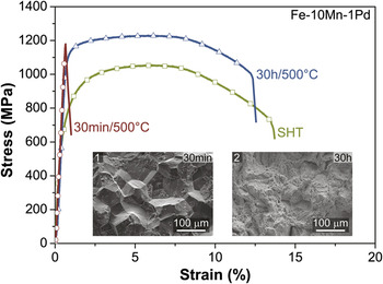
In addition to enhanced degradation rates, high mechanical strength would also be beneficial, so that smaller implant dimensions can be used and thus less material needs to degrade. In the case of Fe-based alloys, maraging steels reveal ultrahigh strength, which is based on the formation of intermetallic precipitates forming in the martensitic matrix during an aging treatment at temperatures between 300 and 500 °C. Reference Decker and Floreen9 Moszner et al. Reference Moszner, Sologubenko, Schinhammer, Lerchbacher, Hänzi, Leitner, Uggowitzer and Löffler10 observed that the martensitic alloy Fe–10Mn–1Pd (in wt.%) exhibits all features characteristic of maraging steels and that considerable age-hardening is achieved through the formation of nanometer-sized precipitates rich in Mn and Pd. For this alloy, an interesting combination of strength and ductility was observed in the overaged state (see Fig. 1). Thus, the potentially attractive electrochemical and biological characteristics, in combination with the suitable mechanical performance, make the material very interesting for biodegradable implant applications. For further alloy optimization, the structural and chemical nature of the precipitates, in particular in the overaged state, is, however, of significant interest.
Engineering stress–strain curves of Fe–10Mn–1Pd in the initial SHT state and after isothermal aging at 500 °C for 30 min (peak-aged) and 30 h (overaged). Reference Moszner, Gerstl, Uggowitzer and Löffler24 A ductile behavior is seen in the SHT condition and in the overaged case. The yield strength of the alloy in the overaged state (1076 ± 6 MPa) is much larger than that of the SHT state (662 ± 13 MPa). Brittle failure results for the material in the peak-aged condition. The insert shows SEM pictures of fracture surfaces from the tensile specimens of the alloy in the peak-aged (1) and overaged (2) conditions. Intergranular fracture, which occurs along prior austenite grain boundaries, is visible in the peak-aged state, and a ductile dimple fracture is apparent in the overaged case.

It is known that the iron-rich martensite alloys of the ternary systems Fe–Mn–Ni and Fe–Mn–Pt also exhibit a maraging behavior. Reference Moszner, Sologubenko, Schinhammer, Lerchbacher, Hänzi, Leitner, Uggowitzer and Löffler10–Reference Tanaka and Suzuki12 Due to its technological relevance, the Fe–Mn–Ni system has been extensively studied, Reference Kardonskii and Perkas11,Reference Squires and Wilson13–Reference Singh and Wayman22 and for this system the precipitation sequence suggested can be summarized as follows: (i) in the initial aging stage the formation of coherent NiMn body-centered-cubic (bcc) zones (A2 or B2) takes place Reference Kardonskii and Perkas11,Reference Yodogawa14,Reference Hossein Nedjad, Nili Ahmadabadi, Furuhara and Maki18 ; (ii) at later stages of aging (overaged region) θ-NiMn intermetallic precipitates with a face-centered tetragonal (fct) structure form Reference Squires and Wilson13–Reference Shiang and Wayman15,Reference Kim and Wayman17,Reference Hossein Nedjad, Nili Ahmadabadi, Furuhara and Maki18,Reference Ray and Seal20 (however, Kardonskii et al. Reference Kardonskii and Perkas11 proposed the formation of Ni3Mn and Singh et al. Reference Singh and Wayman22 the formation of bcc NiMn); and (iii) on prolonged aging the precipitates redissolve and the final equilibrium microstructure only consists of austenite and ferrite (or martensite). Reference Yodogawa14 For the Fe–Mn–Pt system, Tanaka et al. Reference Tanaka and Suzuki12 proposed a similar precipitation sequence, i.e., the formation of coherent bcc PtMn zones (B2) at the initial stages of aging and ordered fct β1-PtMn intermetallic precipitates in the overaged regime. Reference Tanaka and Suzuki12 In contrast to the Fe–Mn–Ni system, the dissolution of the particles on prolonged aging was not observed for the Fe–Mn–Pt alloys, which was ascribed to the higher bonding affinity of Mn with Pt. Reference Tanaka and Suzuki12,Reference Suzuki23 In the Fe–Mn–Pd system, we suggested Reference Moszner, Sologubenko, Schinhammer, Lerchbacher, Hänzi, Leitner, Uggowitzer and Löffler10 the formation of coherent zones rich in Mn and Pd in the initial stages of aging. However, the structure of the precipitates in the overaged state has yet not been clarified, which is our aim in the present work.
For this reason, alloys with a Pd concentration of 1, 3, and 6 wt.% and a fixed Mn content of 10 wt.% were studied. The steels were isothermally aged at 500 °C and the mechanical properties as a function of aging time were investigated. The crystallography of the evolving precipitates was characterized by x-ray diffraction (XRD), and atom probe tomography (APT) was performed to analyze their chemical composition.
II. EXPERIMENTAL
Casts of 80 g were prepared from pure elements (Fe: Armco; Mn: 99.9% pure, Alfa Aesar, Germany; Pd: 99.95% pure, UBS, Switzerland) by vacuum induction-melting under argon atmosphere (purity: 99.998%). Prior to the homogenization treatment, the ingots were encapsulated in quartz tubes also under argon atmosphere. The solution-heat-treatment (SHT) of Fe–10Mn–1Pd was performed at 1250 °C for 12 h, Reference Moszner, Sologubenko, Schinhammer, Lerchbacher, Hänzi, Leitner, Uggowitzer and Löffler10 and for Fe–10Mn–3Pd and Fe–10Mn–6Pd at 1400 °C for 1 h under argon atmosphere, followed by water quenching. The higher SHT temperature for the steels containing 3 and 6 wt.% was chosen because SEM investigations of Fe–10Mn–3Pd revealed that such a temperature is required to obtain a homogenous microstructure. Isothermal aging of the SHT specimens was conducted at 500 °C in air for various durations (up to 1200 h), followed by quenching in water. Prior to aging of the samples for 1200 h at 500 °C, they were sealed in quartz tubes under argon atmosphere to prevent oxidation. To follow the aging process of the heat-treated materials, Vickers hardness measurements were performed using a Brickers 220 hardness tester (Gnehm, Switzerland).
For the preparation of tensile test specimens of Fe–10Mn–1Pd, an ingot of 1 kg was prepared which was forged and swaged to produce rods having a diameter of 6 mm. Reference Moszner, Gerstl, Uggowitzer and Löffler24 The finial SHT of this material was conducted at 1150 °C for 12 h under argon atmosphere followed by water quenching. Stress–strain curves were recorded via standard 25 tensile testing (Schenck-Trebel, Germany), using cylindrical specimens of 2 mm diameter with a gage length of 12 mm at a strain rate of 10−3 s−1.
The microstructure of Fe–10Mn–xPd (x = 1, 3, 6) samples etched with 1 vol% nitric acid in water was investigated using optical microscopy (OM, Reichert-Jung Polyvar Met, Reichert Inc., Depew, NY), and scanning electron microscopy (SEM) was performed with a Hitachi SU-70 (Hitachi High-Technologies Corporation, Tokyo, Japan), equipped with an X-max energy-dispersive x-ray (EDX) detector (Oxford Instruments, Oxford Instruments plc, UK). For phase identification, XRD measurements (PANalytical X’Pert Pro, PANalytical Co., Netherlands) were conducted using Cu Kα (λ = 0.15406 nm) radiation, operating at 45 kV and 40 mA with a step size of 0.03° and a time interval of 12 s, and a diffracted beam monochromator to reduce fluorescence effects. Specimens of all alloys aged for 30 h at 500 °C were analyzed via APT. For this, bars of 0.3 × 0.3 × 15 mm3 were cut from the aged ingots and needle-shaped specimens were obtained by electrochemical polishing via a standard two-step process. Reference Moszner, Sologubenko, Schinhammer, Lerchbacher, Hänzi, Leitner, Uggowitzer and Löffler10,Reference Miller and Forbes26 The APT measurements of Fe–10Mn–1Pd were conducted on a LEAP™ 3000X HR (Cameca, AMETEK Inc., France) in voltage mode using a specimen temperature of ∼80 K, a pulse fraction of 15%, and a constant detection rate of 0.5%. For the alloys Fe–10Mn–3Pd and Fe–10Mn–6Pd, the APT experiments were performed on a LEAP™ 4000X HR (Cameca) in laser mode (355 nm wave length), because premature failure of the specimen occurred with voltage pulsing. The measurements were conducted using a laser energy of 100 pJ, a specimen temperature of ∼24 K, and a constant detection rate of 1%. Data processing was performed using the IVAS 3.6.6 software package (Cameca). Reference Larson, Prosa, Ulfig, Geiser and Kelly27
III. RESULTS AND DISCUSSION
A. Mechanical properties
Figure 1 shows results from tensile tests of Fe–10Mn–1Pd after isothermal aging at 500 °C for different times. In the initial as-quenched condition (solution-heat-treated, SHT), the alloy exhibits a ductile behavior, but brittle failure results in the peak-aged state. However, on prolonged aging, i.e., in the overaged regime, ductility recovers again and the alloy exhibits a similar uniform elongation compared to the SHT condition. The peculiarity of embrittlement and de-embrittlement is discussed in more detail by Moszner et al. Reference Moszner, Gerstl, Uggowitzer and Löffler24 Besides that, the yield strength of the material is considerably increased from 662 ± 13 MPa in the as-quenched condition to 1076 ± 6 MPa in the overaged state. Thus, an interesting combination of strength and ductility results in the overaged condition and smaller implant dimensions can be achieved in that case.
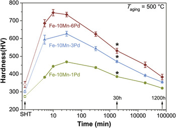
Figure 2 presents age-hardening curves for Fe–10Mn–xPd (x = 1, 2, 6), which were isothermally aged at 500 °C for time periods ranging from 5 min up to 1200 h. All alloys show significant age-hardening. The higher the Pd content of the alloys, the higher is the resulting peak-hardness, which shows clearly that Pd participates in the strengthening process. The hardness evolution of the alloys, i.e., a fast increase in hardness until the peak hardness is reached followed by a slow decrease on prolonged aging, is typical of maraging steels. Reference Decker and Floreen9,Reference Moszner, Sologubenko, Schinhammer, Lerchbacher, Hänzi, Leitner, Uggowitzer and Löffler10
Hardness as a function of aging time at 500 °C, revealing age-hardening behavior of the alloys investigated, which is typical of maraging steels. The heat-treatment states used for the XRD and APT measurements are indicated by black arrows and stars, respectively.

B. Optical microscopy
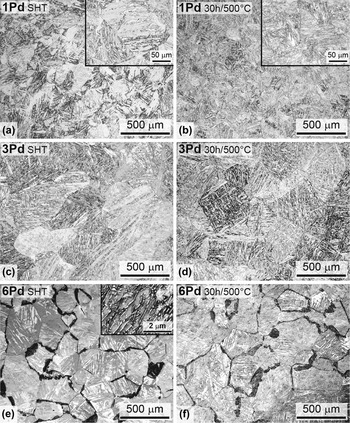
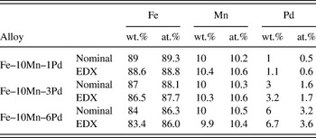
For comparison, Fig. 3 presents optical micrographs of the materials in the SHT condition and after aging for 30 h at 500 °C (overaged state). The SHT specimens containing 1 and 3 wt.% Pd [Figs. 3(a) and 3(c)] exhibit the typical lath-martensitic microstructure known from binary Fe–Mn alloys containing 4–10 wt.% Mn. Reference Roberts28,Reference Bolton and Petty29 SEM investigations of Fe–10Mn–1Pd revealed that the SHT temperature of 1250 °C was sufficient to produce a supersaturated solid solution with a homogenous single-phase microstructure. Reference Moszner, Sologubenko, Schinhammer, Lerchbacher, Hänzi, Leitner, Uggowitzer and Löffler10 However, for the steel containing 3 wt.% Pd, the formation of a single-phase microstructure required a homogenization treatment at 1400 °C [Fig. 3(c)]. For Fe–10Mn–6Pd annealed at 1400 °C, a coarse two-phase microstructure is revealed in the OM images [Fig. 3(e)]. The dominating phase exhibits the characteristic lath-martensitic structure similar to Fe–10Mn–1Pd or Fe–10Mn–3Pd, whereas the second phase (dark-contrast in Fig. 3(e)) was only found along some prior austenite grain boundaries. The inset to Fig. 3(e) shows a backscattered electron image of that phase, also revealing a chemical contrast. A lamellar-like structure is visible, and the bright areas are enriched in Pd and Mn. The image indicates partial remelting of the austenite grains due to overheating slightly above solidus, i.e., quenching from the austenite–liquid two-phase field. Reference Püttgen, Hallstedt, Bleck and Uggowitzer30,Reference Schumann and Oettel31 Nevertheless, a homogenous distribution of the alloying elements and a high level of Pd supersaturation can be assumed in the dominating martensitic phase. This results in a higher degree of hardness upon isothermal aging (Fig. 2) and hence a higher volume fraction of the evolving precipitates, which also generates stronger signals from the precipitates in XRD measurements facilitating their phase identification. Compared to the specimens in the SHT condition, a finer microstructure inside the prior austenite grains was observed during over-aging for the alloys investigated [Figs. 3(b), (d), (f)]. This can be ascribed to the formation of reverted austenite Reference Shiang and Wayman15 and was in particular apparent for Fe–10Mn–1Pd (compare insets to Figs. 3(a) and 3(b)). Table I summarizes the chemical compositions of the alloys determined via EDX-point measurements.
Optical micrographs of the alloys in the SHT condition and after aging for 30 h at 500 °C: (a, b) Fe–10Mn–1Pd (SHT at 1250 °C); (c, d) Fe–10Mn–3Pd (SHT at 1400 °C); (e, f) Fe–10Mn–6Pd (SHT at 1400 °C), all etched with 1% HNO3. The typical lath-martensitic microstructure is visible for the alloys containing 1 and 3 wt.% Pd. For Fe–10Mn–6Pd, a two-phase microstructure is visible with the lath-martensitic structure dominating. The insets in (a, b) show magnified sections, where a finer microstructure inside the prior austenite grains is apparent for the specimens in the overaged state. The inset in (e) shows a backscattered electron image of the intergranular phase with dark contrast in the optical micrograph.

Nominal and measured (EDX-point measurements) chemical compositions in weight (wt.%) and atomic percent (at.%).

C. Atom probe tomography
The APT experiments were conducted to analyze the chemical composition of the precipitates and hence to support the results of the XRD measurements (see section D). Specimens of the alloys in the overaged regime (after aging for 30 h at 500 °C) were investigated via APT. This condition was chosen in particular, as Moszner et al.
Reference Moszner, Sologubenko, Schinhammer, Lerchbacher, Hänzi, Leitner, Uggowitzer and Löffler10
observed for Fe–10Mn–1Pd that in this state the volume fraction of the precipitates (f
V) did not increase any more with progressive aging-time, indicating that f
V has approached an equilibrium value. In addition, the size of the particles was still small enough to fully enclose a few of them in the volume detectable by APT. To illustrate the APT results, cylinders of the same dimension (Ø = 38 nm, height = 40 nm) were cut out of the individual reconstructed volumes. Figure 4(a) presents the atomic positions of Fe (green), Mn (red), and Pd (blue). Enrichment of Mn and Pd at the same areas is clearly visible for all specimens investigated. The composition of the precipitates was determined using the proximity histogram (or proxigram) method.
Reference Hellman, Vandenbroucke, Rusing, Isheim and Seidman32
Isoconcentration surfaces
Reference Hellman, Vandenbroucke, Rusing, Isheim and Seidman32
(surfaces enclosing a region containing a higher concentration of a selected element compared to a chosen threshold value) were created to delineate the precipitates and to serve as reference surface for the proxigram calculation [Fig. 4(a)]. In a second step, discreet shells with widths of 0.1 nm inside and outside of the isoconcentration surfaces were generated. Each point in the proxigram then corresponds to the concentration of a shell at a defined distance from the isoconcentration surface. By convention, the concentrations at negative distances correspond to shells located outside the isoconcentration surface, which can be used to evaluate the matrix composition. Vice versa, data points at positive distances are related to concentrations inside the isoconcentration surface and hence can be used to assess the composition of the precipitates. Figure 4(b) presents the results of the proxigrams (only precipitates fully enclosed within the reconstructed volume were considered) for the investigated specimens aged for 30 h at 500 °C. The proxigrams were calculated using an isoconcentration surface of Mn + Pd set to a threshold value of 35 at.% (average inflection point of the Mn + Pd concentration taking into account the three datasets). The black dashed lines in the proxigrams indicate the data points used to calculate the concentration of the elements within the matrix (C
Matrix) and within the precipitates (C
Prec), and the corresponding values are depicted in each graph. According to Miller et al.
Reference Miller and Forbes26
the concentration in at.% of each element (C
i
) was calculated by C
i
= N
i
/N
t, where N
i
is the number of atoms of the corresponding species i and N
t is the total number of atoms considered. The errors were estimated by the standard deviation
 ${\rm{\sigma }}_i = \sqrt {{{C_i \left( {1 - C_i } \right)} \mathord{\left/ {\vphantom {{C_i \left( {1 - C_i } \right)} {N_t }}} \right. \kern-\nulldelimiterspace} {N_{\rm{t}} }}}$
. Similar concentration profiles were obtained for all three alloys. The Pd content of the matrix nearly decreases to zero, reflecting the fact that most of the Pd is confined within the particles. The precipitates are nearly free of Fe and mainly consist of Mn and Pd, thus indicating a Mn–Pd intermetallic character of the hardening phase. According to the binary Mn–Pd phase diagram,
Reference Okamoto33
two intermetallic phases, β and β1, can be considered here with respect to the measured composition of the precipitates. Both phases exist over a wide compositional range (β = 39 ≤ Pd at.% ≤ 66, β1 = 47 ≤ Pd at.% ≤ 68), at which β only occurs above 540 °C, whereas β1 also exists below that temperature. However, Okamoto
Reference Okamoto33
noted that the true equilibrium phase diagram is still unknown and hence the exact boundaries of the phases need to be considered with care.
(a) 3D reconstruction of Fe–10Mn–xPd alloys (x = 1, 3, 6) after aging for 30 h at 500 °C, showing the atomic positions of Fe (green), Mn (red), Pd (blue), and isoconcentration surfaces for regions containing >35 at.% Mn + Pd (orange). (b) Proxigrams for Fe, Mn, and Pd corresponding to a Mn + Pd isoconcentration of 35 at.%. The dashed black lines indicate the data points used to evaluate the composition of the matrix (C
Matrix) and of the precipitates (C
Prec). The total numbers of atoms considered for the concentration measurements of the matrix
 $\left( {N_{\rm{t}}^{{\rm{Matrix}}}} \right)$
and of the precipitates
 $\left( {N_{\rm{t}}^{{\rm{Prec}}}} \right)$
are also given. For all alloys, the precipitates are nearly free of Fe and mainly consist of Mn and Pd, indicating that a Mn–Pd intermetallic phase has formed.

D. X-ray diffraction
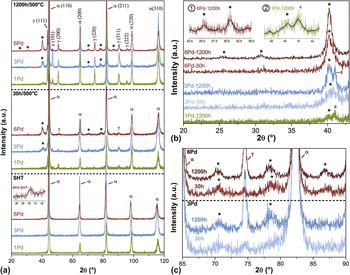
Figure 5 displays the XRD patterns of the alloys investigated in the SHT state, and subjected to aging at 500 °C for 30 h and 1200 h. In the SHT condition, the samples containing 1 and 3 wt.% Pd only reveal the presence of bcc diffraction lines. For Fe–10Mn–6Pd, in addition to strong bcc reflections, a weak peak at around 40.2° is visible (insert in Fig. 5(a)), which can be ascribed to the observed intergranular second phase [Fig. 3(c)]. Since the specimen was rapidly quenched from 1400 °C, the weak x-ray peak can be attributed to the high-temperature phase β-MnPd, which may also have undergone a phase transformation to β1-MnPd. It can be calculated that the reflection with the highest intensity of the β- and β1-phase must in both cases be located at around 40°. However, no further peaks of the intergranular phase were distinguished in the XRD spectra of the SHT condition, which hampers clear identification.
XRD results of Fe–10Mn–xPd alloys (x = 1, 3, 6) in the SHT condition and after aging for 30 h and 1200 h at 500 °C. (a) Overview; (b) and (c) detailed sections at small and high angles. The spectra were normalized with respect to the highest value of each measurement; the stars indicate the positions related to evolving precipitates; the black lines in the inserts represent guidelines for the eye.

For the specimens subjected to aging at 500 °C, peaks related to austenite (γ), epsilon martensite (ε), and also to the evolved precipitates were clearly observable besides bcc diffraction lines (Fig. 5). Considering first the formation of γ and ε, it is known from Ni-based maraging steels that during aging treatment partial reversion from martensite (α′) to γ occurs in the α + γ phase region. Reference Decker and Floreen9,Reference Schnitzer, Radis, Nöhrer, Schober, Hochfellner, Zinner, Povoden-Karadeniz, Kozeschnik and Leitner34 This type of γ is often referred to as reverted γ, and the formation mechanism is associated with a diffusional process leading to an enrichment of Ni within the reverted γ. Reference Schnitzer, Radis, Nöhrer, Schober, Hochfellner, Zinner, Povoden-Karadeniz, Kozeschnik and Leitner34 The martensite start temperature (M s) depends strongly on composition, and it decreases with increasing Ni content. Reference Servant and Lacombe35 Consequently, due to Ni enrichment, the reverted γ gets stabilized upon quenching to room temperature (RT). The Fe–Mn system features transformation characteristics similar to those of Fe–Ni alloys. Reference Bolton and Petty29,Reference Schumann36 However, in the Fe–Mn system, ε-martensite occurs in the compositional range between 10 and 27 wt.% Mn. Reference Schumann36,Reference Martínez, Cotes, Cabrera, Desimoni and Fernández Guillermet37 Hence, it can be concluded for the alloys investigated here that during aging at 500 °C reverted γ enriched in Mn evolves, and upon quenching to RT is partially transformed to ε-martensite. Peaks related to the evolving precipitates can be observed in the XRD patterns of Fe–10Mn–3Pd and Fe–10Mn–6Pd (indicated by stars in Fig. 5) after just 30 h of aging, and on further aging for 1200 h they become even more clearly visible. Figures 5(b) and 5(c) show enlarged sections of the corresponding angular ranges. For Fe–10Mn–1Pd only a weak peak corresponding to the precipitates is observable (insert 2 in Fig. 5(b)) after 1200 h of aging. APT measurements reveal that the precipitates consist mainly of Mn and Pd and, due to their composition, can be related to the β- or β1-MnPd intermetallic phase.
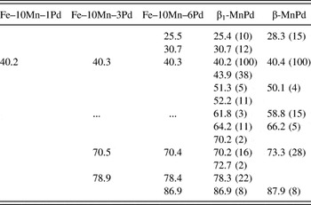
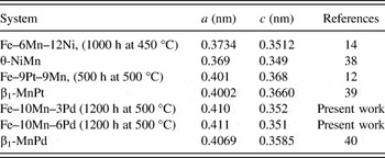
Table II summarizes the peak positions of the precipitates observed in the XRD patterns of the specimens aged for 1200 h at 500 °C, and also lists the 2θ values for β and β1. The observed reflections of the precipitates only match the β1 phase. Some of the expected reflections of the β1 phase were, however, not revealed, as they either showed an overlap with the α, γ, or ε phases or because their relative intensity is very low. The combined results of the APT and XRD measurements confirm that in the Fe–Mn–Pd maraging steels, as also in Fe–Mn-based systems containing Ni or Pt, the precipitates in the overaged state exhibit an ordered fct structure which corresponds to the intermetallic phase near the equiatomic composition of the respective Mn–X (X = Ni, Pd, Pt) binary phase diagrams. Table III summarizes the lattice parameters of the precipitates in the different ternary maraging steels, and those of the corresponding intermetallic binary phases. A good agreement is observed and the lattice parameters of the precipitates differ by an average of only 1% from the equilibrium values of the corresponding binary phases.
Peak positions (2θ in°) of the precipitates observed in the XRD spectra of specimens aged for 1200 h at 500 °C (Fig. 5) and calculated for the fct β1-MnPd phase with a = 0.411 nm and c = 0.351 nm, and for the bcc β-MnPd phase with a = 0.315 nm. The relative intensities are given in parentheses.

Comparison of lattice parameters determined via XRD for the intermetallic particles in the different ternary maraging systems, and those of the fct phase of the respective binary phase diagrams.

IV. SUMMARY
In this study, the precipitates that form during aging at 500 °C in Fe–Mn–Pd maraging steels were characterized using APT and XRD. Alloys containing 1, 3, and 6 wt.% Pd with a fixed Mn content of 10 wt.% were investigated. The steels exhibit considerable hardening upon aging. APT results in combination with XRD measurements reveal that the precipitates in the overaged state consist mainly of Mn and Pd and can be related to the fct β1-MnPd intermetallic phase, which is similar to what has been observed for Fe–Mn–Ni and Fe–Mn–Pt maraging steels.
ACKNOWLEDGMENTS
The authors thank N. Kränzlin for support with the XRD measurements and M. Schinhammer and E. Fischer for help with the sample preparation; and acknowledge the support of the Electron Microscopy Center of ETH Zurich (EMEZ). The authors appreciate the financial support received in the framework of the project “Bio Compatible Materials and Applications – BCMA” initiated by the Austrian Institute of Technology GmbH (AIT).