Introduction
Human strongyloidiasis is a common helminth infection caused by the intestinal nematode Strongyloides stercoralis. This nematode is distributed worldwide, mainly in tropical and subtropical regions (Olsen et al., Reference Olsen, van Lieshout, Marti, Polderman, Polman, Steinmann, Stothard, Thybo, Verweij and Magnussen2009; Paula & Costa-Cruz, Reference Paula and Costa-Cruz2011). The resulting helminthiasis can cause a wide range of clinical manifestations, ranging from asymptomatic and/or mild gastrointestinal disease to hyperinfection and fatal disseminated infection (Grove, Reference Grove1996; Mejia & Nutman, Reference Mejia and Nutman2012).
Parasitological diagnosis of strongyloidiasis is difficult, because the parasite load is usually low and larval output is irregular (Grove, Reference Grove1996). Consequently, infection rates tend to be underestimated. Immunological methods such as the enzyme-linked immunosorbent assay (ELISA), indirect fluorescent antibody test and Western blotting show variable sensitivity and specificity (Costa-Cruz et al., Reference Costa-Cruz, Bullamah, Gonçalves-Pires, Campos and Vieira1997; Feliciano et al., Reference Feliciano, Gonzaga, Gonçalves-Pires, Gonçalves, Rodrigues, Ueta and Costa-Cruz2010; Gonzaga et al., Reference Gonzaga, Ribeiro, Feliciano, Manhani, Silva, Ueta and Costa-Cruz2011). Techniques based on DNA detection are increasingly in use for the diagnosis of parasitic infections and have been proposed for the diagnosis of strongyloidiasis (ten Hove et al., Reference ten Hove, van Esbroeck, Vervoort, van den Ende, van Lieshout and Verweij2009). Polymerase chain reaction (PCR) is generally characterized by high sensitivity, specificity and reproducibility. PCR assays may therefore represent useful alternatives to the commonly used parasitological methods, by increasing the detection rate of infected individuals (Verweij et al., Reference Verweij, Canales, Polman, Ziem, Brienen, Polderman and van Lieshout2009; Paula et al., Reference Paula, Sitta, Malta, Gottardi, Corral, Gryschek and Chieffi2013; Sitta et al., Reference Sitta, Malta, Pinho, Chieffi, Gryschek and Paula2014).
Strongyloides venezuelensis is a parasitic nematode of rodents used as an experimental model for human infections (Machado et al., Reference Machado, Ueta, Gonçalves-Pires, Oliveira, Faccioli and Costa-Cruz2003) and it is particularly useful in the development of new immunological techniques for the diagnosis of strongyloidiasis (Gonçalves et al., Reference Gonçalves, Silva, Ueta and Costa-Cruz2012). In the present study, we used an experimental model of strongyloidiasis to evaluate the efficacy of molecular diagnosis when employed in parallel with parasitological and serological techniques.
Materials and methods
Experimental infections
Four-week-old male rats (Rattus norvegicus) were obtained from the Faculdade de Medicina da Universidade de São Paulo. Animals were maintained under standard laboratory conditions in the Instituto de Medicina Tropical da Universidade de São Paulo (IMT/USP).
Infective third-stage larvae (L3) of S. venezuelensis were obtained from charcoal cultures of infected rat faeces. L3 larvae were washed in 0.01 mol/l phosphate-buffered saline (PBS), pH 7.2, and counted. For each infection, 2000 S. venezuelensis L3 were inoculated subcutaneously into the abdominal region of rats.
Rats were divided into groups of 54 infected and six control rats. From each rat, blood and faecal samples were collected for parasitological, immunological and molecular diagnosis. On days 2, 5, 8, 13, 21, 28, 35, 45 and 60 post infection (pi), faecal samples were collected from six rats and were divided into two aliquots; the first aliquot was analysed using parasitological methods and the second aliquot was stored at − 20°C prior to molecular diagnosis. On the same day as faecal samples were collected, rats were anaesthetized and blood samples were collected through cardiac puncture. Rats were subsequently euthanized in chambers precharged with CO2. Blood samples were centrifuged and the serum was stored at − 20°C prior to serological diagnosis. After euthanasia, the small intestine was removed for the recovery and counting of adult worms of S. venezuelensis (Nakai & Amarante, Reference Nakai and Amarante2001). For non-infected rats (controls), similar procedures were employed for collection of faeces and blood samples.
Diagnosis
Parasitological diagnosis was performed by counting the number of eggs per gram of faeces, using a modified Cornell McMaster method (Gordon & Whitlock, Reference Gordon and Whitlock1939) and charcoal culture (Looss, Reference Looss1911). For the charcoal culture, 1 g of each faecal sample was mixed in distilled water containing activated charcoal, and samples were incubated at 28°C for 2 days. After concentration using the Rugai method (Rugai et al., Reference Rugai, Mattos and Brisola1954), samples were analysed using an optical microscope.
For the serological diagnosis, alkaline extracts were prepared using S. venezuelensis infective L3 larvae (Machado et al., Reference Machado, Ueta, Gonçalves-Pires, Oliveira, Faccioli and Costa-Cruz2003). Briefly, 1 ml of 0.15 mol/l NaOH was added to ~300,000 S. venezuelensis infective L3 and the mixture was gently shaken for 6 h at 4°C. After addition of 0.3 mol/l HCl until pH 7.0, the preparation was centrifuged at 12,400 g for 30 min at 4°C. The protein content of the supernatant was determined by the Lowry method (Lowry et al., Reference Lowry, Rosebrough, Farr and Randall1951).
The ELISA assay was performed as described previously (Gonçalves et al., Reference Gonçalves, Silva, Ueta and Costa-Cruz2012) with minor modifications. Preliminary experiments were carried out to determine the optimal ELISA conditions. Briefly, high-binding microtitre plates (Corning-Costar, New York, USA) were coated overnight at 4°C with 10 μg/ml of S. venezuelensis alkaline extract in 0.06 m carbonate–bicarbonate buffer (pH 9.6). Next, microplates were blocked with PBS containing 0.05% Tween 20 (v/v) plus 3% non-fat milk (w/v) for 45 min at 37°C. Plates were incubated with serum samples (1:10) for 45 min at 37°C and then with the secondary antibody consisting of peroxidase-labelled goat anti-rat IgG (Sigma-Aldrich, St. Louis, Missouri, USA) at a dilution of 1:8000 for 45 min at 37°C. The assay was developed by adding the enzyme substrate (hydrogen peroxide and orthophenylenediamine in 0.1 mol/l citrate phosphate buffer, pH 5.5) for 15 min. The reaction was stopped by addition of 2 n H2SO4 and the optical density determined at 490 nm in a plate reader (Thermo Fischer Scientific, Waltham, Massachusetts, USA).
Molecular analysis
For DNA extraction, approximately 1 g of each faecal sample was mixed with 70% ethanol and shaken vigorously. The mixture was centrifuged and the pellet washed twice with PBS. DNA from each faecal sample was extracted from the pellet by using the commercial QIAamp® DNA stool MiniKit (Qiagen, Hilden, Germany) with slight modifications, including the addition of lysis buffer containing proteinase K solution, followed by incubation at 56°C overnight. The DNA was eluted using 100 μl of elution buffer and quantified in a NanoDrop ND-1000 UV-VIS spectrophotometer v.3.2.1 (NanoDrop Technologies, Wilmington, Delaware, USA).
The QIAamp DNA mini kit (Qiagen) was also used to extract DNA from S. venezuelensis L3 larvae (positive control), faecal samples of uninfected rats, adult individuals of Syphacia muris and Hymenolepis diminuta (PCR controls). The PCR mix without a DNA template was used as a negative control to confirm the absence of environmental contamination.
PCR sensitivity was determined using a 1 g faecal sample from an uninfected animal, spiked artificially with 10, 5, 2 and 1 S. venezuelensis L3 larvae. PCR reactions were performed by using the primer pair (forward 5'-AAAGATTAAGCCATGCATG-3' and reverse 5'-GCCTGCTGCCTTCCTTGGA-3') to amplify a 340-bp fragment from the RNA gene coding for the small ribosomal subunit. This primer set was obtained from S. venezuelensis (accession AJ417026) sequences (Dorris et al., Reference Dorris, Viney and Blaxter2002). The reaction was carried out in 10 μl containing 10 mm deoxyribonucleoside triphosphates (dNTPs), 20 μm of each primer, 1.5 mm MgCl2, 50 mm KCl pH 8.4, 0.1 U of Platinum® Taq DNA polymerase (Applied BioSystems, Life Technologies, Foster City, California, USA) and 4 μl of DNA. Cycling conditions comprised an initial denaturation step at 95°C for 5 min, followed by 40 cycles each at 95°C for 30 s (denaturation) and 60°C for 30 s (annealing), and a final extension step at 72°C for 30 s. PCR was performed in the Mastercycler® ep Gradient S Thermocycler (Eppendorf, Hamburg, Germany). Amplification products were loaded on 2% agarose gel and were submitted to electrophoresis in 1 × Tris–acetate–EDTA (TAE) buffer containing ethidium bromide. Stained DNA bands were visualized under ultraviolet light. Each sample was tested in duplicate.
Data analysis
Statistical analyses were performed using the GraphPad Prism software version 5.0 (Graph Pad Software Inc., San Diego, California, USA). The cut-off value for the ELISA test was determined using receiver operating characteristic (ROC) curve analysis (Sopelete, Reference Sopelete, Mineo, Silva, Sopelete, Leal, Vidigal, Tápia and Bacchin2005). Results were considered significant at P < 0.05.
Results
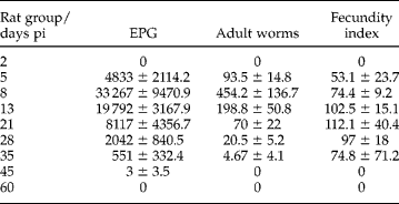
Parasite eggs were detected in the faeces from 5 to 45 days pi and the highest number of eggs per gram of faeces (33,267) was observed at 8 days pi. The number of eggs decreased throughout the experiment (table 1). The highest number of adult worms was recovered at 8 days pi and the highest fecundity index was observed on day 21 pi.
The mean recovery (±SE) of eggs/g of faeces (EPG), adult worms and fecundity index of Strongyloides venezuelensis in experimentally infected rats up to 60 days post infection (pi); all uninfected control rats showed zero values.
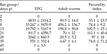
For the parasitological methods, it was observed that the counting of eggs per gram of faeces allowed their detection from day 5 until day 45 pi. The culture method showed infective larvae from day 5 up to 35 days pi.
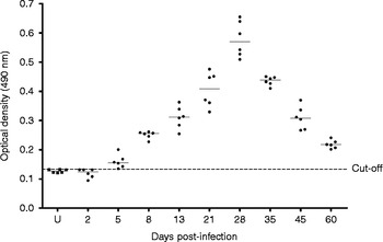
The levels of parasite-specific IgG antibodies in infected rats increased up to 28 days pi and subsequently decreased until 60 days pi. Nevertheless, they remained positive throughout the experiment (fig. 1).
Mean IgG levels in blood samples of rats experimentally infected with Strongyloides venezuelensis, with a cut-off value of 0.138.

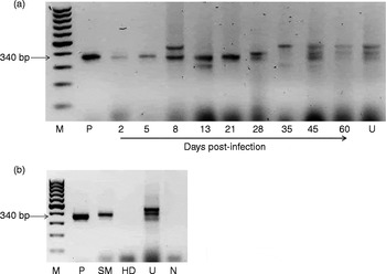
PCR sensitivity was useful to detect one larva/g stool (data not shown). PCR results from control samples: amplification products of differing sizes were observed for S. muris adults and in faecal samples from uninfected animals. No amplification was observed for H. diminuta samples (fig. 2b).
PCR assays of (a) faecal samples of rats infected with Strongyloides venezuelensis up to day 60 pi; and (b) control/uninfected rats and samples infected with the nematode Syphacia muris (SM) and the cestode Hymenolepis diminuta (HD). M,100 bp ladder; P, positive control; N, negative control; U, uninfected samples.

Strongyloides venezuelensis DNA was detected in faecal samples collected from infected animals throughout the experiment, particularly from day 5 to day 21 pi. Non-specific bands of 300, 380 and 420 bp were amplified for some of the faecal samples (fig. 2a).
Strongyloides venezuelensis DNA amplification coincided with the increase in parasite faecal egg output (>4000 EPG, on day 5 to day 21 pi) (table 1 and fig. 2). However, no correlation could be observed comparing the ELISA results versus the number of eggs per gram of faeces. ELISA and PCR findings revealed that at 5 day pi it is possible to confirm the infection by the molecular method, whereas the optical density of the serum samples was near to the established cut-off (fig. 1 and fig. 2a).
Discussion
The PCR assay represents a useful alternative for the identification of Strongyloides spp., allowing molecular analysis, diagnosis and epidemiological investigation (ten Hove et al., Reference ten Hove, van Esbroeck, Vervoort, van den Ende, van Lieshout and Verweij2009; Verweij et al., Reference Verweij, Canales, Polman, Ziem, Brienen, Polderman and van Lieshout2009; Sitta et al., Reference Sitta, Malta, Pinho, Chieffi, Gryschek and Paula2014). The present study is the first to evaluate the performance of molecular diagnosis, when used in parallel with parasitological and serological methods, in experimental S. venezuelensis infection.
Parasite eggs were detected in faeces from day 5 to day 45 pi, and the highest number of eggs was observed at day 8 pi. These results are in agreement with those of Nakai & Amarante (Reference Nakai and Amarante2001), demonstrating that the faecal egg output is proportional to the number of adult worms recovered from the small intestine.
Studies have reported that the beginning of the infection is characterized by an increase in the number of eggs in the faeces and high number of adult worms in the small intestine. In contrast, in advanced infection a decrease in the number of eggs in the faeces is observed which coincides with a low number of adult worms in the small intestine (Baek et al., Reference Baek, Islamb, Kimb, Limb, Hur, Oluoch, Kima and Kakoma2003; Chiuso-Minicucci et al., Reference Chiuso-Minicucci, Marra, Zornella-Pezavento, França, Ishikawa, Amarante, Amarante and Sartori2010; Marra et al., Reference Marra, Chiuso-Minicucci, Machado, Zorzella-Pezavento, França, Ishikawa, Amarante, Sartori and Amarante2011). Our results showed similar kinetics: at day 8 pi an increase in the number of eggs in the faeces was observed, and by day 35 pi (advanced stage of infection) a decrease in the number of parasites had occurred.
Previous investigations have demonstrated that culture techniques show a higher sensitivity for the diagnosis of strongyloidiasis than do conventional methods (Grove, Reference Grove1996). In the present study, the counting of eggs per gram of faeces was positive until 45 days pi, whereas the culture method showed positive results until 35 days pi. This apparent inconsistency between the present data and previous findings may be explained by the standardization of our culture protocol for 1 g of faeces. Similar results were obtained by Paula et al. (Reference Paula, Sitta, Malta, Gottardi, Corral, Gryschek and Chieffi2013), using charcoal culture to analyse faecal samples from rats infected with 2000 L3 S. venezuelensis. Kramme et al. (Reference Kramme, Nicole, Soblik, Erttmann, Tannich, Fleischer, Pannig and Brattig2011) evaluated rats infected with 2500 L3 S. ratti and detected a positive culture until 22 days pi.
Chiuso-Minicucci et al. (Reference Chiuso-Minicucci, Marra, Zornella-Pezavento, França, Ishikawa, Amarante, Amarante and Sartori2010) reported that specific antibody levels were significantly higher during the advanced phase compared to those found in the acute phase. In addition, Gonzaga et al. (Reference Gonzaga, Ribeiro, Feliciano, Manhani, Silva, Ueta and Costa-Cruz2011) demonstrated that the serological methods remained positive for longer periods, even after complete elimination of the parasite. In the present study, levels of parasite-specific IgG antibodies increased gradually until 28 days pi and decreased subsequently. Taken together, these results demonstrate that positivity of the serological methods might not indicate current infection.
Molecular methods were shown previously to be sensitive for the detection of experimental S. venezuelensis infection in Lewis rats (Marra et al., Reference Marra, Chiuso-Minicucci, Machado, Zorzella-Pezavento, França, Ishikawa, Amarante, Sartori and Amarante2010; Paula et al., Reference Paula, Sitta, Malta, Gottardi, Corral, Gryschek and Chieffi2013). Our data show a direct association between S. venezuelensis DNA detection and amount of parasite eggs in faecal samples. These results are in agreement with those of Marra et al. (Reference Marra, Chiuso-Minicucci, Machado, Zorzella-Pezavento, França, Ishikawa, Amarante, Sartori and Amarante2011). In addition, Paula et al. (Reference Paula, Sitta, Malta, Gottardi, Corral, Gryschek and Chieffi2013) showed high sensitivity of PCR for detection of DNA from faecal samples of rats infected with S. venezuelensis. Therefore, the molecular diagnostic technique is more suitable for the initial stage of infection (from 5 to 21 days pi).
The course of infection is accompanied by a decrease in the number of female worms and eggs. This induces the subsequent reduction in the amount of S. venezuelensis DNA in faecal material and may lead to non-specific amplification, especially after 21 days pi. In the present study, a 380-bp band was detected in some faecal samples; a possible explanation for this is a contamination of rats with the parasite S. muris, which is frequently found in the gut of laboratory animals. The appearance of several bands may have occurred because the primer amplified a ubiquitous region of the small subunit ribosomal DNA that was present in mixed samples (Marra et al., Reference Marra, Chiuso-Minicucci, Machado, Zorzella-Pezavento, França, Ishikawa, Amarante, Sartori and Amarante2010).
In summary, the present results indicate that serological and molecular methods show different applicability when used to measure experimental infection with S. venezuelensis. In conclusion, our findings represent the first step towards improving the diagnosis of experimental strongyloidiasis.
Acknowledgements
We are grateful to Professor William de Castro Borges (Universidade Federal de Ouro Preto, Minas Gerais, Brazil) for help with English language and critical revision of the manuscript.
Financial support
This work was supported by the Fundação de Amparo à Pesquisa do Estado de São Paulo (FAPESP 2013/03304-0) and the Coordenação de Aperfeiçoamento de Pessoal de Nível Superior (CAPES) through a scholarship to P.D.M.
Conflict of interest
None.
Ethical standards
All the experiments in this study were approved by and conducted in accordance with the ethical guidelines adopted by the Comissão de Ética no Uso de Animais (CEUA) of IMT/USP (protocol CPE-IMT 2012/152).