Introduction
Nematodes of the genus Mammomonogamus (Ryzhikov, 1948), known also under the common name of gapeworms, are strongylid parasites of the respiratory system of ungulates, elephants, carnivores, non-human primates and humans mainly in tropical areas of the world (Railliet, Reference Railliet1899; von Linstow, Reference von Linstow1899; Gedoelst, Reference Gedoelst1924; Mönnig, Reference Mönnig1932; Vuylsteke, Reference Vuylsteke1935; van den Berghe, Reference van den Berghe1937; Nosanchuk et al., Reference Nosanchuk, Wade and Landolf1995; Červená et al., Reference Červená2017). Five of the 13 known species of Mammomonogamus were described from the members of the family Felidae, including domestic cats, making the felids the most frequent hosts of Mammomonogamus (Tables 1–2; Figs 1–2).
Morphology of cranial extremities of four Mammomonogamus species known from Malay tiger (M. felis) and domestic cats (M. auris, M. ierei, M. mcgaughei). The pictures are adjusted from the original descriptions (Cameron, Reference Cameron1931; Buckley, Reference Buckley1934; Faust and Tang, Reference Faust and Tang1934; Seneviratne, Reference Seneviratne1954). Note the presence/absence of the longitudinal ribs in the buccal capsule. F, female; M, male; lat, lateral view; dv, dorso-ventral view.

Map of distribution of Mammomonogamus spp. in wild and domestic felids. Square is for determined helminths, the species is written in the map. Grey circles are for undetermined Mammomonogamus. 1 – Diesing (Reference Diesing1857), 2 – Cameron (Reference Cameron1931), 3 – Buckley (Reference Buckley1934), 4 – Faust and Tang (Reference Faust and Tang1934), 5 – Guilbride (Reference Guilbride1953), 6 – Seneviratne (Reference Seneviratne1954), 7 – Sakamoto et al. (Reference Sakamoto, Kaneda and Nakagawa1971), 8 – Cuadrado et al. (Reference Cuadrado, Maldonado-Moll and Segarra1980), 9 – Lindquist and Austin (Reference Lindquist and Austin1981), 10 – Sugiyama et al. (Reference Sugiyama1982), 11 – Asato et al. (Reference Asato1986), 12 – Hasegawa (Reference Hasegawa1992), 13 – Patton and Rabinowitz (Reference Patton and Rabinowitz1994), 14 – Magnaval and Magdeleine (Reference Magnaval and Magdeleine2004), 15 – Tudor et al. (Reference Tudor2008), 16 – Krecek et al. (Reference Krecek2010); Gattenuo et al. (Reference Gattenuo, Ketzis and Shell2014).

Overview of Mammomonogamus spp. described from felid carnivores

* The locality on the left is the place, where the parasite was diagnosed and on the right, there is the country of origin.
Morphology of Mammomonogamus spp. found in felids (based on the original descriptions)

a The measurements are given in millimetres if not stated differently.
b Distance of vulva from cranial end.
c Ratio of the distance of vulva from cranial end and the length of the body.
d See Fig. 1. M, male; F, female.
e Ribs were not mentioned in the original description; however, the drawing of buccal capsule shows some structures resembling the ribs (Fig. 1) and their presence was later confirmed by Sugiyama et al. (Reference Sugiyama1982).
In fact, Mammomonogamus dispar (Diesing, Reference Diesing1857), found in a puma Puma concolor (Linnaeus, 1751), in Brazil was the very first syngamid observed in any mammalian host. Unfortunately, the description of the parasite is deficient, providing only the body length and body width of the nematode, along with simple, little detailed drawings (Diesing, Reference Diesing1857; Tables 1–2). Similarly to most of the Mammomonogamus species, M. dispar has never been reported again. The second species known from wild felids, Mammomonogamus felis (Cameron, Reference Cameron1931), was reported only twice. Interestingly, both animals were captive and imported from Thailand (Tables 1–2; Figs 1–2).
The remaining three Mammomonogamus species are known only from domestic cats. Mammomonogamus ierei (Buckley, Reference Buckley1934) is quite a common parasite of the nares and nasopharynx of cats on the Caribbean islands (Table 1, Fig. 2) and Mammomonogamus auris (Faust and Tang, Reference Faust and Tang1934) parasitizes the feline middle ear, which is a quite exceptional localization for a helminth. The latter species was first described in China (Faust and Tang, Reference Faust and Tang1934) and has been reported on several Asian islands over time (Table 1; Fig. 2). Finally, Seneviratne (Reference Seneviratne1954) described Mammomonogamus mcgaughei (Seneviratne, Reference Seneviratne1954) in the pharynx and frontal and nasal sinuses of domestic cats in Sri Lanka. In addition, the same author reported finding M. auris in the middle ear of four other cats, distinguishing the two species by the vulva position and size of the spicules. However, M. mcgaughei has not been reported ever since and its possible conspecificity with other species remains to be investigated.
Eggs of Mammomonogamus were detected in feces or sputum of both domestic cats and free-ranging felids in Asia, Africa, Central and South America (Table 1; Fig. 2). Unfortunately, the exact determination of Mammomonogamus species based solely on the egg morphology is not possible, leaving the true identity of the parasite unrevealed. Even though the species of Mammomonogamus from the Iriomote cats, Prionailurus bengalensis iriomotensis (Imaizumi, 1967), from Japan (Hasegawa, Reference Hasegawa1992) was not determined, based on the locality and site of infection one can assume that it was probably M. auris. Lindquist and Austin (Reference Lindquist and Austin1981) reported a domestic cat originating from Nigeria sneezing out a pair of Mammomonogamus nematodes after episodes of sneezing and coughing spasms; unfortunately, identification was not made to the species level. Interestingly, it is the only case of Mammomonogamus infection in a felid host known from the African continent.
To date, only a little is known about the transmission of Mammomonogamus spp. in felines. The eggs are shed in feces or in nasal discharge and sputum; larvae develop within 10 days to the third stage and emerge from the eggs. The free larvae are not capable of skin penetration; however, they seem more active when the environment temperature is raised (Buckley, Reference Buckley1934; Cuadrado et al., Reference Cuadrado, Maldonado-Moll and Segarra1980). Buckley (Reference Buckley1934) attempted to induce infection in adult cats and kittens using hatched larvae of M. ierei, unfortunately without any success. For this reason, he suggested involvement of an intermediate or paratenic host in the life cycle. A similar strategy is known from other strongylids infecting the respiratory system of felids, such as Aelurostrongylus abstrusus (Railliet, 1898) or Troglostrongylus spp., which use molluscs as intermediate and vertebrates as paratenic hosts (Gerichter, Reference Gerichter1949; Anderson, Reference Anderson2000; Bowman et al., Reference Bowman, Bowman, Hendrix, Lindsay and Barr2002).
The diagnosis of Mammomonogamus is usually based on the clinical signs such as nasal discharge, coughing and sneezing, loss of weight (M. ierei) or head shaking (M. auris) and subsequent observation of adults, e.g. through the tympanic membrane during otoscopic examination in case of M. auris, or occasionally, the adult worms are expelled by the host (Guilbride, Reference Guilbride1953; Cuadrado et al., Reference Cuadrado, Maldonado-Moll and Segarra1980; Lindquist and Austin, Reference Lindquist and Austin1981; Tudor et al., Reference Tudor2008). The eggs can be detected in feces or sputum (Cuadrado et al., Reference Cuadrado, Maldonado-Moll and Segarra1980; Krecek et al., Reference Krecek2010; Gattenuo et al., Reference Gattenuo, Ketzis and Shell2014). Even though the genus Mammomonogamus is traditionally assigned to the family Syngamidae, the eggs do not have polar plugs as found in Syngamus spp. and their outer shell surface is finely striated. Although the eggs of Mammomonogamus superficially resemble those of hookworms, they can be easily distinguished from the latter based on their larger size and the thicker striated shell (Cuadrado et al., Reference Cuadrado, Maldonado-Moll and Segarra1980; Sugiyama et al., Reference Sugiyama1982; Patton and Rabinowitz, Reference Patton and Rabinowitz1994). The egg morphology within the species of Mammomonogamus found in felids is quite uniform, differing only slightly in size (Table 2). Also eggs of Mammomonogamus found in herbivores have very similar morphology differing only in the pattern of shell striations (Graber et al., Reference Graber1971; Červená et al., Reference Červená2017). Treatment of ear infections usually consists of removing the adults after tympanotomy or topical application of selamectin or a combination of thiabendazole, dexamethasone and neomycin (Tudor et al., Reference Tudor2008). Fenbendazole was successfully used to treat M. ierei infection in a cat (Gattenuo et al., Reference Gattenuo, Ketzis and Shell2014).
In domestic cats, two of the known species, namely M. ierei and M. auris, are detected on a regular basis. The differences in morphology of both species are rather minor (Table 2). Tudor et al. (Reference Tudor2008) suggested the capability of Mammomonogamus to move between the nasal sinuses, larynx, trachea and middle ear via the Eustachian tube, nasopharynx and laryngopharynx, opening a question of the conspecificity of M. ierei and M. auris. To test such hypothesis, we compared the sequences of mitochondrial and nuclear markers obtained from Mammomonogamus adults or eggs collected from domestic cats in three geographically distant areas covering area of occurrence of both species. The resulting phylogenetic analyses showed monophyletic status of the genus Mammomonogamus, confirmed the validity of M. ierei and M. auris and suggested the existence of one more species infecting domestic cats.
Material and methods
Material from cats
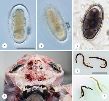
Adults of M. ierei were collected from carcasses of domestic cats submitted for necropsy to Ross University School of Veterinary Medicine, Saint Kitts (West Indies). The helminths were found in the nasal cavity firmly attached to the mucosa, as is typical for the species (Fig. 3) and immediately preserved in 96% ethanol and 10% formalin. The adults of M. auris were collected from the middle ear of a domestic cat referred to Paradise Island Animal Hospital, Saipan (Northern Mariana Islands). As 96% ethanol was not available in Saipan, the helminths were placed in Bacardi 151 (Bacardi Limited, Hamilton, Bermuda; 75.5% of alcohol) and moved to 96% ethanol after arrival at the University of Veterinary and Pharmaceutical Sciences Brno, Czech Republic (ca 2 weeks after their collection). Two couples of M. ierei and two couples of M. auris were examined using an Olympus AX70 microscope equipped with Nomarski interference contrast and a DP 70 digital camera. Basic morphological features were observed and measurements taken. Subsequently, the helminths were separated, washed with physiological saline and the DNA from adults and eggs was isolated using the Genomic DNA Mini Kit GT300 (Tissue) (Geneaid, New Taipei City, Taiwan). Paragenophores of M. ierei and cranial and caudal extremities of M. auris specimens were deposited in the Helminthological collection of the Institute of Parasitology, Biology Centre of the Czech Academy of Sciences, České Budějovice, Czech Republic under the numbers IPCAS N-1158 and IPCAS N-1159.
Eggs and adults of examined Mammomonogamus spp. in domestic cats. (A) Egg of Mammomonogamus sp. from feces of a cat, Fatu Hiva, French Polynesia. (B) Same egg, focus on the surface, showing fine striation of the outer wall. (C) Egg of Mammomonogamus ierei from feces of a cat, Saint Kitts. (D) A cross-section through skull of necropsied cat from Saint Kitts with clearly visible adult of M. ierei emerging from the nasal cavity (encircled). (E) Two pairs of Mammomonogamus auris from middle ear of a cat from Saipan. (F) Two pairs of M. ierei from a cat from Saint Kitts. A–C in same scale, scale bar = 50 um; E–F in same scale, scale bar = 10 mm.

Feces of two domestic cats were collected during a sterilization campaign in Fatu Hiva (French Polynesia) and preserved in 96% ethanol. The cat feces were examined by modified Sheather's flotation and simple sedimentation. The detected eggs of Mammomonogamus were collected and mechanically disrupted before the DNA was isolated as described in Červená et al. (Reference Červená2017). All procedures leading to the collection of material were performed under approved Institutional Animal Care and Use Committee (IACUC) protocols.
Comparative material
To allow comparison of genetic variability and to extend the phylogenetic analyses, we used the following comparative material: (i) DNA sequences of Mammomonogamus, most probably M. loxodontis and M. nasicola obtained from typical eggs isolated from fecal samples of western lowland gorillas Gorilla gorilla gorilla (Savage and Wyman, 1847) and African forest elephants Loxodonta cyclotis (Matschie, 1900) and from African forest buffaloes Syncerus caffer nanus (Boddaert, 1785), respectively (Červená et al., Reference Červená2017, Reference Červená2018), originating from several African localities; (ii) DNA sequences obtained from adults of Mammomonogamus laryngeus (Railliet, Reference Railliet1899) collected from a water buffalo Bubalus bubalis (Linnaeus, 1758) at a slaughterhouse in the municipality of Soure (Pará State, Brazil); and finally (iii) DNA sequences from two individuals of Syngamus spp. nematodes collected from Turdus merula (Linnaeus, 1758) and Corvus corone (Linnaeus, 1758) in the Czech Republic; and (iv) one Cyathostoma cf. bronchialis (Mühling, 1844) from a domestic goose from Slovakia. More details about this comparative material can be found in Červená et al. (Reference Červená2018) and the GenBank accession numbers are listed in the Supplementary Table S1.
DNA amplification
Primers presented in Table 3 were used to amplify partial sequences of 18S rDNA, internal transcribed spacer 1 (ITS1), 28S rDNA and gene for cytochrome c oxidase subunit I (cox1). Fragments of 18S rDNA, ITS, 28S rDNA and cox1 originating from adult helminths were amplified in 25 µL PCR reactions containing 12.5 µL of PPP Master Mix (Top-Bio, Prague, Czech Republic), 0.8 µM of each primer, 7.5 µL PCR H2O (Top-Bio, Prague, Czech Republic) and 1 µL of template DNA. PCR conditions were identical for all markers: initial denaturation at 94 °C for 1 min, followed by 35 cycles of 94 °C denaturation for 40 s, 50 °C annealing for 40 s, 65 °C elongation for 40 s, followed by a 5 min post-amplification extension at 72 °C. To amplify 18S rDNA and cox1 from eggs, multiplex PCR was used. In the first round, 25 µL reaction mixtures contained 12.5 µL of Phusion® High-Fidelity PCR Master Mix HF Buffer (New England Biolabs, Ipswich, UK), 0.5 µ m of each of primers NC18SF1, NC5BR, HCO2198, coxmamF (Table 3) and 3 µL of template DNA. PCR conditions were as follows: initial denaturation at 98 °C for 30 s followed by 35 cycles of 98 °C denaturation for 10 s, 55 °C annealing for 30 s and 72 °C elongation for 1 min with final post-amplification elongation for 7 min at 72 °C. One microlitre of the PCR product was then used as DNA template for the reamplification PCR round using the primers NC18SF1 and NC5BR for 18S rDNA and HCO2198, coxmamF for cox1 separately with the same polymerase and conditions as in the first round. Annealing temperature was 54 and 58 °C for 18S rDNA and cox1, respectively. The ITS1 and 28S rDNA were amplified in multiplex PCR as well with the NC2R, NC28R, NC16 and NC2 primers using the same protocol described above, only the annealing temperature was 60 °C in both rounds of PCR. PCR products were separated in 1.2–2% agarose, visualized using GoodView™ Nucleic Acid Stain (Ecoli s.r.o., Bratislava, Slovakia) and detected using an UV illuminator. DNA was purified directly from the PCR product or from the bands cut from the gel using Gel/PCR DNA Fragments Extraction Kit (Geneaid, New Taipei City, Taiwan). Products were commercially sequenced in both directions by Macrogen (Amsterdam, The Netherlands). As the ITS1 sequences were mostly of low quality, PCR products were cloned using p-GEM®-T Easy Vector System (Promega, Madison, Wisconsin, USA), and single clones were sequenced separately. Sequences obtained in this study were submitted to GenBank under the accession numbers MF668043–MF668083 (Supplementary Table S2).
Primers used for the amplification of nuclear and mitochondrial DNA of Mammomonogamus spp.

Phylogenetic analyses
DNA sequences of 18S rDNA, ITS1, 28S rDNA and cox1 were trimmed, assembled and manually edited in BioEdit 7.2.3 (Hall, Reference Hall1999). Alignment of nuclear DNA sequences was done in ClustalW implemented in BioEdit (Larkin et al., Reference Larkin2007); the nucleotide sequences of cox1 were aligned guided by CLUSTALW amino acid alignment implemented in Geneious (http://geneious.com; Kearse et al., Reference Kearse2012). Pairwise sequence distances for both nucleotides and amino acids (cox1) were calculated using Geneious. Concatenated phylogenetic analysis of 18S and 28S sequences was carried out using Bayesian inference (BI) in MrBayes 3.2.2 (Hastings, Reference Hastings1970; Ronquist and Huelsenbeck, Reference Ronquist and Huelsenbeck2003). GenBank sequences of selected strongylids (Chilton et al., Reference Chilton2006; Supplementary Table S1) were added into the alignment of 18S rDNA and 28S rDNA for comparison. BI was done in two simultaneous runs of four Metropolis-couple Monte Carlo Markov chains of one million generations sampled each 100 generations and 25% generations discarded as burn-in. The GTR + I + Γ model of sequence evolution used in BI for both nuclear and mitochondrial DNA was chosen with Modeltest 3.7 (Posada and Crandall, Reference Posada and Crandall1998).
Results
Morphology
The morphometry of the adult helminths is summarized in Table 4. Based on the morphology, geographical locality and site of infection, the helminths from Saint Kitts were determined as M. ierei and specimens from Saipan as M. auris. This determination corresponds well with their within-host localization. The eggs of Mammomonogamus were detected in one of two fecal samples originating from Fatu Hiva. Their size varied from 95–115 × 50–60 µ m and their thick shell was covered in fine parallel wavy line striations (Fig. 3). There was no evident difference when compared with the eggs of M. ierei originating from Saint Kitts (Fig. 3).
Morphometry of adults Mammomonogamus ierei and Mammomonogamus auris available for our study

a The measurements are given in millimetres if not stated differently.
b Distance of vulva from cranial end.
c Ratio of the distance of the vulva from cranial end and the body length.
d The spicules were not observed in M. ierei.
Nuclear markers: 18S and 28S rDNA, ITS1
Partial sequence of 18S rDNA (~1217 bp) was identical in all eight adults of M. ierei and M. auris. The sequence obtained from one Mammomonogamus egg from Fatu Hiva differed from the others only in a single nucleotide. A fragment of ~748 bp of 28S rDNA was identical in all four individuals of M. ierei, while the two couples of M. auris differed in one nucleotide. The pairwise sequence distances among the Mammomonogamus spp. from cats are summarized in Table 5. The sequences originating from cats differed from the sequences from African herbivores from 6.78 to 8.51% and from M. laryngeus from 6.66 to 7.32%.
Pairwise sequence distances of 28S rDNA (A), nucleotide (B) and amino acid sequence (C) of cox1 for Mammomonogamus ierei (Saint Kitts), Mammomonogamus auris (Saipan) and Mammomonogamus sp. (Fatu Hiva) found in cats

The ~599 bp alignment of ITS1 sequences originating from four adults of M. auris and four adults of M. ierei showed two obviously different patterns (Supplementary Fig. S3). Two areas of short tandem repeats resulting in length variability were found in both species. Firstly, the trinucleotide repeat CGT was present in 8–30 repetitions in M. ierei and in 10–21 repetitions in M. auris (Supplementary Fig. S4). Secondly, CA dinucleotide was present in 9–16 repetitions in M. ierei, while in M. auris it was only 8–9 repetitions. Mammomonogamus auris had one more short area of 5–6 CA repeats, which was not present in M. ierei.
The tree resulting from BI of concatenated alignment of 2035 bp of 18S rDNA and 28SrDNA showed a monophyletic clade comprising all Mammomonogamus sequences. The sequences of Mammomonogamus spp. formed three distinct subclades in cats based on the geographic origin and host species (Fig. 4). The remaining syngamids, represented by Syngamus and Cyathostoma sequences, cluster in another, well-supported clade rather distant from that of Mammomonogamus spp.
Phylogenetic tree based on Bayesian inference of concatenated 18S rDNA and 28S rDNA sequences of Mammomonogamus spp. originating from domestic cats (M. ierei, M. auris, Mammomonogamus sp. from Fatu Hiva), water buffalo (M. laryngeus) and African herbivores, syngamids from birds and other strongylids. Sequences of Metastrongylus elongatus, Protostrogylus rufescens and Aelurostrongylus abstrusus used as an outgroup are not displayed. Node-supporting values refer to BI posterior probability (values above 0.9 displayed only). Red line marks the samples from Saint Kitts, blue line the samples from Fatu Hiva and green line is for the samples from Saipan. CZ, Czech Republic; CAR, Central African Republic; F, female; M, male.

Mitochondrial marker: cox1
The ~420 bp fragment of cox1 was obtained from all eight adults of M. ierei and M. auris and from one egg of Mammomonogamus sp. from Fatu Hiva. The pairwise nucleotide sequence distances are summarized in Table 5B. While all individuals of M. auris had an identical cox1 sequence, the two couples of M. ierei differed in six nucleotides. The distance between the feline Mammomonogamus spp. and Mammomonogamus from African herbivores varied from 15.71 to 22.43%, and when compared with the sequence of M. laryngeus, the distance ranged from 24.52 to 24.76%. The pairwise amino acid sequence distance within the Mammomonogamus spp. from domestic cats is summarized in Table 5C.
The tree resulting from BI of 420 bp of cox1 of Mammomonogamus and Necator americanus, used as an outgroup, showed three distinct branches within the Mammomonogamus spp. originating from cats based on the geographic localities (Fig. 5).
Phylogenetic tree resulting from Bayesian inference of 420 bp of cox1 gene sequences of Mammomonogamus spp. originating from domestic cats (M. ierei, M. auris, Mammomonogamus sp. from Fatu Hiva), water buffalo (M. laryngeus) and African herbivores. Based on the topology of the tree resulting from the analysis of nuclear DNA, Necator americanus was used as an outgroup. Node-supporting values refer to BI posterior probability (values above 0.9 displayed only). Red line marks the samples from Saint Kitts, blue line the samples from Fatu Hiva and green line is for the samples from Saipan. F, female; M, male; CAR, Central African Republic.

Discussion
Since the middle of the 19th century, five species of Mammomonogamus-infecting felid carnivores have been described. The two of these nominal taxa, namely M. dispar and M. felis, were recorded from free-ranging felids and have not been recorded since (Diesing, Reference Diesing1857; Cameron, Reference Cameron1931). The three remaining species, M. mcgaughei, M. ierei and M. auris, are known as parasites of domestic cats; the latter two species are locally detected on a regular basis (Buckley, Reference Buckley1934; Faust and Tang, Reference Faust and Tang1934; Seneviratne, Reference Seneviratne1954; Cuadrado et al., Reference Cuadrado, Maldonado-Moll and Segarra1980; Sugiyama et al., Reference Sugiyama1982; Tudor et al., Reference Tudor2008; Gattenuo et al., Reference Gattenuo, Ketzis and Shell2014). Reports of unidentified Mammomonogamus sp. from wild felids in the areas of occurrence of known Mammomonogamus species, especially M. ierei and M. auris, raise questions about their possible conspecificity and the role of wild felids as natural reservoirs of Mammomonogamus (Hasegawa, Reference Hasegawa1992; Patton and Rabinowitz, Reference Patton and Rabinowitz1994; Magnaval and Magdeleine, Reference Magnaval and Magdeleine2004).
While the reports of Mammomonogamus in free-ranging felids are restricted to the mainland of South America and Asia, including one Japanese island (Diesing, Reference Diesing1857; Hasegawa, Reference Hasegawa1992; Patton and Rabinowitz, Reference Patton and Rabinowitz1994; Magnaval and Magdeleine, Reference Magnaval and Magdeleine2004), domestic cats are known to host Mammomonogamus spp. on numerous islands in the Caribbean, Southeast Asia and Pacific Ocean islands, with a single case known from continental China (Faust and Tang, Reference Faust and Tang1934; Guilbride, Reference Guilbride1953; Seneviratne, Reference Seneviratne1954; Cuadrado et al., Reference Cuadrado, Maldonado-Moll and Segarra1980; Sugiyama et al., Reference Sugiyama1982; Asato et al., Reference Asato1986; Tudor et al., Reference Tudor2008; Krecek et al., Reference Krecek2010; Gattenuo et al., Reference Gattenuo, Ketzis and Shell2014). It appears that M. auris is restricted to the Far East, while M. ierei occurs only in the Caribbean region, suggesting the speciation of M. ierei after the migration of felids to the Americas (O'Brian and Johnson, Reference O'Brian and Johnson2007). We examined the material collected from domestic cats in two localities, where Mammomonogamus typically occurs and, in addition, we found a single cat infected with Mammomonogamus in a totally new geographic area. In fact, the finding of Mammomonogamus in Fatu Hiva, Marquesas Islands in the French Polynesia, represents the first record of Mammomonogamus in domestic cat from the Southern Hemisphere.
Until now, the taxonomy of the genus Mammomonogamus at the specific and higher level has been assessed based only on the morphology. Traditionally, this genus is referred to as a member of the family Syngamidae, due to similar, rather basic, morphological features, site of infection and the state of permanent copulation (Ryzhikov, Reference Ryzhikov1949; Baruš and Tenora, Reference Baruš and Tenora1972). That was also the reason why we included comparative sequences from Cyathostoma and Syngamus spp. in our dataset. Our study is the first analysing the DNA sequences of Mammomonogamus spp. infecting domestic cats and shows clear monophyly of the genus, involving also the sequences from African herbivores and M. laryngeus from water buffalo. Surprisingly, the syngamids infecting birds formed another clade rather distant from Mammomonogamus. The tree topology together with differences in egg morphology suggest that the biological and morphological traits used for the definition of Syngamidae are not true synapomorphies and the current grouping of syngamids seems to be rather artificial. Under such a scenario, permanent copulation and affinity to the respiratory tract, including the auditory system, could have evolved independently among strongylids parasitizing avian and mammalian hosts. Poly- or paraphyletic characters are common also in other families within the order Strongylida, as presented by Chilton et al. (Reference Chilton2006). This issue and its consequences to the taxonomy of the group should be addressed in the future.
Topology of the phylogenetic trees resulting from the analyses of nuclear and mitochondrial DNA confirm M. ierei and M. auris as two distinct species (Figs 4–5). The identity of the Mammomonogamus isolate from Fatu Hiva, French Polynesia remains questionable. While the cox1 sequence does cluster with M. ierei, the nuclear DNA sequence clusters with M. auris but the node is not strongly supported in either case. The pairwise sequence distances comparison of the Fatu Hiva haplotype to the other two feline Mammomonogamus species suggests its distinctiveness at species level, as the values are similar to those for interspecific comparison between M. ierei and M. auris (Table 5) and between African Mammomonogamus spp. (Červená et al., Reference Červená2018).
The origin of Mammomonogamus infections in domestic cats represents intriguing question. The areas of its typical occurrence in the Far East and Caribbean region are far away from the domestication centres of the domestic cat in the Near East (Hu et al., Reference Hu2014; Vigne et al., Reference Vigne2016) and these parasites can hardly be classified as heirloom parasites of the Felis silvestris lineage leading to the modern domestic cat. Therefore, the spillover from a wildlife reservoir, presumably felid carnivores, represents a plausible explanation. The existence of two geographically well separated Mammomonogamus species in domestic cats strongly suggests two centres of ‘domestication’ of Mammomonogamus nematodes in cats. Importantly, in both geographic areas, wild felid species are known to host Mammomonogamus nematodes, thus representing potential candidates for reservoir hosts. Mammomonogamus ierei was first described in a domestic cat from the island of Trinidad, where the ocelots Leopardus pardalis (Linnaeus, 1758), known hosts of Mammomonogamus, are present (Murray and Gardner, Reference Murray and Gardner1997; Magnaval and Magdeleine, Reference Magnaval and Magdeleine2004). Similarly, Mammomonogamus infection is reported from leopard cats P. bengalensis in the Far East (Hasegawa, Reference Hasegawa1992; Patton and Rabinowitz, Reference Patton and Rabinowitz1994). Interestingly enough, both these wild cat species were historically commonly kept as pets in the areas of their occurrence, which facilitates contact with domestic cats. On the contrary, the Mammomonogamus infections nowadays typically occur in the populations of domestic cats living in island environments where the wild felid species are absent. Spread of the infection over the islands can be explained by the movement of domestic cats in recent times. Such a scenario suggests either a direct life cycle or the presence of rather unspecific intermediate and/or paratenic hosts. The saurian reptiles (geckoes, skinks and anoles) are among the most abundant terrestrial vertebrates in the areas of occurrence of Mammomonogamus in cats and are invariably hunted by cats during outdoor activity. The involvement of these hosts in the life cycle deserves more attention, especially because the mode of transmission is not known yet.
Supplementary material
The supplementary material for this article can be found at https://doi.org/10.1017/S0031182018000768
Acknowledgements
We would like to thank the government of the Central African Republic, the World Wildlife Fund, the Ministère de l'Education Nationale, de l'Alphabétisation, de l'Enseignement Supérieur, et de la Recherche for granting permission and providing permits to conduct our research in the Central African Republic and the Primate Habituation Programme for logistical support in the field. We are thankful to the Centre National de la Recherche Scientifique et Technologique (CENAREST) and the Agence National des Parcs Nationaux (ANPN) for research authorization in Gabon. We thank SFM Safari Gabon for hosting our research and seeing the value of health monitoring as part of the development of ape tourism programmes, especially we thank Matthew H. Shirley and Emilie Fairet as well as our field assistants Pierre Bukosso and Kharl Remanda for their significant support in the field. We express our thanks to the Ministère des Fôrets et de la Faune and Ministère de la Recherche Scientifique et de l'Innovation in Cameroon for permitting our research. We thank Projet Grands Singes, Centre for Research and Conservation, Royal Zoological Society of Antwerp and WWF Kudu-Zumbo Programme for providing their material and logistical support in the field. We would like to thank all trackers and research assistants from all study sites, especially to Arlette Tchankugni Nguemfo and Charmance Irene Nkombou for helping with sample collection. We thank Jana Bulantová and her team from the Department of Parasitology, Faculty of Science, Charles University, Prague for providing us with specimens of Syngamus sp. collected from a crow. We thank Diedra Metzler and Katie Neuville for their help with the collection of Mammomonogamus ierei, Marcela Figuerêdo Duarte Moraes, Ivan Moura Lapera, Ana Cláudia Alexandre de Albuquerque, Andressa de Souza Pollo for their help with the collection, identification and processing of Mammomonogamus laryngeus samples.
Financial support
This work derives from the Laboratory for Infectious Diseases Common to Humans and (non-Human) Primates from Czech Republic (HPI-Lab) and was co-financed by the European Social Fund and the state budget of the Czech Republic (project OPVK CZ.1.07/2.3.00/20.0300); the Czech Science Foundation (15-05180S); ‘CEITEC’ – Central European Institute of Technology (CZ.1.05/1.100/02.0068); the European Regional Development Fund; and the Institute of Vertebrate Biology, Czech Academy of Sciences (RVO: 68081766); supported by the project LO1218 with financial support from the Ministry of Education, Youth and Sports of the Czech Republic under the NPU I program. We acknowledge a grant for the development of research organization from the Ministry of Agriculture of the Czech Republic (RVO: RO0516).
Conflicts of interest
None.
Ethical standards
All procedures leading to the collection of material were performed under approved Institutional Animal Care and Use Committee (IACUC) protocols.