Introduction
Application of chilled semen is noteworthy due to recent growth in its use in clinical practice during artificial insemination (Hesser et al., Reference Hesser, Darr, Gonzales, Power, Scanlan, Thompson, Love, Christensen and Meyers2017). As a technique, it is a simple method used in several countries for semen transportation to maintain sperm viability for short periods (Hesser et al., Reference Hesser, Darr, Gonzales, Power, Scanlan, Thompson, Love, Christensen and Meyers2017) thereby reducing animal transport costs. The use of chilling also allows propagation of different genetic materials that permit genetic diversity (Mellanby et al., Reference Mellanby, Ogden, Clements, French, Gow, Powell, Corcoran, Schoeman and Summers2013).
In this context, several protocols have been analyzed aimed to better address sperm cooling time, such as use of different transport containers (Lopes et al., Reference Lopes, Simoes, Ferreira, Martins-Bessa and Rocha2009) or centrifugation protocols (Galvez et al., Reference Galvez, Ortiz, Hidalgo, Morrell and Dorado2015). In these studies, the most popular extender for chilled dog semen was based on an egg yolk−Tris−fructose medium (Rota et al., Reference Rota, Strom and Linde-Forsberg1995; Iguer-ouada and Verstegen, Reference Iguer-ouada and Verstegen2001b; Lopes et al., Reference Lopes, Simoes, Ferreira, Martins-Bessa and Rocha2009). However, as egg yolk is an animal product, selection of this component has multiple disadvantages such as standardization difficulty, due to its variable composition, and risk of contamination (i.e., salmonellosis) (Beccaglia et al., Reference Beccaglia, Anastasi and Luvoni2009b; Reed et al., Reference Reed, Ezeh, Hamic, Thompson and Caperton2009; Kmenta et al., Reference Kmenta, Strohmayer, Muller-Schlosser and Schafer-Somi2011). Therefore, development of an alternative extender that can replace egg yolk and still maintain an ideal semen quality after chilling, is required.
Soy lecithin is a promising option as a substitute for egg yolk, due to ease of component standardization, readily availability and reduced potential risk of contamination (Fukui et al., Reference Fukui, Kohno, Togari, Hiwasa and Okabe2008). Lecithin has a similar composition (i.e. low-density lipoprotein) as egg yolk, and may provide protection to the sperm plasma membrane during cold-shock (Forouzanfar et al., Reference Forouzanfar, Sharafi, Hosseini, Ostadhosseini, Hajian, Hosseini, Abedi, Nili, Rahmani and Nasr-Esfahani2010).
In dogs, the use of lecithin as an extender for sperm preservation has been applied in cryopreservation protocols, with some promising results on post-thaw semen quality (Axner and Lagerson, Reference Axner and Lagerson2016; Hidalgo et al., Reference Hidalgo, Bayon, Galvez, Urbano, Ortiz and Dorado2014). In cooling procedures, Beccaglia et al. (Reference Beccaglia, Anastasi, Chigioni and Luvoni2009a) described a protective effect of lecithin (concentration of 0.04%) similar to egg yolk until 4 days of storage at 5ºC. Kmenta et al. (Reference Kmenta, Strohmayer, Muller-Schlosser and Schafer-Somi2011) also observed that 0.8% of lecithin associated with catalase was able to improve the sperm longevity of canine semen in cold-stored conditions for 8 days. In addition, Kasimanickam et al. (Reference Kasimanickam, Kasimanickam, Memon and Rogers2012) reported that 0.4% of soy lecithin in extender improved sperm parameters when compared with the concentration of soy lecithin at 0.04% and egg yolk in samples stored at 5ºC for 10 days. However, narrower intervals showed differences when several concentrations of lecithin were used.
Despite previously reported effects of lecithin, the ideal concentration to use and its consequences for chilled sperm over time have not been detailed. Therefore, this study aimed to compare several concentrations of soy lecithin (0.01, 0.05 and 0.1%) in egg-yolk extenders used to cool (5ºC) canine semen over 5 days.
Materials and methods
Chemical and media
Unless otherwise mentioned, chemicals and media used were purchased from Sigma-Aldrich (St. Louis, MO, USA).
Animals and sample collection
Twelve 1−6-year-old dogs of distinct body weights (30.0 ± 3.3 kg) and breeds were used. After semen collection, macroscopic and microscopic analyses were performed. Samples were discarded if contaminated (i.e., urine and blood) or if sperm motility (subject) was lower than 70%.
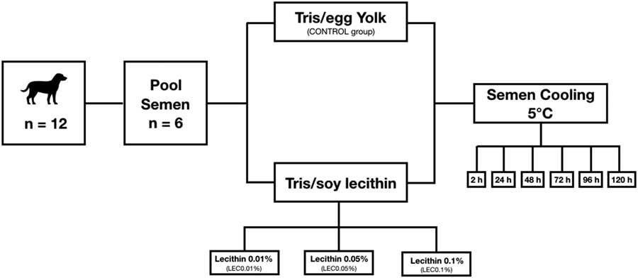
One ejaculate from two different randomly chosen dogs was pooled (n = 6). The pooled semen (n = 6) was considered the experimental unit. Subsequently, an aliquot of the sample was diluted 1:200 in formal saline solution for sperm counting in a Neubauer chamber; the result was expressed as million sperm per total volume. Next, samples were diluted in different extenders and maintained at 5°C over a cooling-curve period (0.2ºC/min until 5ºC was reached, over 2 h). Samples were evaluated at 2, 24, 48, 72, 96 or 120 h (Fig. 1).
Experimental design of the cooling procedure in dog sperm with egg yolk or soy lecithin as extenders.

Semen cooling
Four extenders were used for semen cooling: Tris−citric acid−egg yolk (Control group) composition was following recommendations for dogs (Rota et al., Reference Rota, Strom and Linde-Forsberg1995) (0.26 mol Tris-hydroxymethylaminomethane, 0.14 mol citric acid monohydrate, 60 mmol d-fructose, 20% egg yolk, 20 mmol of gentamicin in distilled water, 1179 mOsm/l, pH 6.95). The lecithin tested was Solec F (fine soy lecithin powder, Solae Company, St. Louis, MO, EUA). The lecithin extender composition was similar to the Control, but egg yolk was replaced by 0.01% (LEC0.01%), 0.05% (LEC0.05%) or 0.1% (LEC0.01%). Lecithin concentrations were selected based a previous study conducted by our group (Dalmazzo et al., 2018). Diluted samples were maintained at 5ºC until evaluation. The experimental design is illustrated in Fig. 1.
Computer-assisted sperm analysis
The computer-assisted sperm analysis (CASA; HTM-IVOS Ultimate 12.3; Hamilton Thorne Biosciences, Beverly, MA, USA) was performed as described for dogs (Iguer-ouada and Verstegen, Reference Iguer-ouada and Verstegen2001a). Briefly, 7 µl of sample was placed on a previously warmed (37°C) microscope slide and covered with a coverslip. Ten fields were selected ar random and the following variables were assessed: (1) percentage total motility (TM, %); (2) progressive motility (PROG, %); (3) average pathway velocity (VAP, μm/s); (4) straight-line velocity (VSL, μm/s); (5) curvilinear velocity (VCL, μm/s); (6) amplitude of lateral head displacement (ALH, μm); (7) beat cross frequency (BCF, Hz); (8) straightness (STR,%); and (9) linearity (LIN, %). Sperm were also classified into groups based on velocity: (1) rapid (RAP, VAP > 50 μm/s, %); (2) medium (MED, 30 μm/s < VAP < 50 μm/s, %); (3) slow (SLOW, VAP < 30 μm/s or VSL < 15 μm/s, %); or (4) static (STATIC, %).
Plasma membrane integrity
Eosin/nigrosin staining was performed for the sperm plasma membrane integrity analysis (Lagergren, Reference Lagergren1953), as previously applied for dogs (Angrimani et al., Reference Angrimani, Lucio, Veiga, Silva, Regazzi, Nichi and Vannucchi2014). In brief, 5 µl of the sample was mixed with 5 µl of prepared stain on a previously warmed (37ºC) glass slide. Smears were evaluated at ×1000 magnification on a light microscopy (Nikon, Eclipse E200, Japan). Intact cells were observed with no stain; sperm with plasma membrane injury were stained a rose colour. The results were expressed as percentage (%) after counting 200 sperm cells.
Acrosome integrity
Fast green/rose Bengal stain was used for acrosome integrity analysis, according to Pope et al. (Reference Pope, Zhang and Dresser1991), and adapted for dogs (Brito et al., Reference Brito, Lucio, Angrimani, Losano, Dalmazzo, Nichi and Vannucchi2017). A staining solution containing 1% (w/v) rose Bengal, 1% (w/v) fast green, and 40% ethanol in McIlvaine’s citrate phosphate buffer was used. For staining, 5 μl diluted semen was mixed with 5 μl stain solution on a previously warmed slide (37ºC). After 60 s, smears were made and evaluated under a light microscope at ×1000 magnification (Nikon, Eclipse E200, Japan). Acrosome integrity was inferred when the acrosomal region was stained either purple or darker than the post-acrosomal region. Conversely, damage was inferred when the acrosomal region was brighter than the post-acrosomal area or unstained. The results were expressed as a percentage (%) after counting 200 sperm cells.
Mitochondrial activity
For mitochondrial activity analysis, a 3′3-diaminobenzidine (DAB) assay (Hrudka, Reference Hrudka1987) was performed. Semen samples were diluted in DAB solution (1 mg/ml) in PBS (pH 7.0). The mixture was then incubated at 37°C for 1 h, protected from light. Subsequently, smears were prepared on glass slides, and fixed in 10% formalin buffer for 15 min. Evaluation occurred at ×1000 magnification using a microscope (Nikon, Eclipse E200, Japan). Results were expressed as percentage (%) after counting 200 sperm cells. Cells was classified into four categories: high mitochondrial activity (DAB class I − 100% of the midpiece stained); medium mitochondrial activity (DAB class II − more than 50% of the midpiece stained); low mitochondrial activity (DAB class III − less than 50% of the midpiece stained); or absence of mitochondrial activity (DAB class IV – no staining in the midpiece).
Assay of the sperm chromatin structure
For chromatin analysis with a fluorescent probe, the Guava EasyCyte™ Mini System (Guava® Technologies, Hayward, CA, USA) was used, with a 488 nm argon laser and the following filters (photodetector): PM1 (583 nm) for yellow fluorescence; PM2 (680 nm) for red fluorescence; and PM3 (525 nm) for green fluorescence. Each assay evaluated 20,000 spermatozoa, and data were analyzed using FlowJo v8.7 software (Flow Cytometry Analysis Software – Tree Star Inc., Ashland, Oregon, USA). To assess chromatin susceptibility to acid-induced denaturation, a previous protocol based in the sperm chromatin structure assay (SCSA) (Evenson and Jost, Reference Evenson and Jost2000), adapted for dogs by Lucio et al. (Reference Lucio, Silva, Regazzi, Angrimani, Nichi, Assumpcao and Vannucchi2016), was used. Evaluation for chromatin instability was performed by flow cytometry, in which the quantity of metachromatic shift from green (double-stranded DNA) to red (denatured single-stranded DNA) of acridine orange fluorescence was observed.
For this process, each sample was diluted in 100 μl of TNE buffer (0.01 mol Tris–HCl, 0.15 mol NaCl, 1 mmol EDTA, pH 7.4) and mixed with 400 μl of an acidified detergent solution (8 mmol HCl, 0.1% Triton X-100, and 0.15 mol NaCl, pH 1.2). After 30 s, staining was performed by mixing 600 μl of acridine orange solution (37 mmol citric acid, 0.126 mol Na2HPO4, 1.1 mmol disodium EDTA, and 0.15 mol NaCl, pH 6.0). After 5 min, samples were evaluated by flow cytometry. In addition, DNA fragmentation was calculated based on the percentage of spermatozoa external to the main population in a histogram of αT (ratio between red fluorescence and total fluorescence) using FlowJo software (Version Mac) (Evenson and Jost, Reference Evenson and Jost2000; Minervini et al., Reference Minervini, Guastamacchia, Pizzi, Dell’Aquila and Barile2013).
Sperm resistance to oxidative stress
To induce lipid peroxidation, a combination of 4 mmol ferrous sulphate (100 µl) and 20 mmol sodium ascorbate (100 µl) was added to 400 µl of sperm (100 × 106 spermatozoa/ml), and the samples were incubated for 90 min at 37°C (Gomez et al., Reference Gomez, Irvine and Aitken1998). Evaluation of thiobarbituric acid reactive substances (TBARS) was performed according to Ohkawa et al. (Reference Ohkawa, Ohishi and Yagi1979) and Nichi et al. (Reference Nichi, Goovaerts, Cortada, Barnabe, De Clercq and Bols2007). After 90 min, 500 µl of the mixture was added to 1000 µl 10% trichloroacetic acid for protein precipitation. Samples were then centrifuged (18,000 g, for 15 min, at 15ºC). Subsequently, 500 µl of the supernatant was mixed with 500 µl 1% (v/v) thiobarbituric acid in 0.05 N sodium hydroxide, in a glass tube and placed into a boiling water bath (100ºC) for 20 min. After this period, the chemical reaction was immediately cooled in an ice bath (0ºC) to stop the reaction. Thiobarbituric acid reactive substances (TBARS) were quantified using a spectrophotometer (U-2001 spectrophotometer, Hitachi High Technologies America, Inc., San Jose, CA, USA) at a wavelength of 532 nm. The results were compared with a standard curve previously prepared with a standard solution of malondialdehyde (Lucio et al., Reference Lucio, Silva, Regazzi, Angrimani, Nichi, Assumpcao and Vannucchi2016). The TBARS concentration was determined using the value of 1.56 × 10⁵ × M/ml as the MDA extinction coefficient (Buege and Aust, Reference Buege and Aust1978). The lipid peroxidation index was described as ng TBARS/106 spermatozoa.
Statistical analysis
All data were evaluated using SAS System for Windows (SAS Institute Inc., Cary, NC, USA). Effects of group (CONT, LEC0.01%, LEC0.05% and LEC0.1%), evaluation times (2, 24, 48, 72, 96 or 120 h), and interactions between the factors were estimated by analysis of variance (ANOVA) for repeated measures (SAS MIXED). Differences between treatments were analyzed using parametric and non-parametric tests according to residual normality (Gaussian distribution) and homogeneity of variance. In the absence of significant interactions, the effect of the groups was analyzed by merging all evaluation times, taking into account any Bonferroni correction (PROC GLIMMIX). Conversely, the effect of the evaluation times was analyzed using data from all groups. The results were described as mean and standard error. Significance level was 5% (P < 0.05).
Results
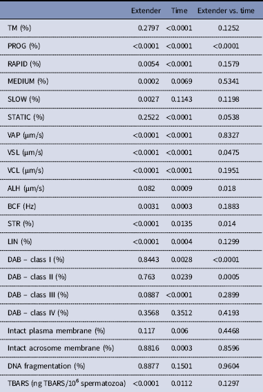
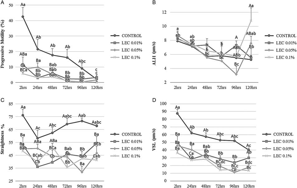
There were significant interactions (P<0.05) between the times of evaluation (2, 24, 48, 72, 96 and 120 h) and the experimental groups (CONT, LEC0.01%, LEC0.05% and LEC0.1%) for sperm progressive motility, sperm velocity straight line, amplitude of lateral head displacement, sperm straightness, and medium and high mitochondrial activity variables (Table 1). Moreover, we observed a decrease in progressive motility in the Control, LEC0.01% and LEC0.1% groups 24 h after cooling and the LEC0.05% group at 72 h (Fig. 2). All samples had their lowest progressive values at 120 h (Fig. 2). Furthermore, the Control group showed superior percentages of progressive motility compared with other groups at 2, 24, 48, 72 and 96 h (Fig. 2).
Probability values for main effects of extender and times (2, 24, 48, 72, 96 or 120 h) and their interaction with sperm parameters

Progressive motility (PROG; A), amplitude of lateral head displacement (ALH; B), straightness (STR; C) and straight-line velocity (VSL; D) in different groups (Control vs. LEC0.01% vs. LEC0.05% vs. LEC0.1%) during the experimental period (2, 24, 48, 72 and 120 h). A–CIndicate significant differences between groups and a–cindicate significant differences between time (P < 0.05).
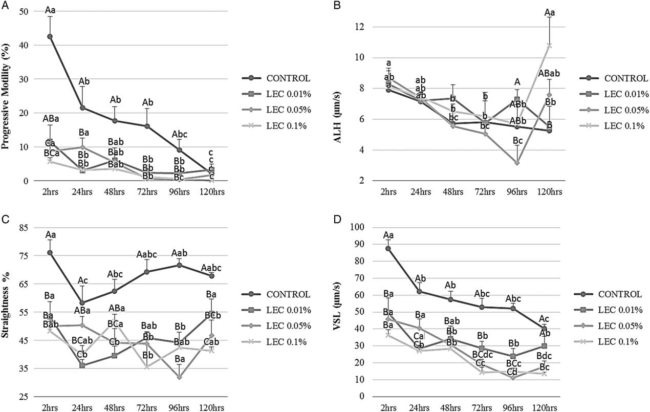
For amplitude of lateral head displacement analysis after 120 h, the LEC0.1% group had the highest values compared with the Control and LEC0.01% groups (Fig. 2). Nevertheless, progressive ALH reduction occurred in the Control samples during evaluation (Fig. 2). In addition, the Control group had the higher percentages of straightness compared with the lecithin groups at 2 h until 120 h of cooling (Fig. 2). For strait-line velocity, a decrease in the Control and LEC0.1% groups was observed after 24 h of chilling (Fig. 2); however, after 120 h, all groups presented significantly lower values (Fig. 2). The Control group presented higher values of VSL at 2, 24, 48, 72 and 96 h (Fig. 2).
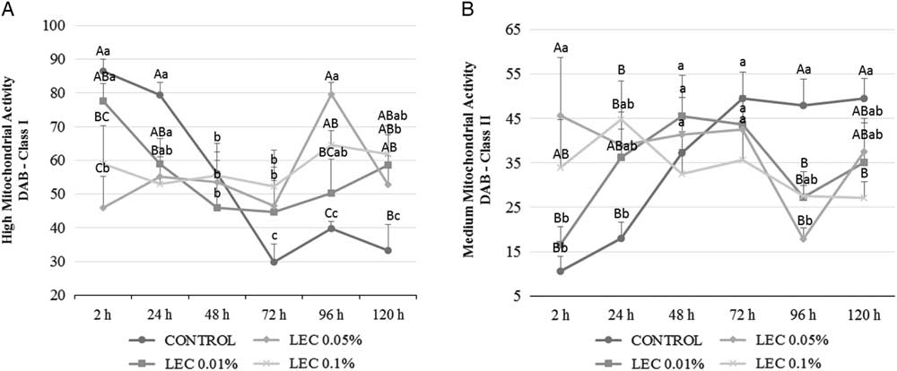
For high mitochondrial activity analysis, a decrease in the Control group values occurred after 48 h of cooling, and remained significantly lower for the lecithin groups at 96 and 120 h (Fig. 3). Conversely, an increase in the medium mitochondrial analysis occurred in Control and LEC0.01% samples after 48 h of cooling (Fig. 3). Moreover, at 2 h, LEC0.05% had the highest percentage of DAB – Class II. However, after 120 h of cooling, the Control group showed superior values compared with the LEC0.1% group (Fig. 3).
High mitochondrial activity (DAB – Class I; A) and medium mitochondrial activity (DAB – Class II; B) in different groups (Control vs. LEC0.01% vs. LEC0.05% vs. LEC0.1%) during the experimental period (2, 24, 48, 72 and 120 h). A–CIndicate significant differences between groups and a–cindicate significant differences between time (P < 0.05).

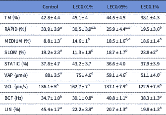
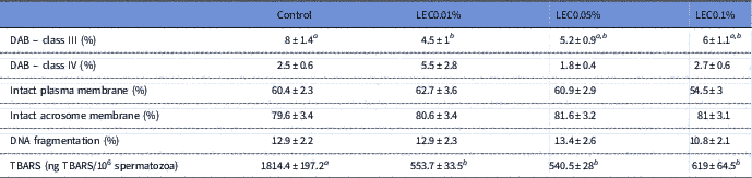
For variables for which there was no observed interaction, the time of cooling and extender factors were analyzed separately. In this way, for differences between treatments, higher values were observed in the Control group for LIN, VAP and TBARS (Tables 2 and 3). Rapid sperm numbers were lower in the LEC0.1% group compared with the Control group (Table 2). Lower percentages of slow spermatozoa and low mitochondrial activity were related to LEC0.01% (Tables 2 and 3). Conversely, VCL levels were higher in the LEC0.01% group compared with the other groups (Table 2). Furthermore, sperm BCF and percentages of medium velocity were lower in the Control compared with the groups treated with lecithin (Table 2).
Mean ± standard error of the sperm kinetics in the different groups (Control, LEC0.01%, LEC0.05% and LEC0.1%)

a–c Indicates significant differences between groups (P < 0.05).
Mean ± standard error of the sperm functional tests in the different groups (Control, LEC0.01%, LEC0.05% and LEC0.1%)

a,b Indicate significant differences between groups (P < 0.05).
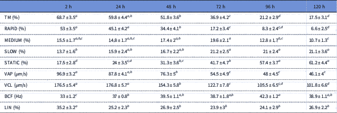
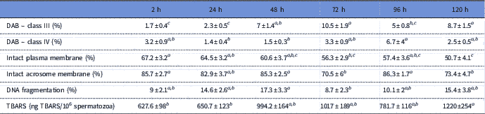
For time effects on variables without interaction, it was possible to describe a progressive reduction between 2 h and the remaining cooling times for motility, rapid and medium sperm velocities, VAP, VCL, LIN, and percentages of intact plasma membranes and acrosomes (Tables 4 and 5). However, the percentage of static sperm, low sperm velocity, BCF, TBARS concentration and low mitochondrial activity increased during the cooling times (Tables 4 and 5). The absence of mitochondrial activity and percentage of DNA fragmentation presented some differences during the cooling time, however these were similar between 2 and 120 h (Table 5).
Mean ± standard error of the sperm kinetic parameters on the times 2, 24, 48, 72, 96 and 120 h

a–d Indicate significant differences between groups (P < 0.05).
Mean ± standard error of sperm functional tests on the time (2, 24, 48, 72, 96 and 120 h

a–c Indicate significant differences between groups (P < 0.05).
Discussion
The effects of different concentrations of soy lecithin (0.01, 0.05 and 0.1%) in the extender composition were tested over several different cooling times (2, 24, 48, 72, 96 or 120 h) and a comparison of conventional egg-yolk extender and soy lecithin was performed.
Based on our data, the lowest values of sperm kinetics (i.e., PROG, VSL, LIN, VAP and STR) occurred in the groups treated with lecithin when compared with egg-yolk extender. Conversely, Beccaglia et al. (Reference Beccaglia, Anastasi, Chigioni and Luvoni2009a), described that, in canines, an extender based on soy lecithin (0.04%) presented similar results to egg yolk after 4 days of chilling. In this study, we used lecithin at different concentrations (0.01, 0.05 and 0.1%) that were similar to those described in Beccaglia et al. (Reference Beccaglia, Anastasi, Chigioni and Luvoni2009a), but used a different type of soy lecithin (Solec F). Therefore, any differences could be due to lecithin composition.
Furthermore, according to Kasimanickam et al. (Reference Kasimanickam, Kasimanickam, Memon and Rogers2012), the addition of 0.4% soy lecithin extender reduced the rate of damage in sperm parameters in dogs. A lecithin concentration of 1% in rams (Toker et al., Reference Toker, Alcay, Gokce and Ustuner2016) and dogs (Axner and Lagerson, Reference Axner and Lagerson2016) during cryopreservation protocols showed good sperm quality thawing. These authors used high soy lecithin concentrations compared with our study (0.01, 0.05 and 0.1%), and results were similar after sperm cryopreservation in dogs to those in a previous study using egg yolk and lecithin (Dalmazzo et al., 2018). Therefore, we concluded that, for sperm cooling, high concentrations of soy lecithin, greater than 0.1%, are necessary to maintain sperm quality (i.e., motility kinetics). However, during sperm cryopreservation, soy lecithin concentrations seem to have no influence on sperm quality post-thawing.
In addition, Cross (Reference Cross1994) described that phosphatidylcholine, the main composition of soy lecithin, could induce sperm hyperactivation and acrosome reactions, triggering capacitation. Studies have reported that the main feature of hyperactivation is a lower percentage of PROG, LIN and STR associated with a higher ALH (Katz et al., Reference Katz, Yanagimachi and Dresdner1978; Setyawan et al., Reference Setyawan, Kim, Oh, Kim, Jo, Lee, Choi and Lee2016). Therefore, it is possible that an extender based on soy lecithin induces hyperactivation in dogs, leading to a higher percentage of ALH and lower PROG, LIN and STR. Our results indicated that soy lecithin promotes hyperactivation, as PROG and STR values were lower and ALH values were higher than in the egg yolk group. Nevertheless, to test this hypothesis, future studies are encouraged using different techniques (i.e., FITC-PNA and fluo-4 AM probes) to assess capacitation in dog spermatozoa (Leemans et al., Reference Leemans, Gadella, Stout, Nelis, Hoogewijs and Van Soom2015).
Groups that received soy lecithin extender presented higher mitochondrial activity compared with those that received egg yolk. We note that the high mitochondrial activity (DAB – class I), at 0.1% concentration did not change over the 120 h of cooling at 5°C. Such data appear to be contradictory, as the same group presented high rates of DAB – class I and low percentages of sperm motility. Mitochondria are necessary for ATP production through oxidative phosphorylation and consequently promote sperm motility (Fariello et al., Reference Fariello, Pariz, Spaine, Cedenho, Bertolla and Fraietta2012). Losano et al. (Reference Losano, Angrimani, Dalmazzo, Rui, Brito, Mendes, Kawai, Vannucchi, Assumpcao, Barnabe and Nichi2017a) described that glycolysis appears to be as important as oxidative phosphorylation for ovine sperm kinetics, being capable of maintaining motility in cells with low mitochondrial function. Similar effects have been observed previously in mice (Mukai and Okuno, Reference Mukai and Okuno2004), bovine (Losano et al., Reference Losano, Padin, Mendez-Lopez, Angrimani, Garcia, Barnabe and Nichi2017b) and humans (Nascimento et al., Reference Nascimento, Shi, Tam, Chandsawangbhuwana, Durrant, Botvinick and Berns2008). In this context, sperm motility in dogs could be related mainly to glycolysis, as observed in other mammals, and this could explain the oscillation in sperm kinetics, but with the preservation of mitochondrial function. Furthermore, it is important to report a beneficial effect of soy lecithin to protect mitochondrial activity after long cold storage, in contrast with samples stored in egg-yolk extender. Therefore, in future studies using soy lecithin as a cooling extender, extenders with mitochondrial stimulatory treatments for example, with glycolysis substrates, might maintain motility kinetics, as previously described in other species (Losano et al., Reference Losano, Angrimani, Dalmazzo, Rui, Brito, Mendes, Kawai, Vannucchi, Assumpcao, Barnabe and Nichi2017a; Nascimento et al., Reference Nascimento, Shi, Tam, Chandsawangbhuwana, Durrant, Botvinick and Berns2008).
Although the egg yolk presented benefits for motility, higher values in TBARS concentrations for samples stored with this extender were also observed. This same effect has been described previously in bulls (Crespilho et al., Reference Crespilho, Nichi, Guasti, Freitas-Dell’Aqua, Sa, Maziero, Dell’Aqua and Papa2014), rams (Sicherle et al., Reference Sicherle, Maia, Bicudo, Rodello and Azevedo2011) and wild cats (Angrimani et al., Reference Angrimani, Barros, Losano, Cortada, Bertolla, Guimaraes, Correa, Barnabe and Nichi2017). In the present study, after adding ferrous sulphate and sodium ascorbate, we induced oxidative stress, as described by Nichi et al. (Reference Nichi, Goovaerts, Cortada, Barnabe, De Clercq and Bols2007). Angrimani et al. (Reference Angrimani, Barros, Losano, Cortada, Bertolla, Guimaraes, Correa, Barnabe and Nichi2017) described that this phenomenon, in samples preserved with egg yolk, may influence the findings on oxidative status. Therefore, further studies in canine sperm are required to evaluate the influence of egg-yolk extender on the production of lipid peroxidation metabolites, as our data did not show any deleterious effects on the remaining sperm parameters analyzed (i.e., DNA fragmentation and plasma membrane and acrosome integrity).
Moreover, after 120 h of cooling, it was possible to observe an increase in the production of lipid peroxidation metabolites. Therefore, the results may indicate a higher production of reactive oxygen species (ROS) during long cold storage time, promoting oxidative stress. Oxidative stress is extremely harmful to sperm cells, as spermatozoa have limited amounts of antioxidant and a plasma membrane rich in polyunsaturated fatty acids that may be oxidized by ROS (Irvine et al., Reference Irvine, Twigg, Gordon, Fulton, Milne and Aitken2000). Among the main effects of oxidative stress in sperm, damage to the plasma membrane affects motility kinetics and DNA fragmentation (Aitken and De Iuliis, Reference Aitken and De Iuliis2010). Our data showed an increase in TBARS concentrations and lesions in the plasma and acrosomal membranes that could be associated with a decrease in sperm kinetics. Therefore, we suggest further studies in dogs to test extenders based on soy lecithin and antioxidants for sperm cooling, as described by Kmenta et al. (Reference Kmenta, Strohmayer, Muller-Schlosser and Schafer-Somi2011), who found increased sperm longevity using 0.8% lecithin associated with catalase.
In conclusion, soy lecithin at 0.01, 0.05 or 0.1% were not able to protect sperm during cooling. However, soy lecithin shows a mitochondrial protective effect that can be explored for the development of new extenders. Furthermore, after a long storage time, samples presented structural sperm damage and had higher production of lipid peroxidation metabolites. Therefore, in dogs, the association of extender and antioxidants appears to be an option for a long cooling times.
Acknowledgements
Authors thank the Brazilian Army for providing the animals used in this experiment and the Solae Company for the soy lecithin. Moreover, authors thank Dr Camilla M Mendes and Prof. Mayra E O A Assumpção for support in flow cytometry analysis.
Financial support
This research was financially supported by FAPESP (process no. 2011/0922-0) and CAPES.
Conflicts of interest
None
Ethical standards
This study was approved by the Bioethics Committee of the School of Veterinary Medicine and Animal Science, University of São Paulo (protocol no. 2329/2011). Moreover, unless otherwise mentioned, chemicals and media used were from Sigma-Aldrich (St. Louis, MO, USA).