Dilated cardiomyopathy (DCM) is characterised by an enlarged left ventricular chamber and reduced systolic ejection without a concomitant increase in left ventricular wall thickness. Reference Maron, Towbin and Thiene1–Reference Lee, Hsu and Kantor4 It is the most common form of cardiomyopathy in children Reference Towbin, Lowe and Colan5,Reference Bozkurt, Colvin and Cook6 and is a major cause of progressive heart failure and death. Reference Towbin, Lowe and Colan5,Reference Daubeney, Nugent and Chondros7,Reference Wilkinson, Landy and Colan8 In the paediatric setting, some cases have evidence of viral infection or myocarditis, Reference Lee, Hsu and Kantor4,Reference Bozkurt, Colvin and Cook6,Reference Griffin, Kearney and Ni9-Reference Venugopalan12 mutations in myocardial proteins, inborn errors of metabolism,or myocardial toxins. Reference Bozkurt, Colvin and Cook6 Etiology, nevertheless, remains unknown, that is, idiopathic, for a broad majority. Reference Kirk, Dipchand and Rosenthal2,Reference Towbin, Lowe and Colan5,Reference Bozkurt, Colvin and Cook6,Reference Hsu and Canter13,Reference Pahl, Dipchand and Burch14 Cardiac transplantation is an accepted treatment of last resort for chronic heart failure due to DCM that has failed conventional medical therapies. Reference Venugopalan12,Reference Blume, Naftel, Bastardi, Duncan, Kirklin and Webber15 DCM is the predominant diagnosis precipitating cardiac transplantation in children and adolescents. Reference Kirk, Dipchand and Rosenthal2,Reference Pahl, Dipchand and Burch14,Reference Kirk, Naftel and Hoffman16,17
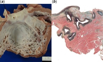
Endocardial fibroelastosis (EFE) is a non-specific and chronic reaction to myocardial wall stress that usually becomes more severe over time. Reference Venugopalan12 Pathologically, it is characterised by a proliferation of collagen and elastic fibre rich connective tissue within the endocardial lining of cardiac chambers (Fig 1). Reference Lurie18 The term “endocardial fibroelastosis” was first coined in 1943. Reference Weinberg and Himelfarb19 For decades thereafter, “primary EFE” was considered to be a distinct diagnostic entity, typically applied to the dilated, failing heart of any infant in which EFE was present in the absence of a congenital anomaly or an alternative assignable cause. Reference Lurie18,Reference Noren, Adams and Anderson20-Reference Seki, Patel, Ashraf, Perens and Fishbein22 The concept of primary EFE has begun to lose favour as EFE’s association with various paediatric cardiac conditions, both congenital and acquired, has been increasingly recognised. Reference Maron, Towbin and Thiene1,Reference Lurie18,Reference Lurie23 Although no longer a formal disease classification, the notion of “primary EFE” continues to incite controversy. Reference Lurie18,Reference Seki, Patel, Ashraf, Perens and Fishbein22,Reference Lurie23
(a) LV EFE in explanted heart from 1-year-old female DCM patient. (b) Whole mount section of LV wall from heart explant of 4-month-old female DCM patient demonstrating black stained markedly thickened fibro-elastotic endocardium and patchy green-gray stained fibrosis of the ventricular myocardium (Elastic trichrome stain).

There have been several reports detailing paediatric DCM’s pathologic features. Reference Seki, Patel, Ashraf, Perens and Fishbein22,Reference Chen, Nouri, Balfour, Jureidini and Appleton24-Reference Crossland, Edmonds and Rassl29 Despite recognition of a DCM–EFE relationship in infants and children, Reference Maron, Towbin and Thiene1,Reference Chen, Nouri, Balfour, Jureidini and Appleton24-Reference Arola, Tuominen, Ruuskanen and Jokinen28,Reference Griffin, Hernandez and Martin30-Reference Arya, Karpawich and Gupta35 comprehensive pathologic analysis of the frequency and nature of this relationship has rarely been reported. The objective of our study, therefore, was to analyse the surgical pathology reports of explanted idiopathic DCM hearts at The Hospital for Sick Children (HSC) in order to: (1) characterise the pathological spectrum of paediatric DCM and (2) compare and contrast the pathological features, including EFE, amongst paediatric age subgroups.
Methods
Since 1986, all explanted hearts from transplant recipients at HSC have undergone a detailed pathological examination. After fixation in a neutral buffered solution for a minimum of 24 hours, an average of 10 blocks per specimen were obtained. Histology staining consisted primarily of Hematoxylin and Eosin (H & E) with additional special stains (Elastic Trichrome, Masson Trichrome, and Movat Pentachrome) also applied in the majority of cases. We conducted a retrospective review of all heart explant surgical pathology reports from 1986 to 2016 (inclusively) and identified all native explants with a primary pathologic diagnosis of DCM.
Data collection: surgical pathology reports
Surgical pathology reports detailing the results of the pathological examinations were reviewed. Most specimens included a limited amount of atrial tissue; consequently, only data on ventricular pathologic parameters were collected. Listed heart weights and wall thicknesses were recorded and compared to the expected range for intact hearts from decedents of the same age (Schulz, Reference Schulz and Giordano36 for infants and children up to 15 years of age; Kayser Reference Kayser37 and Scholz, Reference Scholz, Kitzman, Hagen, Ilstrup and Edwards38 for patients aged 16–18 years). Gross features, such as chamber dilatation and EFE, and microscopic characteristics, such as myocardial inflammation and fibrosis, were documented and graded semi-quantitatively as “none”, “mild”, “moderate”, or “severe”, based on the original pathologist’s findings. The grading of histologically confirmed EFE was based primarily on the reporting of the gross (naked-eye) examination. Any other significant findings were also noted.
Statistical analysis
Since the retrieved information was a combination of continuous and categorical variables, several strategies were implemented to facilitate statistical analysis. For each measurement of the continuous variables of explant weight and ventricular wall thicknesses, a z-score was calculated to standardise the observation as well as to detect outliers. Next, these z-scores were converted to “yes” or “no” observations for the following pathologic parameters: (1) increased explant weight (cardiomegaly), right ventricular wall hypertrophy, left ventricular wall hypertrophy (z ≥ 2 = “yes”); (2) decreased explant weight, right ventricular wall thinning, left ventricular wall thinning (z ≤ -2 = “yes”); and normal explant weight, normal right ventricular wall thickness, normal left ventricular wall thickness (z between −2 and 2 = “yes”). For the categorical variables of chamber dilatation, EFE, myocardial inflammation, and myocardial fibrosis, the grades were consolidated into two categories based on the following criteria: (1) “none” or “mild” = negative; and (2) “moderate” or “severe” = positive in order to mitigate inherent observational subjectivity. Cases with the “mild” descriptor were grouped with cases in which the examined characteristic was absent because it typically denotes a minimal presence that does not necessarily impact functionality. Our cohort was subsequently divided into “age at the time of transplantation” subgroups akin to those employed by the International Society for Heart and Lung Transplantation (ISHLT) for data analysis. They are (1) < 1-year of age; (2) 1–5 years of age; (3) 6–10 years of age; and (4) ≥ 11 years of age. 17 Scatterplots were used to depict associations between common gross phenotypic features of DCM-induced heart failure (i.e., increased heart weight, dilated left ventricular chamber, and normal left ventricular wall thickness) and EFE amongst the age groups. Detected patterns were then inputted into a binary logistic regression model to confirm any statistical relationship. Anticipating that our sample size may be small and non-normally distributed, we chose statistical tests better suited to analyse these types of data. Pathologic variables were evaluated against the age group classifications using Fisher’s exact test of Independence. If the resulting p-value was ≤ 0.05, a relationship was deemed to be statistically significant. To further understand the magnitude of the difference (odds ratio) for a statistically significant finding, exact logistic regression was used. Data analysis for this paper was generated using SAS software, Version 9.6 of the SAS System for Windows. (Copyright © 2020 SAS Institute Inc. SAS and all other SAS Institute Inc. product or service names are registered trademarks or trademarks of SAS Institute Inc., Cary, NC, United States of America.)
Ethics
This study was approved by The Hospital for Sick Children Research Ethics Board (REB#: 1000026283).
Results
Of the 309 cardiac explant surgical pathology reports reviewed, 89 were from infants and children diagnosed with heart failure due to DCM [Males (M): 47; Females (F): 42] and form the basis of this study. Recipient ages ranged from 2 days to 18 years (mean: 6.8 years). There were 23 aged < 1 year (M: 10; F: 13), 22 between 1 to 5 years (M: 9; F: 13), 10 between 6 to 10 years (M: 5; F: 5), and 34 aged 11 years or older (M: 23; F: 11).
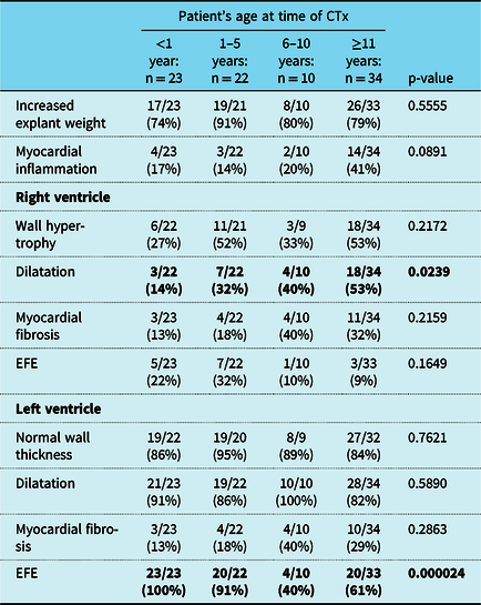
Notable pathologic characteristics of the DCM-cardiac transplantation explants are summarised in Table 1. These hearts were phenotypically characterised by increased heart weight (cardiomegaly) even with most of the atria missing, a dilated left ventricular chamber, and left ventricular wall thickness within the expected range for a patient’s age at the time of cardiac transplantation. Right ventricular wall hypertrophy was common, ranging from 27 to 53% of hearts amongst the four age groups. Right ventricular dilatation frequency gradually increased from 14% in the youngest to 53% in the oldest age group. Myocardial fibrosis, which was present in 26% of the explants, was often biventricular. Myocardial inflammation was most commonly found in the oldest age group (41%). Only 4 cases of the 67 left ventricular EFE positive (left ventricular EFE +ve) DCM hearts (6%) had histopathology suggestive of active or remote myocarditis. Left ventricular EFE was observed in all transplantees aged < 1 year, 91% of those aged 1–5 years, 40% in those aged 6–10 years, and 61% of those aged 11 years or older. The frequency of right ventricular EFE was greatest in those 1 to 5 years of age (32%).
Pathological parameters of DCM explants (n = 89). CTx = cardiac transplantation

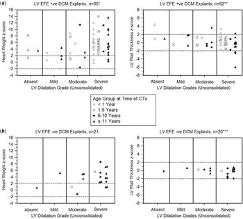
The results of scatterplot analysis of the degree of left ventricular chamber dilatation with heart weight and left ventricular wall thickness determinations in left ventricular EFE +ve and left ventricular EFE -ve hearts are shown in Figure 2. Scatterplot distributions were similar irrespective of the presence or absence of EFE with the majority of the explants in both groups characterised by severe left ventricular chamber dilatation, increased heart weight, and a normal left ventricular wall thickness.
(a) Age group distribution of DCM explants according to presence of LV EFE (LV EFE +ve), LV dilatation grade, heart weight z-score and LV wall thickness z-score. (b) Age group distribution of DCM explants according to absence of LV EFE (LV EFE -ve), LV dilatation grade, heart weight z-score and LV wall thickness z-score. Note: For all scatterplots, a z-score between -2 and 2 indicated that either the heart weight or LV wall thickness was within normal range for the patient’s age at time of CTx. *2 cases had incomplete information. **5 cases had incomplete information. ***1 case had incomplete information.

Binary logistic regression was applied to investigate whether age at the time of cardiac transplantation or any of the pathological characteristics observed in the scatterplots (severe left ventricular chamber dilatation, cardiomegaly, or normal left ventricular wall thickness) could predict the presence of left ventricular EFE. Since all recipients < 1 year had left ventricular EFE, data separation would occur if the age groupings previously described were used as predictor variables. Therefore, age at the time of cardiac transplantation was dichotomised at 5 years for entry into the model. The only statistically significant correlation identified was between patients aged ≤ 5 years at cardiac transplantation and left ventricular EFE incidence in their explants (p = 0.0002). These recipientsʼ native hearts had 22 times the odds (OR = 21.56; CI = 4.37–106.44) of left ventricular EFE being present than those who underwent surgery at age 6 years or older.
The Fisher calculations were not statistically significant for all pathological parameters, with two exceptions: right ventricular dilatation and left ventricular EFE. In the case of right ventricular dilatation, the p-value = .0239; for left ventricular EFE, p = 0.000024 (Table 1). Observation of either of these two features was therefore statistically related to a DCM transplantee’s age at the time of cardiac transplantation.
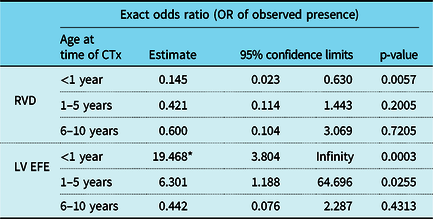
Exact logistic regression results for right ventricular dilatation and left ventricular EFE data are summarised in Table 2. In our model, the oldest age group was used as the reference variable for comparison. The association of right ventricular dilatation and the youngest transplant age category was statistically significant (p = 0.0057). For this group, the odds of finding right ventricular dilatation were 15% of the odds of finding it in patients aged 11 or older, that is, right ventricular dilatation was more likely to be found in the oldest patients (85% odds). Regarding left ventricular EFE, there were two age groups with significant results: the < 1-year-olds and the 1–5-year-olds (p = 0.0003 and p = 0.0255, respectively). Recipients aged 1–5 had a sixfold increase in the odds of observing left ventricular EFE in their explants, while for those aged < 1, the odds rose by 19.
Results of the exact logistic regression analysis for RVD and LV EFE. Recipients aged ≥11 years at time of CTx used as reference for comparison. *A median unbiased estimate and a one-sided p-value

Discussion
EFE
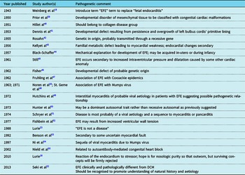
The term “endocardial fibroelastosis” was first coined by Weinberg and Himelfarb in their 1941 paper to describe the thickened pearly white endocardial lining they found in the hearts of infants who died in congestive heart failure. Reference Weinberg and Himelfarb19 Since this seminal publication, multiple aetiologies have been proposed for EFE as a primary disease (Table 3).Over the ensuing years, the concept that left ventricular EFE represented a distinct form of cardiomyopathy lost favour, in large part due to broader awareness of its association with a variety of paediatric conditions, both congenital and acquired. Reference Maron, Towbin and Thiene1,Reference Arola, Tuominen, Ruuskanen and Jokinen28,Reference Chou, Wang and Chou31,Reference Takahashi, Kanetake, Moriya and Funayama33-Reference Arya, Karpawich and Gupta35 At present, EFE is most commonly thought to be a reaction to heart chamber stresses of variable aetiology and not a primary disease entity, a view first advocated by Black-Schaffer Reference Black-Schaffer44 and strongly supported by Lurie Reference Lurie18,Reference Lurie23 among others. Reference Venugopalan12,Reference Still45,Reference Fishbein, Ferrans and Roberts52,Reference Benson, Wilson, Freedom, Freedom, Benson and Smallhorn53 Despite this, the concept of primary EFE as a specific diagnostic entity distinct from DCM persists. Reference Seki, Patel, Ashraf, Perens and Fishbein22 We concur with Seki and colleagues Reference Seki, Patel, Ashraf, Perens and Fishbein22 that further investigative attention to this pathological phenomenon is warranted to promote a greater understanding of its natural history and aetiology.
Historical survey of major EFE publications

Paediatric DCM with EFE
There is now wider recognition of the association of EFE with paediatric DCM. Reference Maron, Towbin and Thiene1,Reference Chen, Nouri, Balfour, Jureidini and Appleton24-Reference Matitiau, Perez-Atayde and Sanders26,Reference Arola, Tuominen, Ruuskanen and Jokinen28,Reference Griffin, Hernandez and Martin30-Reference Arya, Karpawich and Gupta35 Furthermore, Aiello and Higuchi have shown that the frequency and severity of EFE in dilated hearts from children were inversely correlated with their age. Reference Aiello and de Lourdes Higuchi25 Nevertheless, the scientific statement published by the American Heart Association (AHA) in 2016 on Current Diagnostic and Treatment Strategies for Specific Dilated Cardiomyopathies makes only minimal reference to EFE Reference Bozkurt, Colvin and Cook6 and the 2019 AHA statement on Cardiomyopathies in Children does not refer to it at all. Reference Lipshultz, Law and Asante-Korang56 This lack of attention to EFE as a noteworthy pathological finding persists even though several reports have shown that histological evidence of EFE in the context of paediatric DCM was associated with worse patient outcomes. Chen et al, Reference Chen, Nouri, Balfour, Jureidini and Appleton24 in a review of 23 paediatric patients with congestive cardiomyopathy, reported that all five children with pathologically confirmed EFE (either by biopsy or autopsy) died. Four presented with symptoms at ≤ 5 months of age and the post-diagnosis survival period ranged from 0.2 to 1.7 years. Matitiau et al, Reference Matitiau, Perez-Atayde and Sanders26 in their survey of 24 children with infantile DCM, reported that a histological finding of EFE in a patient with DCM was statistically associated with poorer survival (p< 0.02). In the Finnish paediatric DCM population investigated by Arola et al, Reference Arola, Tuominen, Ruuskanen and Jokinen28 31 (50%) succumbed to the disease. Of the 29 who were autopsied, 12 (41%) had EFE. This DCM and EFE group differed from the remaining cohort in that all subjects were diagnosed before the age of 2 years and died soon after presentation. Based on both univariate and multivariate survival analyses, it was concluded that EFE, as a predictor variable in the setting of DCM, was the most critical indicator of death. Although these data suggest the possibility that in the context of paediatric DCM, EFE is a marker of poor prognosis, major selection bias inherent in its diagnosis at autopsy has limited the clinical utility of these studies. Reference Alvarez, Wilkinson and Lipshultz32
Our study and its implications
The DCM explants in our study were phenotypically characterised by increased weight (cardiomegaly) and left ventricular chamber dilatation, with left ventricular wall thickness within the expected range for a patient’s age at the time of cardiac transplantation. The only pathological characteristics statistically associated with age were right ventricular dilatation and left ventricular EFE. Right ventricular dilatation was less common in patients aged < 1 year when compared to those aged ≥ 11 years. Since DCM-related right-sided chamber dilatation tends to worsen over time, the difference between the two age groups may be linked to the longer time course of disease in older children.
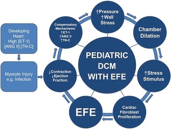
Left ventricular EFE was much more prevalent in the two younger subsets and was present in all hearts from recipients < 1 year of age (OR = 6 and OR = 19, respectively). The reason for this striking association between EFE and the youngest age group and, to a lesser extent, the 1- to 5-year-old age group is unclear. Hearts of infants typically have elevated concentrations of Endothelin-1 (ET-1), Angiotensin II (ANG II), and Tenascin-C (TN-C) due to the role they play in growth and development at these crucial stages. Reference Brutsaert57,Reference Imanaka-Yoshida and Aoki58 They are also vital components of the body’s pathophysiological response to heart failure entailing increased biosynthesis and secretion of these proteins, which influences cardiac fibroblast proliferation as part of myocardial remodelling. Reference Venugopalan12,Reference Imanaka-Yoshida and Aoki58-Reference Knudson and Cabrera62 Most fibroblasts in EFE are proposed to originate from embryonic epicardial-derived mesenchymal cells. Reference Zhang, Huang and Liu63 We postulate that during infancy, when compensatory mechanisms are activated to counteract cardiac dysfunction as a consequence of myocyte injury, there is a concurrent impact on the endocardium, which promotes fibro-elastotic proliferation (Fig 3). Genetic variation in signalling pathway elements might explain why some individuals develop left ventricular EFE deposits, while others undergoing similar stress do not. Reference Lurie18
Pathophysiologic evolution of pediatric DCM with EFE. ET-1: Endothelin-1; ANG II: Angiotensin II; TN-C: Tenascin-C.

A retrospective analysis of cardiac explants cannot definitively address left ventricular EFE’s prognostic significance in the setting of DCM. The high frequency of EFE in our series of hearts does raise the possibility that it may portend a poor prognosis as previously suggested by previous authors. Reference Chen, Nouri, Balfour, Jureidini and Appleton24,Reference Matitiau, Perez-Atayde and Sanders26,Reference Arola, Tuominen, Ruuskanen and Jokinen28 Cardio-imaging assessment challenges have greatly hampered EFE’s clinical utilisation as a prognostic marker. The diagnosis continues to be rendered primarily “after the fact” upon surgical pathological assessment of explants Reference Seki, Patel, Ashraf, Perens and Fishbein22 or at autopsy. Reference Chen, Nouri, Balfour, Jureidini and Appleton24-Reference Matitiau, Perez-Atayde and Sanders26,Reference Arola, Tuominen, Ruuskanen and Jokinen28 The use of left ventricular endomyocardial biopsy as a diagnostic modality Reference Chen, Nouri, Balfour, Jureidini and Appleton24,Reference Matitiau, Perez-Atayde and Sanders26,Reference Arola, Tuominen, Ruuskanen and Jokinen28 is limited by technical considerations, notably in the very young where the diagnostic yield would be the greatest, and the small sample obtained, which may not be representative of the ventricle as a whole. For patients in whom a left ventricular assist device is indicated, open transmural left ventricular biopsy may be an option.
That DCM hearts with and without EFE were otherwise phenotypically similar, in our view, argues against the notion of primary EFE as a distinct diagnostic entity. Whether a primary entity or not, left ventricular EFE is an important distinctive pathological feature commonly found in paediatric DCM, the presence of which deserves to be terminologically highlighted. Thus, we advocate for the descriptive designation, "DCM with EFE", as previously proposed by Benson et al Reference Benson, Wilson, Freedom, Freedom, Benson and Smallhorn53 to differentiate these cases from those without significant EFE and to facilitate future scientific investigation.
Study limitations
Selection bias was inherent in our cohort since this was a retrospective analysis of paediatric DCM patients whose severity of myocardial dysfunction necessitated cardiac transplantation. Another limitation was our study’s sample size. While the number of specimens overall was suitable for statistical evaluation, division into the four subgroups for in-depth analyses produced less than ideal sample sizes, possibly limiting our conclusions regarding paediatric DCM with EFE. Through non-parametric statistics, which were more appropriate given the smaller, non-normally distributed datasets, we were able to control for this potential weakness. Finally, a third limitation concerned the transformation of a pathologist’s subjective observations into values which were then statistically tested via grade consolidation. Specifically, equating the “mild” descriptor of a pathologic feature with it being “negative” (i.e., absent) may have contributed to an underestimation of its actual presence in the explant. Because quantification was required to analyse the data appropriately, it was nonetheless essential.
Conclusion
Our analysis of 89 paediatric DCM-cardiac transplantation explantsʼ pathologic characteristics demonstrated that left ventricular EFE is significantly more common in hearts from recipients aged < 5 years, and even more so, in those under the age of 1 year. Cardiac specimens with and without EFE were otherwise phenotypically similar, arguing against the notion of “primary EFE” as a distinct diagnostic entity. Regardless of these nosological considerations, we do concur with Seki and colleagues Reference Seki, Patel, Ashraf, Perens and Fishbein22 that further investigative attention into this pathological phenomenon is warranted in order to promote a greater understanding of its natural history and aetiology. We, therefore, propose that the descriptive clinco-pathological designation of “Dilated Cardiomyopathy with Endocardial Fibroelastosis (DCM with EFE)” should be adopted so as to facilitate future investigation, particularly as it may relate to the potential prognostic/therapeutic significance of left ventricular EFE.
Acknowledgements
The authors would like to thank Cory Borkhoff, Clinical Epidemiologist, and Bryan Maguire, Biostatistician, The Hospital for Sick Children, for their assistance with the statistical analyses used in this paper. Their help and advice were invaluable.
Financial support
This research received no specific grant from any funding agency, commercial or not-for-profit sectors.
Conflicts of interest
None.