





Introduction
In 2019, antimicrobial resistance (AMR) was estimated to be responsible for 1.27 million deaths and to contribute to 4.95 million deaths worldwide. Reference Murray, Ikuta and Sharara1 Global AMR surveillance efforts continue to evolve, but remain constrained by limited structural and laboratory capabilities in low- and middle-income countries. Reference Lim, Ashley and Hamers2 Even well-resourced surveillance relies on clinical isolates from symptomatic or screened patients, potentially missing asymptomatic colonization and emerging threats. Reference Tacconelli, Sifakis and Harbarth3 Hospitals are recognized hotspots for AMR selection and dissemination, concentrating antimicrobial use, vulnerable patients, and resistant organisms. Reference Hocquet, Muller and Bertrand4,Reference Hassoun-Kheir, Stabholz and Kreft5 Humans shed resistant bacteria and antimicrobial resistance genes (ARG) into hospital plumbing, while antibiotics and disinfectants are introduced, exerting selective pressure. Reference Uluseker, Kaster and Thorsen6 These agents can persist in biofilms within drains and pipes, potentially distorting temporal wastewater signals, before entering municipal systems and the wider environment. Reference Hocquet, Muller and Bertrand4,Reference Uluseker, Kaster and Thorsen6
Wastewater-based epidemiology (WBE) provides near-comprehensive, pooled biological samples of entire populations, independent of socioeconomic status, or healthcare access. Reference Boehm, Hughes and Duong8 Initially established for viral pathogens (poliovirus, SARS-CoV-2, respiratory viruses, measles), Reference Kilaru, Hill and Anderson7,Reference Poretsky, Dhiman and Hendricks9 WBE is increasingly applied to AMR. Reference Hassoun-Kheir, Stabholz and Kreft5,Reference Uluseker, Kaster and Thorsen6,Reference Kilaru, Hill and Anderson7 WBE has profiled resistance genes across cities and countries, with national programs now quantifying AMR alongside traditional surveillance. Reference Knight, Webster and Perry10,Reference Hendriksen, Munk and Njage11
Hospital wastewater offers key advantages compared to municipal wastewater: higher concentrations of resistant bacteria and resistance genes, Reference Hassoun-Kheir, Stabholz and Kreft5 and a smaller, defined catchment with minimal agricultural or stormwater input, reducing environmental noise. Reference Hassoun-Kheir, Stabholz and Kreft5 Wastewater can be analyzed using culture-based methods (recovering viable, susceptibility-testable isolates) or culture-independent molecular methods (PCR, sequencing, metagenomics). However, correlation between hospital wastewater signals and traditional AMR surveillance remains unknown. We performed this scoping review to assess concordance between hospital wastewater-based AMR surveillance and traditional patient-based surveillance.
Methods
Search strategy
The search strategies were drafted by an experienced librarian from the Stanford Lane Medical Library. We retrieved publications from Medline (PubMed), Embase (Elsevier), Biosis (EBSCO), Web of Science, and preprints (biorxiv.org) from database inception to May 5, 2026. The search strategy is detailed in Supplementary Table 1. Search results were exported to Covidence (Melbourne, Australia), where duplicates were automatically removed by comparing titles, years, volumes, and authors, and then corrected manually. 12
In this study, “traditional hospital AMR surveillance” was defined as monitoring of AMR using patient-derived data. We categorized studies by clinical data type: 1) active surveillance – prospective, systematic screening conducted as part of research studies, concurrent with wastewater sampling; 2) routine diagnostics – clinical isolates from standard diagnostic processes during routine care, or 3) database/registry – data from existing surveillance systems, electronic health records, or antibiograms, potentially historical. Surveillance scope describes data breadth: (1) comprehensive patient-level surveillance – systematic tracking with clinical outcomes and infection/colonization status; (2) population-level isolates – clinical isolates with limited patient context, or (3) limited/convenience isolates– small numbers (<20) selected for comparison.
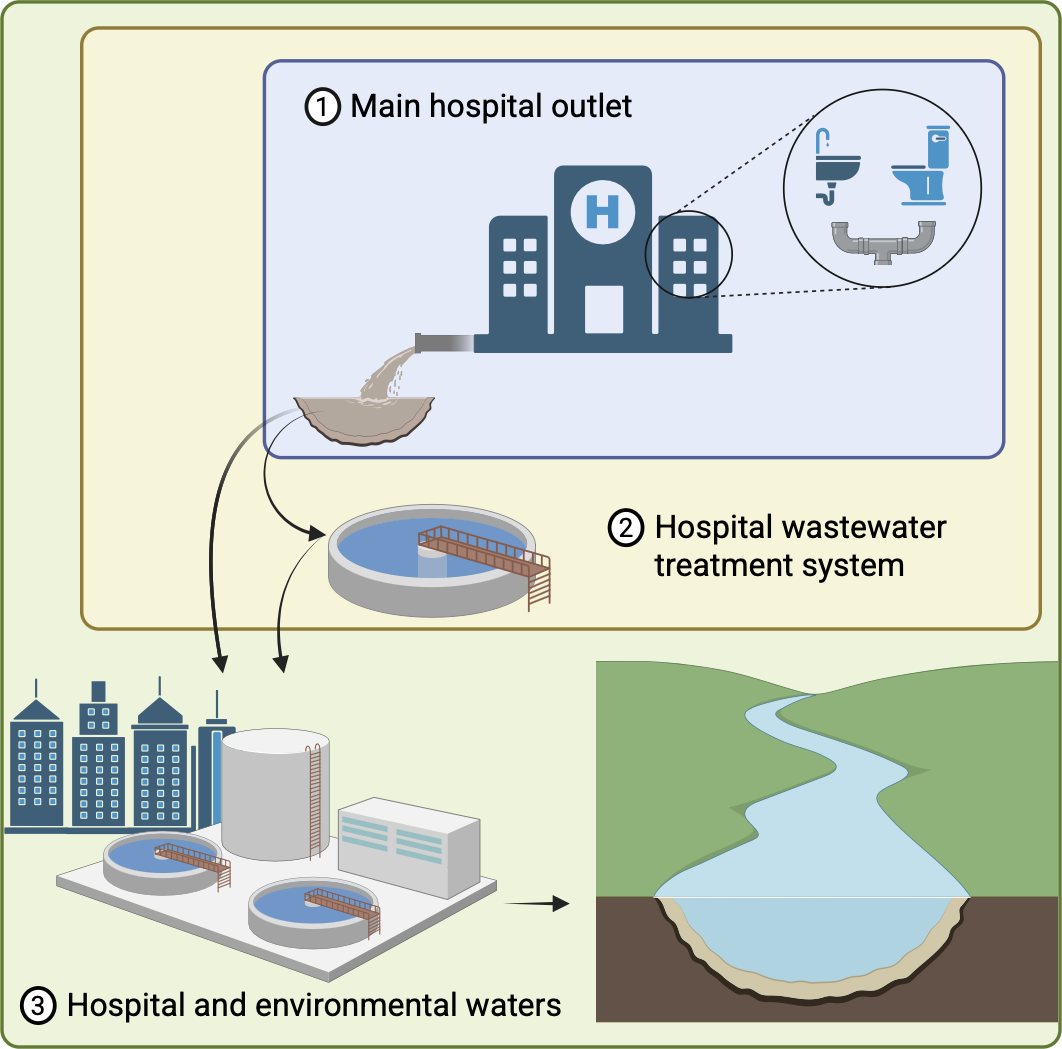
Hospital WBE was defined as the sampling and analysis of liquid waste streams originating within hospital plumbing (including ward-level drains, internal sewer lines, terminal hospital outlets, and dedicated hospital wastewater treatment facilities) for the detection or quantification of AMR bacteria, AMR genes, or both (Figure 1).
Hospital wastewater sampling locations along the discharge pathway from hospital outlet to environmental release.

We used the PICO framework to formulate the research question and guide search strategy development (Supplementary Table S2). Reference Eriksen and Frandsen13 Concordance between hospital wastewater and traditional surveillance was classified into four mutually exclusive categories (Supplementary Table S3).
Study selection
We included studies conducted exclusively in acute care hospitals or with surrounding communities. To be included, a study needed to focus on the potential of hospital WBE and its concordance with traditional hospital AMR surveillance. Studies were excluded if they did not include any form of traditional hospital AMR surveillance or focused exclusively on viral pathogens. Only primary research studies were included (Supplementary Table 4). The reporting follows the PRISMA Extension for Scoping Reviews (PRISMA-ScR). Reference Tricco, Lillie and Zarin14
Our search returned 6,934 records. After deduplication, 4,695 records underwent title and abstract screening by two independent reviewers (MJ, EVC); 165 full texts were then assessed for eligibility, and 36 studies met the inclusion criteria (Figure 1). Discrepancies at each stage were resolved by a third reviewer (GRN).
Two independent reviewers (MJ and EVC) abstracted data for each article using a standardized form (Supplementary Table 5). Extracted data were cross-checked by three distinct people to minimize individual bias.
Data analysis and visualization
Results were visualized in summary tables. A map was created using the “world” package, and a plot using the “UpSetR” package in RStudio 2026.01.1 + 403. Reference Conway, Lex and Gehlenborg15,Reference Hassoun-Kheir, Stabholz and Kreft16,17 The geographical classification of studies was determined by their sampling sites. Countries in which the studies were conducted were grouped according to the World Bank income classification into four categories: high-income, upper-middle-income, lower-middle-income, and low-income countries. 18
Results
From the 4,695 studies screened, a total of 36 met the inclusion criteria and were included in this scoping review (Figure 2). Reference Acosta, Lee and Bautista19,Reference Zong and Zhang54 They were conducted in 22 countries between 2008 and 2026 (Figure 3), 44% (16/36) were published in 2024–2026 (Table 1).
PRISMA flowchart of study selection process for hospital wastewater antimicrobial resistance surveillance studies.

Geographic distribution of hospital wastewater antimicrobial resistance surveillance studies across 22 countries, 2008–2026 (N = 36 studies).

Characteristics and temporal alignment of hospital wastewater and clinical antimicrobial resistance surveillance across 36 studies from 22 countries, 2008–2026

Table 1. Long description
The table has 36 rows and 10 columns. The columns are: Author, year of publication, Country name (HMI), Hospital wastewater sampling (Method, Source, Design, Period details), Traditional AMR surveillance (Clinical data type, Surveillance scope, Period details), and Temporal code. The rows list individual studies with specific details under each column. For example, Row 1: Acosta et al, 2025, Canada (HMI), 24-hour composites, Hospital outlet, Cross-sectional, Not mentioned, Database/registry, Comprehensive patient-level, Single year antibiogram, =. Row 2: Al-Bayati et al, 2025, Iraq (UMI), Not mentioned, Not mentioned, Cross-sectional, -, Routine diagnostics, Population-level isolates, 2 months, =. The table provides a detailed comparison of various studies on hospital wastewater and AMR surveillance.
Class: (HI) High income, (UMI) Upper-middle income, (LMI) lower-middle income. Source: (1) Hospital outlet = main and/or internal discharge point hospital building(s), (2) Hospital wastewater treatment system, (3) Hospital and environmental waters = hospital plus community systems such as municipal treatment plants and surface waters. Clinical data type: Active surveillance = prospective, systematic screening or sampling of patients conducted for the study; Routine diagnostics = clinical isolates obtained through standard diagnostic laboratory processes as part of routine patient care; Database/registry = antimicrobial resistance data extracted from existing hospital surveillance systems, electronic health records, or antibiograms; Historical isolates/data = clinical specimens, isolates, or surveillance data collected before the wastewater sampling period. Surveillance Scope: Comprehensive patient-level = systematic tracking of patients with clinical outcomes and infection/colonization status; Population-level isolates = collection of clinical isolates with limited patient context; Limited/convenience isolates = small number of isolates (typically <20) selected for comparison purposes. ww=wastewater collection period. Temporal code: Relationship between clinical and wastewater sampling periods: = concurrent; < clinical before wastewater; > clinical after wastewater; ± mixed; ? temporal relationship unclear or not specified.
Study designs and hospital wastewater sampling
Study designs and wastewater sampling strategies varied widely across the literature. Most studies used a longitudinal design (75%, 27/36); the remainder were cross-sectional, based on a single sampling event (25%, 9/36). Among longitudinal studies, surveillance durations ranged from 3 weeks to 41 months, with weekly sampling (n = 7/36, 19.4%) and monthly sampling (n = 3) being the most common, although 10 longitudinal studies reported a surveillance period without specifying the sampling frequency. Wastewater was most often collected at hospital sewer outlets (53%, 19/36) - the terminal point where the hospital outlet enters the municipal sewer - followed by combined sampling of hospital outlet and downstream environmental waters (28%, 10/36), and dedicated on-site hospital wastewater treatment systems (14%, 5/36). The sampling site was not reported in 6% (2/36). Grab sample (a single sample collected at one time point) was the most frequent method (39%, 14/36), followed by composite samples (36%, 13/36), of which 24-hour composites accounted for 22% (8/36). The sampling method was not reported in 25% (9/36).
Traditional hospital AMR surveillance
Traditional hospital AMR surveillance relies heavily on clinical data, which were most often derived from routine diagnostic isolates obtained during standard care (58.3%, 21/36) Reference Al-Bayati, AboKsour, Taha, Hussein and Haider20,Reference Ayzanoa, Castillo-Vilcahuaman and Salvatierra21,Reference Cai, Sun and Yao25 Reference Caplin, Hanlon and Taylor26,Reference Chang, Moshi and Nguyen28–Reference Davidova-Gerzova, Lausova and Sukkar31,Reference Duran-Bedolla and Bocanegra-Ibarias33,Reference Gheorghe-Barbu, Surleac and Barbu35,Reference Koudoum, Founou and Founou39–Reference Masalane, Bester, Ismail, Essack and Mbanga42,Reference Perry, Lepper and McNally44–Reference Rolbiecki, Krawczyk, Korzeniewska, Sawicki and Harnisz46,Reference Wu, Dong and Cai50–Reference Zhang, Su and Meng53 or from existing antibiograms, electronic health records, or registries (25%, 9/36), Reference Acosta, Lee and Bautista19,Reference Baba, Kuroda, Sekizuka and Kanamori22,Reference Babler, Sharkey and Arenas23 Reference Carlsen and Christner27,Reference Drieux, Haenn, Moulin and Jarlier32,Reference Flach, Hutinel, Razavi and Larsson34,Reference King, Schmidt and Essack38,Reference Siri, Bumyut, Precha, Sirikanchana, Haramoto and Makkaew47,Reference Velazquez-Meza, Galarde-López and Cornejo-Juárez49 fewer studies used historical isolates collected before the wastewater period (13.8%, 5/36), Reference Gu, Wu and Chen36,Reference Jakobsen, Sandvang and Hansen37,Reference Mbanga, Kodzai and Oosthuysen43,Reference Varela, Ferro and Vredenburg48,Reference Zong and Zhang54 and only one study (2.7%) performed prospective active patient screening concurrent with wastewater sampling. Reference Bian, Wang and Ding24 The depth of clinical information likewise varied: 25% (9/36) provided comprehensive patient-level data including infection status and outcomes, Reference Acosta, Lee and Bautista19–Reference Babler, Sharkey and Arenas23,Reference Cai, Sun and Yao25,Reference Chang, Moshi and Nguyen28,Reference Flach, Hutinel, Razavi and Larsson34,Reference Jakobsen, Sandvang and Hansen37,Reference Siri, Bumyut, Precha, Sirikanchana, Haramoto and Makkaew47,Reference Velazquez-Meza, Galarde-López and Cornejo-Juárez49 58.3% (21/36) reported clinical isolates with limited patient context, Reference Al-Bayati, AboKsour, Taha, Hussein and Haider20–Reference Baba, Kuroda, Sekizuka and Kanamori22,Reference Carlsen and Christner27,Reference Chaudhury, Oransathit and Peerapongpaisarn29–Reference Duran-Bedolla and Bocanegra-Ibarias33,Reference Gheorghe-Barbu, Surleac and Barbu35,Reference Gu, Wu and Chen36,Reference King, Schmidt and Essack38,Reference Koudoum, Founou and Founou39,Reference Masalane, Bester, Ismail, Essack and Mbanga42,Reference Perry, Lepper and McNally44,Reference Rolbiecki, Krawczyk, Korzeniewska, Sawicki and Harnisz46,Reference Varela, Ferro and Vredenburg48,Reference Wu, Dong and Cai50,Reference Yang, Lin and Liao52–Reference Zong and Zhang54 and 16.6% (6/36) used limited or convenience samples (typically<20 isolates). Reference Caplin, Hanlon and Taylor26,Reference Kovacic, Seruga Music and Dekic40,Reference Liedhegner, Bojar, Beattie, Cahak, Hristova and Skwor41,Reference Mbanga, Kodzai and Oosthuysen43,Reference Rodriguez-Nava, Zulli and Grieshop45,Reference Xie, Huang, Zhang, Xu, Zhang and Li51 Temporal alignment was frequently incomplete: clinical and wastewater data were collected concurrently in only 39% (14/36) of studies, Reference Acosta, Lee and Bautista19,Reference Al-Bayati, AboKsour, Taha, Hussein and Haider20,Reference Baba, Kuroda, Sekizuka and Kanamori22,Reference Bian, Wang and Ding24,Reference Chaudhury, Oransathit and Peerapongpaisarn29,Reference Drieux, Haenn, Moulin and Jarlier32,Reference Flach, Hutinel, Razavi and Larsson34,Reference Koudoum, Founou and Founou39,Reference Masalane, Bester, Ismail, Essack and Mbanga42,Reference Perry, Lepper and McNally44,Reference Rodriguez-Nava, Zulli and Grieshop45,Reference Velazquez-Meza, Galarde-López and Cornejo-Juárez49,Reference Xie, Huang, Zhang, Xu, Zhang and Li51,Reference Yang, Lin and Liao52 28% (10/36) used mixed designs in which longer clinical surveillance overlapped shorter wastewater campaigns, Reference Ayzanoa, Castillo-Vilcahuaman and Salvatierra21,Reference Babler, Sharkey and Arenas23,Reference Cai, Sun and Yao25,Reference Carlsen and Christner27,Reference Chang, Moshi and Nguyen28,Reference Duran-Bedolla and Bocanegra-Ibarias33,Reference Jakobsen, Sandvang and Hansen37,Reference Rolbiecki, Krawczyk, Korzeniewska, Sawicki and Harnisz46 –Reference Varela, Ferro and Vredenburg48 11% (4/36) collected clinical data entirely before or after wastewater sampling, Reference Davidova-Gerzova, Lausova and Sukkar31–Reference Gu, Wu and Chen36–Reference Kovacic, Seruga Music and Dekic40–Reference Zong and Zhang54 and 22% (8/36) provided insufficient detail to determine the temporal relationship, Reference Caplin, Hanlon and Taylor26,Reference Davidova-Gerzova, Lausova and Sukkar30,Reference Gheorghe-Barbu, Surleac and Barbu35,Reference King, Schmidt and Essack38,Reference Liedhegner, Bojar, Beattie, Cahak, Hristova and Skwor41,Reference Mbanga, Kodzai and Oosthuysen43,Reference Wu, Dong and Cai50,Reference Zhang, Su and Meng53 limiting the assessment of concordance between the two surveillance approaches.
AMR surveillance in hospital wastewater
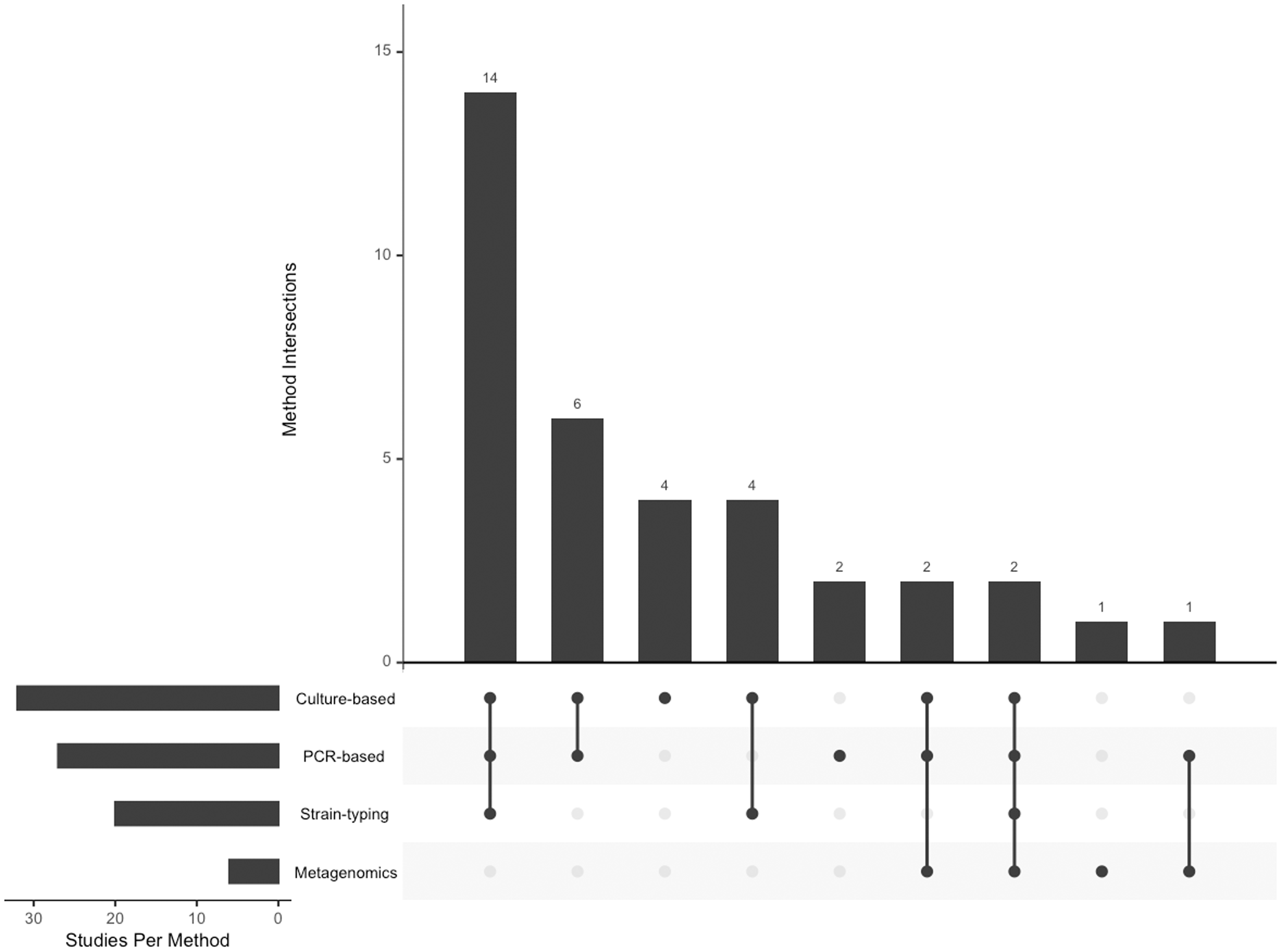
To characterize AMR in wastewater, 89% (32/36) of studies used culture-based methods, 89% (32/36) molecular methods, and 78% (28/36) used both simultaneously. Among molecular methods, PCR was most common (75%, 27/36), followed by whole-genome sequencing (WGS) (42%, 15/36), and metagenomics (17%, 6/36). Strain typing was performed in 53% (19/36) of studies, primarily by WGS, but also by multilocus sequence typing, pulsed-field gel electrophoresis, ERIC-PCR, and Random Amplified Polymorphic DNA (RAPD) fingerprinting (Table 2). The most frequent method combinations were culture with PCR and WGS (33%, 12/36), culture with PCR (22%, 8/36), and culture with other strain-typing methods (14%, 5/36) (Figure 4).
Distribution of individual and combined methodological approaches across 36 hospital wastewater surveillance studies, 2008–2026.

Molecular methods and analytical applications used in hospital wastewater antimicrobial resistance surveillance studies, 2008–2026

Table 2. Long description
A table comparing molecular methods and their applications in hospital wastewater AMR surveillance studies. The table has 36 rows and 12 columns. Column headers are: Authors, Molecular method, ARG detection, Plasmids / MGE detection, Multi-target ARG detection, ARG-microbiome quantification, Resistome profiling, Microbial community profiling, Molecular typing, Strain / clone, Phylogroup, Isolate characterization (ARG / virulence), Genetic context characterization, Genomic comparison. Row 1: Bian et al (2023), HT-qPCR, check, check, check, check, check, check, check, check, check, check, check, check, check. Row 2: Chaudhury et al (2025), Real-time PCR, check, check, check, check, check, check, check, check, check, check, check, check. Row 3: Chaudhury et al (2025), Kovacic et al (2017), Jakobsen et al (2021), Chang et al (2026), Gheorghe-Barbu et al (2024), Multiplex PCR, check, check, check, check, check, check, check, check, check, check, check, check, check. Row 4: Caplin et al (2008), Species-specific PCR, check, check, check, check, check, check, check, check, check, check, check, check. Row 5: Caplin et al (2008), Carlsen et al (2022), Davidova-Gerzova et al (2024), Gheorghe-Barbu et al (2024), MLST, check, check, check, check, check, check, check, check, check, check, check, check, check. Row 6: Drieux et al (2016), Liedhegner et al (2022), Target PCR, check, check, check, check, check, check, check, check, check, check, check, check. Row 7: Davidova-Gerzova et al (2024), Davidova-Gerzova et al (2023), Zhang et al (2025), Gheorghe-Barbu et al (2024), Mbanga et al (2023), Wu et al (2023), Xie et al (2023), Caplin et al (2008), Babic et al (2023), Flach et al (2021), Kovacic et al (2017), Jakobsen et al (2021), Koudoum et al (2025), Rolbiecki et al (2024), Zong et al (2013), Isolate PCR, check, check, check, check, check, check, check, check, check, check, check, check, check. Row 8: Babler et al (2023), Flach et al (2021), Acosta et al (2025), qPCR, check, check, check, check, check, check, check, check, check, check, check, check. Row 9: Koudoum et al (2025), Zong et al (2013), Davidova-Gerzova et al (2024), Gheorghe-Barbu et al (2024), Rolbiecki et al (2024), ERIC-PCR, check, check, check, check, check, check, check, check, check, check, check, check, check. Row 10: Zong et al (2013), Inverse PCR, check, check, check, check, check, check, check, check, check, check, check, check. Row 11: Chang et al (2026), qPCR, check, check, check, check, check, check, check, check, check, check, check, check. Row 12: Liedhegner et al (2022), Clermont PCR, check, check, check, check, check, check, check, check, check, check, check, check. Row 13: Rodriguez-Nava et al (2026), Droplet digital PCR, check, check, check, check, check, check, check, check, check, check, check, check. Row 14: Ayzanzo et al (2026), Carlsen et al (2022), Chang et al (2026), Davidova-Gerzova et al (2024), Gheorghe-Barbu et al (2024), Flach et al (2021), Gu et al (2024), Koudoum et al (2025), Rolbiecki et al (2024), WGS, check, check, check, check, check, check, check, check, check, check, check, check, check. Row 15: Liedhegner et al (2022), WGS, check, check, check, check, check, check, check, check, check, check, check, check. Row 16: Masalane et al (2025), Wu et al (2023), WGS, check, check, check, check, check, check, check, check, check, check, check. Row 17: Rolbiecki et al (2024), WGS, check, check, check, check, check, check, check, check, check, check, check, check. Row 18: Xie et al (2023), Zhang et al (2025), WGS, check, check, check, check, check, check, check, check, check, check, check. Row 19: Chaudhury et al (2025), WGS, check, check, check, check, check, check, check, check, check, check, check, check. Row 20: Chang et al (2026), Flach et al (2021), Gheorghe-Barbu et al (2024), Liedhegner et al (2022), Rolbiecki et al (2024), Xie et al (2023), Zhang et al (2025), Chaudhury et al (2025), MALDI-TOF Mass Spectrometry, check, check, check, check, check, check, check, check, check, check, check, check, check. Row 21: Jakobsen et al (2008), Kovacic et al (2017), PFGE, check, check, check, check, check, check, check, check, check, check, check. Row 22: Varela et al (2013), RAPD fingerprinting, check, check, check, check, check, check, check, check, check, check, check, check. Row 23: Chang et al (2026), Targeted amplicon sequencing, check, check, check, check, check, check, check, check, check, check, check, check. Row 24: Varela et al (2013), Xie et al (2023), Rolbiecki et al (2024), Zong et al (2013), 16S rRNA sequencing, check, check, check, check, check, check, check, check, check, check, check, check, check. Row 25: Acosta et al (2025), Baba et al (2023), Perry et al (2021), Metagenomics, check, check, check, check, check, check, check, check, check, check, check, check. Row 26: Cai et al (2021), Metagenomics, check, check, check, check, check, check, check, check, check, check, check, check. Row 27: Rolbiecki et al (2024), Metagenomics, check, check, check, check, check, check, check, check, check, check, check, check.
The table describes the primary function of each molecular method with representative references for each categories: ARG detection, detection of specific ARGs; Plasmid/MGE detection, detection of plasmids and mobile genetic elements involved in the carriage and potential transfer of ARGs;Multi-target ARG detection, simultaneous detection of multiple ARG targets in a single assay; ARG/Microbial Quantification, quantification of ARGs or microbial abundance; Resistome Profiling, broad assessment of ARG composition and diversity; Molecular Typing, identification of microbial species, strains, or clonal relatedness; Microbial Community Profiling, characterization of microbial taxonomic composition and relative abundance in samples;Isolate Characterization, characterization of ARG profiles in cultured isolates; Genetic Context Characterization, characterization of ARG-associated genetic features, including mutations, allelic variants, genomic context, mobile genetic elements, and plasmid-associated structures; Genomic Comparison, comparative analysis of genomic features, typically between wastewater and clinical isolates, to assess genetic relatedness, strain-level similarity, and potential epidemiological links.
Abbreviations: ARG: Antimicrobial resistance genes, PCR: Polymerase Chain Reaction, isolate PCR: Microbial isolation and PCR, qPCR:Quantitative PCR, HT-qPCR: High-Throughput Quantitative PCR, MLST: Multilocus Sequence Typing, ERIC-PCR: Enterobacterial Repetitive Intergenic Consensus - PCR, WGS: Whole Genome Sequencing, PFGE: Pulsed-Field Gel Electrophoresis, MGE: Mobile Genetic Elements, RAPD: Random Amplified Polymorphic DNA.
Most studies focused on Gram-negative bacteria (83%, 30/36), Reference Acosta, Lee and Bautista19–Reference Baba, Kuroda, Sekizuka and Kanamori22,Reference Bian, Wang and Ding24,Reference Cai, Sun and Yao25,Reference Carlsen and Christner27,Reference Chaudhury, Oransathit and Peerapongpaisarn29–Reference Siri, Bumyut, Precha, Sirikanchana, Haramoto and Makkaew47,Reference Wu, Dong and Cai50–Reference Yang, Lin and Liao52,Reference Zong and Zhang54 with 31% (11/36) examining Gram-positive bacteria, and 6% (2/36) fungi; pathogen categories were not mutually exclusive. Most targeted specific pathogens or resistance phenotypes (81%, 29/36) rather than profiling the entire resistome (19%, 7/36). The most frequently investigated organisms were Escherichia coli (n = 8), Klebsiella spp. (n = 7), Enterococcus spp. (n = 4), and Acinetobacter baumannii (n = 3). Two studies targeted Candida auris. Reference Babler, Sharkey and Arenas23,Reference Chang, Moshi and Nguyen28
Comparison between hospital wastewater and traditional AMR surveillance
Concordance between hospital wastewater and traditional surveillance varied by pathogen and resistance mechanism, and was assessed qualitatively in nearly all studies. Overall patterns showed strongest agreement for ESBL-producing organisms, variable concordance for carbapenem resistance, and inconsistent findings for Gram-positive pathogens.
Carbapenem resistance (9 studies): Two showed strain-level concordance: sewage carbapenem-resistant Acinetobacter baumannii (CRAB) with blaOXA genes shared 100% sequence identity with clinical isolates, Reference Kovacic, Seruga Music and Dekic40 and ST2 CRAB from sewage matched ICU-patient isolates. Reference Gu, Wu and Chen36 Four showed gene-level concordance: carbapenem-resistant Pseudomonas shared carbapenemase genes without strain-level comparison; Reference Al-Bayati, AboKsour, Taha, Hussein and Haider20 sewage carbapenemase genes tracked organism abundance for blaNDM, blaOXA-48-like, and blaKPC but not blaVIM or blaIMP; Reference Flach, Hutinel, Razavi and Larsson34 wastewater and clinical carbapenem-resistant Klebsiella fell into shared phylogenetic clades despite functional differences, Reference Xie, Huang, Zhang, Xu, Zhang and Li51 and correlation between wastewater carbapenem-resistance gene abundance and clinical resistance varied by antimicrobial and bacterial family. Reference Perry, Lepper and McNally44 One study was discordant, recovering greater carbapenemase-producing Enterobacterales diversity in wastewater with few clonal matches. Reference Carlsen and Christner27 Two characterized carbapenemase-carrying organisms without clinical comparison. Reference Duran-Bedolla and Bocanegra-Ibarias33,Reference Zong and Zhang54
ESBL-producing organisms (6 studies): All reported concordance of varying strength. Two demonstrated strain-level concordance: wastewater ESBL E. coli clusters matched patients strains, Reference Drieux, Haenn, Moulin and Jarlier32 and the dominant clinical K. pneumoniae lineage (ST307) was shared with wastewater and traceable to surface water. Reference Davidova-Gerzova, Lausova and Sukkar31 Three showed gene-level concordance: selected E. coli STs (SNP 0–18) across patients, wastewater, and rivers; Reference Davidova-Gerzova, Lausova and Sukkar30 shared resistance genes, plasmids, and mobile genetic; Reference Rolbiecki, Krawczyk, Korzeniewska, Sawicki and Harnisz46 and clonal relatedness by ERIC-PCR. Reference Koudoum, Founou and Founou39 One found concordance only in resistance gene profiles, with similar ESBL genes but divergent phylogenetic backgrounds. Reference Liedhegner, Bojar, Beattie, Cahak, Hristova and Skwor41
For ESKAPE pathogens and vancomycin-resistant enterococci, studies reported concordance in AMR profiles, resistance genes, and clonal lineages. Reference Baba, Kuroda, Sekizuka and Kanamori22,Reference Caplin, Hanlon and Taylor26,Reference Chaudhury, Oransathit and Peerapongpaisarn29,Reference Varela, Ferro and Vredenburg48 For other Gram-positives, findings were inconsistent: vancomycin-resistant staphylococci were detected in wastewater despite clinical absence, Reference Velazquez-Meza, Galarde-López and Cornejo-Juárez49 and methicillin-resistant Staphylococcus aureus (MRSA) findings were inconclusive. Reference Baba, Kuroda, Sekizuka and Kanamori22,Reference Velazquez-Meza, Galarde-López and Cornejo-Juárez49 For Candida auris, results were mixed: one study found no correlation between wastewater concentrations and patient census, Reference Babler, Sharkey and Arenas23 whereas another detected it in wastewater 13–143 days before clinical identification with>90% genomic concordance. Reference Chang, Moshi and Nguyen28
Only one study quantified overall concordance using correlation analysis, reporting strong correlation (Spearman’s rho = .88, P value<.05) between wastewater antibiotic resistance indices and clinical bacterial infection cases. Reference Siri, Bumyut, Precha, Sirikanchana, Haramoto and Makkaew47 At the gene level, blaCTX-M showed consistent prevalence gradients across clinical (74%), hospital environment (80%), and wastewater (85%) samples. Reference Koudoum, Founou and Founou39
Only two studies (6%) explicitly incorporated outbreak contexts: Carlsen et al found a K. pneumoniae outbreak clone persisting in wastewater four years postoutbreak, Reference Carlsen and Christner27 while Chang et al detected emerging C. auris subclades 13–143 days before clinical identification during an active outbreak. Reference Chang, Moshi and Nguyen28 These findings illustrate that wastewater may reflect both current transmission and historical contamination.
Discussion
We reviewed current approaches to hospital wastewater-based AMR epidemiology. Three findings stand out. First, agreement between wastewater and traditional surveillance was inconsistent and pathogen-dependent, with reasonable concordance for ESBL, limited and mostly gene-level concordance for carbapenemase-producing Enterobacterales, but weaker or absent concordance for some gram-positive organisms (e.g., Staphylococcus aureus). Second, the presence of methodological and reporting heterogeneity, especially due to incomplete temporal alignment between clinical and wastewater sampling (concurrent in only 13 out of 36 studies), limits the ability to compare findings across studies. Third, evidence was concentrated in high- and upper-middle-income countries, with almost none from low-income settings where the AMR burden is greatest. Existing hospital WBE approaches for AMR are highly heterogeneous, and no single method can be considered superior; rather, method selection depends on the specific objectives of a study. Most studies have focused on proof-of-concept investigations, while fewer have employed time-series studies incorporating patient-level data.
Current studies demonstrate feasibility but cannot determine surveillance utility. The defining feature of hospital wastewater, aggregating shedding from patients, staff, visitors, and environmental reservoirs, is both a strength and limitation. Reference Hocquet, Muller and Bertrand4,Reference Uluseker, Kaster and Thorsen6 This pooled signal has value only when interpretable in relation to the population generating it, yet such interpretation was rarely possible. Most studies reported microbiological methods in detail but provided limited clinical context: only one study performed prospective active surveillance, only a quarter provided patient-level data, and no study reported epidemiological rates (e.g., resistant infections per 1,000 patient-days). Wastewater measurements could not be normalized to hospital census, and studies compared wastewater findings with laboratory detections rather than with epidemiologically meaningful measures of resistance burden. Whether wastewater signals track real population-level resistance changes remains unknown.
We cannot determine if hospital wastewater provides early AMR warning or track intervention success. Most studies lacked frequent sampling, sustained longitudinal follow-up, or formal lead-lag analyses. Only one detected an organism ahead of clinical identification (C. auris, 13–143 days, with>90% genomic concordance), Reference Chang, Moshi and Nguyen28 and only one statistically correlated wastewater with clinical case counts. Reference Siri, Bumyut, Precha, Sirikanchana, Haramoto and Makkaew47 Organism fate and transport differ by taxon—illustrated by carbapenem-resistant K. pneumoniae sharing phylogenetic clades between wastewater and clinical isolates yet showing reduced serum survival compared to clinical counterparts. Reference Xie, Huang, Zhang, Xu, Zhang and Li51 Some resistant organisms may inhabit wastewater as environmental residents rather than patient-derived shedding, exemplified by carbapenemase genes in environmental Citrobacter spp. Reference Duran-Bedolla and Bocanegra-Ibarias33 and blaNDM-1 in Acinetobacter johnsonii. Reference Zong and Zhang54 Early-warning capability and intervention monitoring are plausible but fundamentally untested.
Differences in sampling, laboratory methods, and analytical approaches likely contribute to inconsistent concordance; a quarter of studies did not report sampling approach at all. Culture-based wastewater surveillance may show greater concordance with clinical data simply because traditional surveillance is culture-based, suggesting apparent concordance for ESBL-producing Enterobacterales may be partly methodological rather than biological. Reference McConn, Kraft and Durso55 Molecular methods capture a broader resistome signals not directly comparable with clinical case counts, but detect unculturable taxa, mobile elements, and emerging genes that culture may miss. Reference Knight, Webster and Perry10,Reference Diamond, Keshaviah and Bento56 No study employed viability-resolving approaches like propidium monoazide treatment. Reference Shi, Abd Rahman and de Rezende57 Culture-based and culture-independent methods are complementary, with appropriate choice depending on surveillance objectives, yet without standardized protocols and validation against clinical outcomes, determining which approaches reliably serve which purposes remains impossible.
Studies concentrated on fecally-excreted Enterobacterales (E. coli, Klebsiella spp.), Reference Mohany, Aslam and Ali58 while Pseudomonas and S. aureus received limited attention despite clinical prominence. Discordance between wastewater and traditional surveillance may not indicate failure: clinical detection depends on healthcare-seeking and testing practices, whereas wastewater captures shedding from diagnosed and undiagnosed individuals. One study recovered diverse carbapenemase-producing Enterobacterales in hospital wastewater with few clinical matches, Reference Carlsen and Christner27 and another detected C. auris weeks to months before clinical identification, Reference Chang, Moshi and Nguyen28 indicating wastewater can capture undetected resistance. However, discordance may also reflect compartment-specific microbial ecology or environmental persistence rather than incomplete clinical ascertainment.
Wastewater-based surveillance may offer greater value where clinical microbiology capacity is constrained, yet evidence derives almost entirely from high- and upper-middle-income countries. The most recent global AMR analysis (1990–2021) attributed the highest mortality to South Asia and sub-Saharan Africa, Reference Naghavi, Vollset and Ikuta59 but only two included studies originated from lower-middle-income countries (Cameroon and Zimbabwe) and none from low-income countries. Reference Koudoum, Founou and Founou39,Reference Mbanga, Kodzai and Oosthuysen43 Hospital wastewater surveillance is particularly needed in high-burden, resource-limited settings where clinical surveillance infrastructure is often weakest. Yet these contexts remain critically underrepresented.
Future studies should evaluate surveillance performance rather than merely characterize wastewater AMR content. We recommend adherence to Environmental Microbiology Minimum Information (EMMI) Guidelines for standardized reporting of sampling methods, analytical approaches, and quality control measures. 60 Beyond technical standardization, surveillance validation requires: (1) prospective longitudinal designs (≥12 months) with concurrent wastewater and clinical sampling; (2) comprehensive patient-level clinical comparators including epidemiological rates; (3) quantitative concordance metrics; and (4) evaluation during outbreaks, interventions, and stable endemic periods to assess whether wastewater signals precede clinical detection, track intervention success, and correlate with baseline prevalence. Studies meeting these criteria, particularly pilot studies in low- and middle-income countries, would provide the evidence needed to determine whether hospital wastewater surveillance offers actionable information beyond traditional AMR monitoring.
This review has limitations. First, most included studies were designed to characterize the wastewater AMR profile rather than to evaluate surveillance performance; outcomes most relevant to surveillance utility (analytical sensitivity, temporal concordance, predictive value, and population-level representativeness) were rarely assessed. Second, studies varied widely in methods, reporting standards, and the pathogens and resistance genes targeted, precluding quantitative synthesis. Third, 86% of studies were conducted in large tertiary care centers, with only two from district or community hospitals. These design limitations, rather than wastewater surveillance itself, constrain our conclusions.
In conclusion, hospital wastewater can detect resistant organisms and genes present in clinical populations, demonstrating feasibility. However, current evidence cannot determine utility, whether this approach reliably reflects clinical AMR burden, provides early warning, or tracks interventions. The extreme methodological heterogeneity precludes generalization and highlights the urgent need for standardized protocols and studies explicitly designed to validate surveillance performance. Until such evidence exists, hospital wastewater remains a promising but unproven complement to traditional AMR surveillance. Supplementary material
Supplementary material
The supplementary material for this article can be found at https://doi.org/10.1017/ice.2026.10558
Author contributions
Conceptualization, G.R.N. and J.L.S.; search strategy, E.W.; literature review, E.V.C. and M.J.; data analysis, E.V.C., M.J.; writing—original draft, E.V.C., G.R.N., A.A, J.L.P. and M.J.; writing—reviewing and editing, J.L.S., E.V.C, A.R.M., A.A., G.R.N., N.P, M.S., J.L.P, J.W., S.C., A.B., A.Z., W.T., and E.M.
Funding statement
This work was supported by Stanford University, School of Medicine, Team Science Seed Grant, the Rosenkranz Prize, the King Center on Global Development, Stanford Center for Innovation in Global Health, and the Center for Digital Health.
Competing interests
All authors report no conflicts of interest relevant to this article.




 Open access

