





Introduction
Schistosomiasis is a neglected tropical disease (NTD) caused by infection with parasitic worms of the genus Schistosoma that are transmitted by freshwater snail vectors (intermediate hosts). An estimated 253.7 million people are infected, with many more at risk, ∼90% of whom live in Africa (WHO, 2026).
The World Health Organization (WHO) guidelines for progressing towards the elimination of schistosomiasis as part of the 2030 NTD goals (WHO, 2022) highlight the need for increased vector-focused interventions such as molluscicide application to combat the disease (WHO, 2017; Garba Djirmay et al. Reference Garba Djirmay, Yadav, Guo, Rollinson and Madsen2024) and using snail surveillance as a predictive tool for assessing transmission of schistosomes and the prevalence of schistosomiasis in the population, facilitating the ultimate goal of parasite elimination (King et al. Reference King, Sturrock, Kariuki and Hamburger2006; Rollinson et al. Reference Rollinson, Knopp, Levitz, Stothard, Tchuem Tchuenté, Garba, Mohammed, Schur, Person and Colley2013; Kamel et al. Reference Kamel, Laidemitt, Lu, Babbitt, Weinbaum, Mkoji and Loker2021). Identifying freshwater bodies where the specific species of snail host are present and observing patent infections, whereby schistosome cercariae are actively being shed from the snail, are the first stages in assessing infection risk (Allan et al. Reference Allan, Dunn, Emery, Stothard, Johnston, Kane, Khamis, Mohammed and Rollinson2013). The distribution of snail vectors and the likelihood of observing patent schistosome infections is determined by a mosaic of abiotic and biotic variables that make identifying areas of transmission challenging (DeWitt, Reference DeWitt1955; Chu et al. Reference Chu, Massoud and Sabbaghian1966; Purnell, Reference Purnell1966; El-Hassan, Reference El-Hassan1974; Pennance et al. Reference Pennance, Person, Muhsin, Khamis, Muhsin, Khamis, Mohammed, Kabole, Rollinson and Knopp2016; Kalinda et al. Reference Kalinda, Chimbari and Mukaratirwa2017a, Reference Kalinda, Chimbari and Mukaratirwa2017b; Rabone et al. Reference Rabone, Gouvras, Gouvras, Hamidou, Hamidou, Webster, Emery, Emery, Garba and Rollinson2019). In addition, closely related snail vector species and larval schistosome species are morphologically cryptic, and the prevalence of patent schistosome infections is generally very low (Allan et al. Reference Allan, Dunn, Emery, Stothard, Johnston, Kane, Khamis, Mohammed and Rollinson2013; Pennance et al. Reference Pennance, Person, Muhsin, Khamis, Muhsin, Khamis, Mohammed, Kabole, Rollinson and Knopp2016, Reference Pennance, Allan, Emery, Rabone, Cable, Garba, Hamidou, Webster, Rollinson and Webster2020a), especially in regions nearing elimination (Pennance et al. Reference Pennance, Ame, Amour, Suleiman, Muhsin, Kabole, Ali, Archer, Allan, Emery, Rabone, Knopp, Rollinson, Cable and Webster2022a). Furthermore, as well as patent schistosome infections, snails that have been exposed to Schistosoma (signifying waterbody contamination with miracidia) can result in other infection outcomes observed at the time of snail collection (as reviewed in Pennance et al. Reference Pennance, Lam, Bigot, Trapp, Spaan, Ogara, Rawago, Andiego, Mulonga, Odhiambo, Mutuku, Loker, Odiere and Steinauer2024): prepatent (early stage of snail infestation before cercarial shedding occurs), latent (encapsulated sporocysts) or failed infections (where the larval schistosome has been unable to establish an infection in the challenged snail host). All these outcomes are missed through monitoring of cercarial shedding; however, they provide important information that Schistosoma transmission is likely occurring (Kamel et al. Reference Kamel, Laidemitt, Lu, Babbitt, Weinbaum, Mkoji and Loker2021). It is therefore proposed that the molecular detection of Schistosoma DNA in snail hosts will be a more efficient and definitive approach to monitor schistosome presence in snails and certify elimination, or interruption, of transmission in endemic areas (Rollinson et al. Reference Rollinson, Knopp, Levitz, Stothard, Tchuem Tchuenté, Garba, Mohammed, Schur, Person and Colley2013; Kamel et al. Reference Kamel, Laidemitt, Lu, Babbitt, Weinbaum, Mkoji and Loker2021).
Molecular xenomonitoring refers to the collection and testing of vectors for the presence of pathogen DNA to monitor the presence of these etiological agents of medical and veterinary importance (Minetti et al. Reference Minetti, LaCourse, Reimer and Storhard2016). The technique is often applied for detection of disease-causing agents transmitted by hematophagous insects, such as lymphatic filarial parasitic worms in mosquito vectors (Schmaedick et al. Reference Schmaedick, Koppel, Pilotte, Torres, Williams, Dobson, Lammie and Won2014; Pilotte et al. Reference Pilotte, Zaky, Abrams, Chadee and Williams2016, Reference Pilotte, Unnasch and Williams2017; Cook et al. Reference Cook, Pilotte, Minetti, Williams and Reimer2017) and trypanosomes in tsetse flies (Cunningham et al. Reference Cunningham, Lingley, Haines, Ndung’u, Torr and Adams2016; Garrod et al. Reference Garrod, Adams, Lingley, Saldanha, Torr and Cunningham2020; Saldanha et al. Reference Saldanha, Aziku, Trima, Drapari, Garrod, Ombanya, Tirados, Betson, Mugenyi, Dunkley, Torr, Hope and Cunningham2026). Several assays have been developed for detecting trematodes in freshwater snails (Kaplan et al. Reference Kaplan, Dame, Reddy and Courtney1995; Born-Torrijos et al. Reference Born-Torrijos, Poulin, Raga and Holzer2014; Rathinasamy et al. Reference Rathinasamy, Hosking, Tran, Kelley, Williamson, Swan, Elliott, Rawlin, Beddoe and Spithill2018; Archer et al. Reference Archer, Yeo, Gadd, Pennance, Cunningham, Juhàsz, Jones, Chammudzi, Kapira, Lally, Namacha, Mainga, Makaula, LaCourse, Kayuni, Musaya, Stothard and Webster2024a), including medically important schistosome species; S. japonicum (Kumagai et al. Reference Kumagai, Furushima-Shimogawara, Ohmae, Wang, Lu, Chen, Wen and Ohta2010; Tong et al. Reference Tong, Chen, Zhang, Yang, Kumagai, Furushima-Shimogawara, Lou, Yang, Wen, Lu, Ohta and Zhou2015), S. mansoni (Hamburger et al. Reference Hamburger, Xu, Ramzy, Jourdane and Ruppel1998a; Reference Hamburger, Ramzy, Jourdane and Ruppel1998b; Melo et al. Reference Melo, Gomes, Barbosa, Werkhauser and Abath2006; Abbasi et al. Reference Abbasi, King, Muchiri and Hamburger2010; Kane et al. Reference Kane, Stothard, Rollinson, Leclipteux, Evraerts, Standley, Allan, Betson, Kaba, Mertens and Laurent2013; Fernández-Soto et al. Reference Fernández-Soto, Gandasegui Arahuetes, Sánchez Hernández, López Abán, Vicente Santiago and Muro2014; Lu et al. Reference Lu, Zhang, Mutuku, Mkoji and Loker2016; Gandasegui et al. Reference Gandasegui, Fernández-Soto, Hernández-goenaga, López-abán, Vicente and Muro2016; Caldeira et al. Reference Caldeira, Jannotti-passos and Dos Santos Carvalho2017; Schols et al. Reference Schols, Carolus, Hammoud, Mulero, Mudavanhu and Huyse2019; Casotti et al. Reference Casotti, Gryschek, Paula, Gomes-gouvêa, Pinho, Tuan, Dias-neto, Luna and do Espírito-santo2020; Pennance et al. Reference Pennance, Lam, Bigot, Trapp, Spaan, Ogara, Rawago, Andiego, Mulonga, Odhiambo, Mutuku, Loker, Odiere and Steinauer2024; Andrus et al. Reference Andrus, Standley, Stothard and Wade2025), S. haematobium (Hamburger et al. Reference Hamburger, He-na, Abbasi, Ramzy, Jourdane and Ruppel2001, Reference Hamburger, Hoffman, Kariuki, Muchiri, Ouma, Koech, Sturrock and King2004; Abbasi et al. Reference Abbasi, King, Muchiri and Hamburger2010, Reference Abbasi, Webster, King, Rollinson and Hamburger2017; Allan et al. Reference Allan, Dunn, Emery, Stothard, Johnston, Kane, Khamis, Mohammed and Rollinson2013; Kane et al. Reference Kane, Stothard, Rollinson, Leclipteux, Evraerts, Standley, Allan, Betson, Kaba, Mertens and Laurent2013; Schols et al. Reference Schols, Carolus, Hammoud, Mulero, Mudavanhu and Huyse2019; Kaiglová et al. Reference Kaiglová, Changoma, Špajdelová, Jakubcová and Bírová2020) and others as reviewed by Kamel et al. (Reference Kamel, Laidemitt, Lu, Babbitt, Weinbaum, Mkoji and Loker2021). The first developed assay for the molecular detection of S. haematobium DNA in Bulinus employed the highly repetitive Dra1 sequence, which has been the marker of choice for many xenomonitoring and diagnostic studies for urogenital schistosomiasis (Hamburger et al. Reference Hamburger, He-na, Abbasi, Ramzy, Jourdane and Ruppel2001). However, the specificity of Dra1 and interpretation of results are not reliable, with false-positive and -negative results often observed (Pennance et al. Reference Pennance, Archer, Lugli, Rostron, Llanwarne, Ali, Amour, Suleiman, Li, Rollinson, Cable, Knopp, Allan, Ame and Webster2020b). Hence, a high-throughput assay using new markers targeting the 5.8S rDNA and ITS2 with high sensitivity and specificity for schistosomes has been developed that needs field applicability testing (Pennance et al. Reference Pennance, Archer, Lugli, Rostron, Llanwarne, Ali, Amour, Suleiman, Li, Rollinson, Cable, Knopp, Allan, Ame and Webster2020b).
Zanzibar, an archipelago with 2 major islands (Unguja and Pemba) in the United Republic of Tanzania, has been targeted with multidisciplinary interventions to eliminate schistosomiasis, with efforts intensifying over the past decade (Knopp et al. Reference Knopp, Person, Ame, Ali, Hattendorf, Juma, Muhsin, Khamis, Mohammed and Utzinger2019a; Trippler et al. Reference Trippler, Knopp, Welsche, Webster, Stothard, Blair, Allan, Ame, Juma, Kabole, Ali, Rollinson and Pennance2023). Although schistosomiasis prevalence has significantly decreased across the islands, focal areas of higher schistosomiasis prevalence (e.g. >10% prevalence locally when the island-wide prevalence is <5%), referred to as persistent transmission hotspots, remain (Pennance et al. Reference Pennance, Person, Muhsin, Khamis, Muhsin, Khamis, Mohammed, Kabole, Rollinson and Knopp2016; Knopp et al. Reference Knopp, Ame, Person, Hattendorf, Rabone, Juma, Muhsin, Khamis, Hollenberg and Mohammed2019b; Trippler et al. Reference Trippler, Ame, Hattendorf, Juma, Abubakar, Ali, Kabole, Rollinson and Knopp2021). Areas across the islands with different Schistosoma transmission intensities provide an ideal system for testing a xenomonitoring assay, since S. haematobium prevalence in Bulinus species is expected to vary with focal transmission intensities. Additionally, the presence of 2 snail vectors for S. haematobium (B. globosus and to a lesser extent B. nasutus, see Pennance et al. Reference Pennance, Ame, Amour, Suleiman, Muhsin, Kabole, Ali, Archer, Allan, Emery, Rabone, Knopp, Rollinson, Cable and Webster2022a), and the occurrence of S. bovis in B. globosus in at least 1 region in Pemba (Pennance et al. Reference Pennance, Ame, Amour, Suleiman, Allan, Rollinson and Webster2018), allows for further investigation into the distribution of these parasite species across the island.
The current study aims to demonstrate the feasibility and capacity of a high-throughput xenomonitoring assay for the detection of S. haematobium and S. bovis in large snail populations collected between 2017 and 2018 across Pemba (Pennance et al. Reference Pennance, Archer, Lugli, Rostron, Llanwarne, Ali, Amour, Suleiman, Li, Rollinson, Cable, Knopp, Allan, Ame and Webster2020b). The assay was investigated in different S. haematobium transmission settings, the inferred highest (persisting schistosomiasis hotspot areas) and lowest (persisting very low-/intermittent-prevalence areas) transmission settings established following community-wide biannual preventive chemotherapy and additional interventions conducted between 2012 and 2016 (Knopp et al. Reference Knopp, Ame, Person, Hattendorf, Rabone, Juma, Muhsin, Khamis, Hollenberg and Mohammed2019b). The investigations across different transmission settings enabled further insights into the distribution of S. haematobium and S. bovis across Pemba and building from previous studies (Stothard et al. Reference Stothard, Loxton, Rollinson, Mgeni, Khamis, Ameri, Ramsan and Savioli2000; Pennance et al. Reference Pennance, Neves, Webster, Gower, Knopp, Khamis, Ame, Ali, Rabone, Emery, Allan, Muhsin, Suleiman, Kabole, Walker, Rollinson and Webster2022b) the role of the different Bulinus spp. (B. globosus and B. nasutus) in their transmission.
Methods
Study area selection and characterization
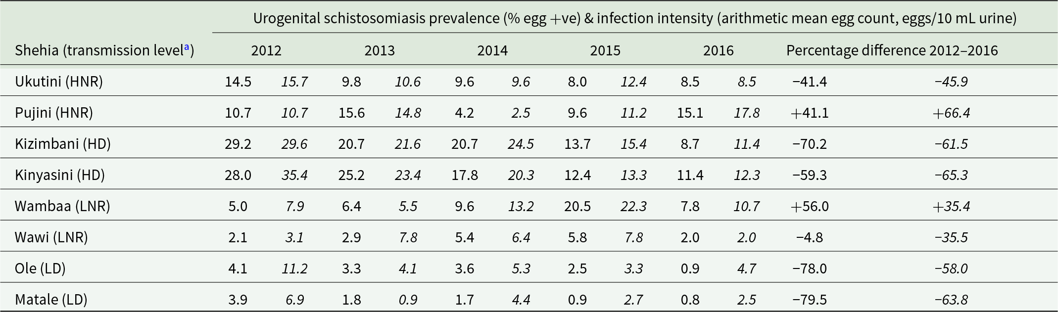
Urogenital schistosomiasis infection prevalence data of 9–12-year-old schoolchildren from Pemba (Knopp et al. Reference Knopp, Mohammed, Ali, Khamis, Ame, Albonico, Gouvras, Fenwick, Savioli and Colley2012; Knopp et al. Reference Knopp, Person, Ame, Ali, Hattendorf, Juma, Muhsin, Khamis, Mohammed and Utzinger2019a, Reference Knopp, Ame, Person, Hattendorf, Rabone, Juma, Muhsin, Khamis, Hollenberg and Mohammed2019b) were used to select shehias to be included in the xenomonitoring study by assessing the change in prevalence from 2012 to 2016. Urogenital schistosomiasis prevalence changes allowed the majority of shehias in Pemba to be categorized into 1 of 4 transmission levels (Table 1). Following the inclusion criteria outlined in Supplementary Figure 1 and feasibility (primarily pre-existing knowledge on access to freshwater bodies for malacological surveys and not having molluscicide application as part of previous control efforts), 2 shehias per transmission category were selected for inclusion in the study (Table 1). In each shehia, 4 surveys were performed in October 2017 (Survey 1), February 2018 (Survey 2), July 2018 (Survey 3) and November 2018 (Survey 4). Surveying in the rainy seasons (between March to May and November to December) was avoided where possible, because many sites are inaccessible, and the high and fluctuating water levels disperse and disrupt freshwater snail distribution.
Shehias of Pemba (Zanzibar, United Republic of Tanzania) selected for inclusion in xenomonitoring study, showing schistosomiasis transmission level, Schistosoma haematobium prevalence (%) and infection intensity (eggs/10 mL urine) shown in italics in 9–12-year-old schoolchildren from each year between 2012 and 2016 (Knopp et al. Reference Knopp, Ame, Person, Hattendorf, Rabone, Juma, Muhsin, Khamis, Hollenberg and Mohammed2019b) and the percentage change of prevalence and infection intensity from 2012 to 2016

Table 1 Long description
Eight Pemba shehias are listed with yearly urogenital schistosomiasis prevalence (percent egg-positive) and average infection intensity (eggs per 10 milliliters of urine) for schoolchildren from 2012 through 2016, plus the percent change from 2012 to 2016. Some high-prevalence shehias in 2012 show declines by 2016: Kizimbani drops from 29.2 percent to 8.7 percent and Kinyasini from 28.0 percent to 11.4 percent, with intensity also decreasing in both. Ukutini declines from 14.5 percent to 8.5 whereas Pujini increases from 10.7 to 15.1 percent. Low-prevalence shehias vary: Matale falls from 3.9 percent to 0.8 percent and Ole from 4.1 percent to 0.9 percent, both with marked intensity reductions. In contrast, Wambaa increases from 5.0 percent to 7.8 percent, while Wawi changes little in prevalence from 2.1 percent to 2.0 percent. Transmission-level labels group shehias by starting prevalence and degree of reduction, so comparisons should consider that categories reflect baseline differences as well as change over time.
a Transmission levels determined from urogenital schistosomiasis prevalence (egg positive) of 9–12-year-old schoolchildren from Pemba (Knopp et al. Reference Knopp, Ame, Person, Hattendorf, Rabone, Juma, Muhsin, Khamis, Hollenberg and Mohammed2019b), here defined as follows: high non-responder (HNR) and high decliner (HD), both having >10% urogenital schistosomiasis prevalence in 2012 and having <50% or >50% reduction in prevalence, respectively, by 2016, and low non-responder (LNR) and low decliner (LD) both having >0% to <5% prevalence in 2012 and having <50% or >50% reduction in prevalence, respectively, by 2016.
Initially, during malacological Survey 1, Uwandani shehia was included as a high non-responder shehia. However, between Surveys 1 and 2, Uwandani was impacted by the activity of a different research group trialling focal molluscicides at freshwater bodies, which significantly reduced snail populations at these sites. Therefore, Uwandani was replaced with Ukutini in Survey 2, and no malacological surveys or collections were made in Ukutini during Survey 1.
Sampling and species identification of Bulinus spp. in Pemba
Bulinus spp. snails were collected from each human–freshwater contact site (HWCS), transported back to the Public Health Laboratory (Chake-Chake, Pemba), and checked for patent schistosome infections, generally within 3–4 h of collecting, as described previously (Pennance et al. Reference Pennance, Ame, Amour, Suleiman, Muhsin, Kabole, Ali, Archer, Allan, Emery, Rabone, Knopp, Rollinson, Cable and Webster2022a). After screening for patent infections, the snails were preserved in glass vials containing 100% ethanol. Shedding snails were preserved individually, and non-shedding snails were grouped by collection site for each survey. Total DNA was extracted from whole snail soft tissues using a Qiagen BioSprint 96 Workstation and Qiagen BioSprint 96 DNA Blood Kit (Qiagen, Hilden, Germany) (see Supplementary File 1) from all shedding snails as well as 3 non-patent snails per site and survey for further molecular characterization to confirm species identifications of either B. globosus or B. nasutus and determine genetic diversity (see Pennance et al. Reference Pennance, Ame, Amour, Suleiman, Muhsin, Kabole, Ali, Archer, Allan, Emery, Rabone, Knopp, Rollinson, Cable and Webster2022a). Based on these results, an assumption was made on the identity of the Bulinus sp. present at each site, as it was not feasible to conduct species identifications on all snails, and the 2 snail species generally follow an allopatric distribution, not sharing or switching freshwater sites.
Selection of snails for analysis and identification of Schistosoma DNA presence
Across Surveys 1 and 4, for 18 HWCSs where >94 snails were collected, total DNA from whole snail soft tissue was extracted from a random subset (n = 95–111) of B. globosus and/or B. nasutus individuals, and for 39 sites where ≤94 snails were collected, all snails were extracted. For Surveys 2 and 3, total DNA from whole snail soft tissue was extracted from 51 to 72 B. globosus and/or B. nasutus individuals collected during each survey from 13 sites, and all snails were extracted from 28 sites with ≤50 individuals collected during each survey. All snail DNA was extracted using a Qiagen BioSprint 96 Workstation and Qiagen BioSprint 96 DNA Blood Kit (Qiagen, Hilden, Germany) (see Supplementary File 1). Note that snail size, an estimation of age, was not considered when randomly selecting samples to process using the xenomonitoring assay. In total, across the 4 surveys, 5465 (of the 9430 collected) non-patent snails were screened using a previously developed high-throughput diagnostic xenomonitoring polymerase chain reaction (PCR) (Pennance et al. Reference Pennance, Archer, Lugli, Rostron, Llanwarne, Ali, Amour, Suleiman, Li, Rollinson, Cable, Knopp, Allan, Ame and Webster2020b), which is capable of a limit of detection of a single larval stage at the initiation of infestation. Schistosoma DNA-positive snails determined from this diagnostic PCR underwent a second Schistosoma-targeted PCR and subsequent Sanger sequencing of a partial Schistosoma nuclear 5.8S-ITS2 region to determine Schistosoma species (S. haematobium and S. bovis), as detailed in Supplementary File 1.
Statistical analysis
All statistical analyses were conducted in R v4.5.3 (R Core Team, 2026). To assess the variation in S. haematobium DNA prevalence in snail vectors, a binomial generalized linear mixed model (GLMM) was fitted using the lme4 v1.38 package. The S. haematobium status of each snail, defined as presence or absence of S. haematobium DNA/cercariae (i.e. incorporating both patent (shedding) and DNA-positive snails determined through molecular screening), was the model response variable with fixed effects of transmission level (see 4 categories in Table 1) and survey (4 time points). To account for the non-independence of samples collected from the same HWCS, each site was included as a random intercept term. The estimated marginal means and pairwise comparisons for effects were calculated using the emmeans v2.0.1 package. Full details of the model parameters are given in Supplementary File 2. GLMMs fitted to investigate the effects of environmental and abiotic factors on snail abundance and S. haematobium DNA presence/absence in snails were reported previously (Pennance, Reference Pennance2020).
Results
Malacological surveys: establishing patent Schistosoma infections and Schistosoma DNA-positive snails
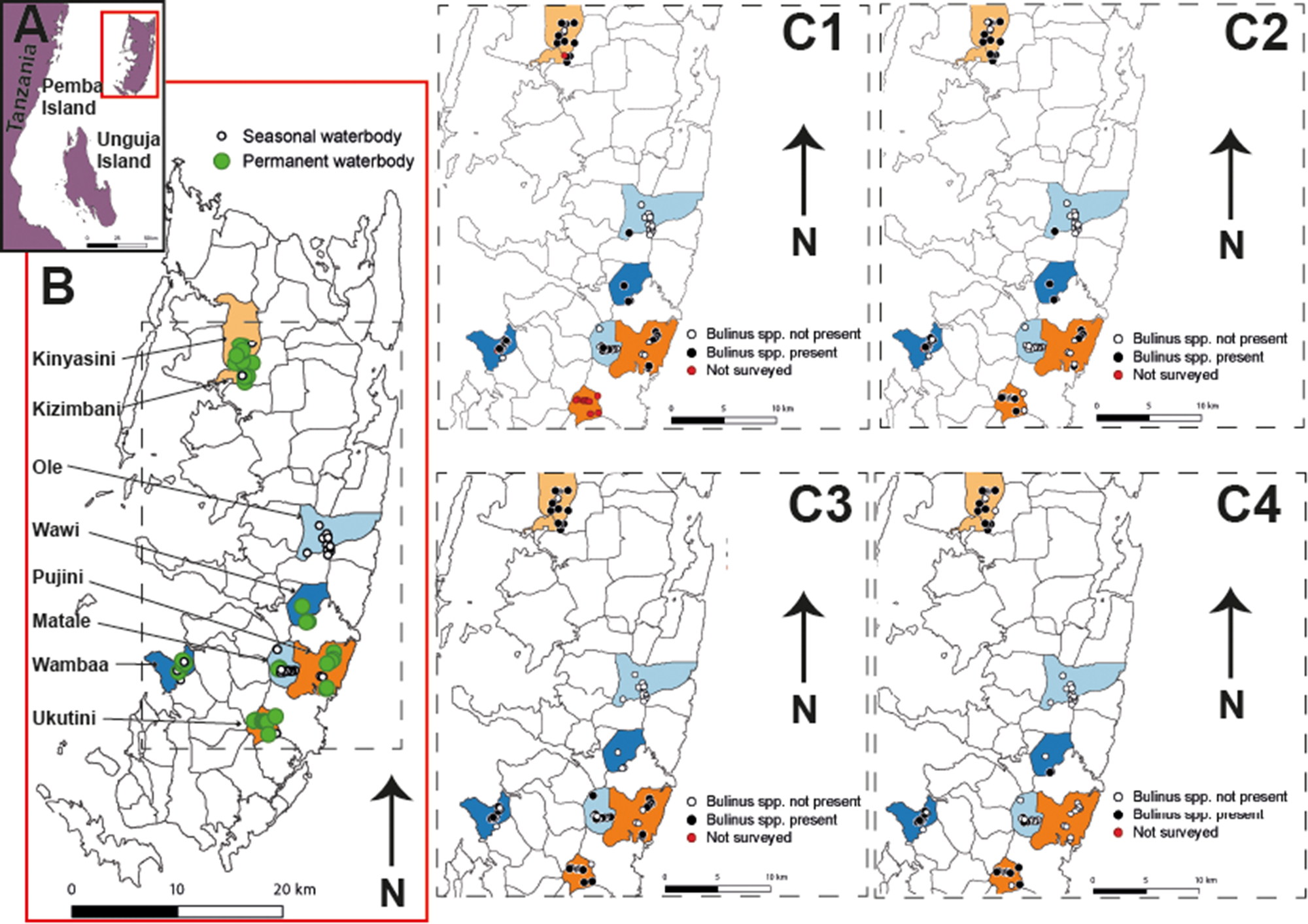
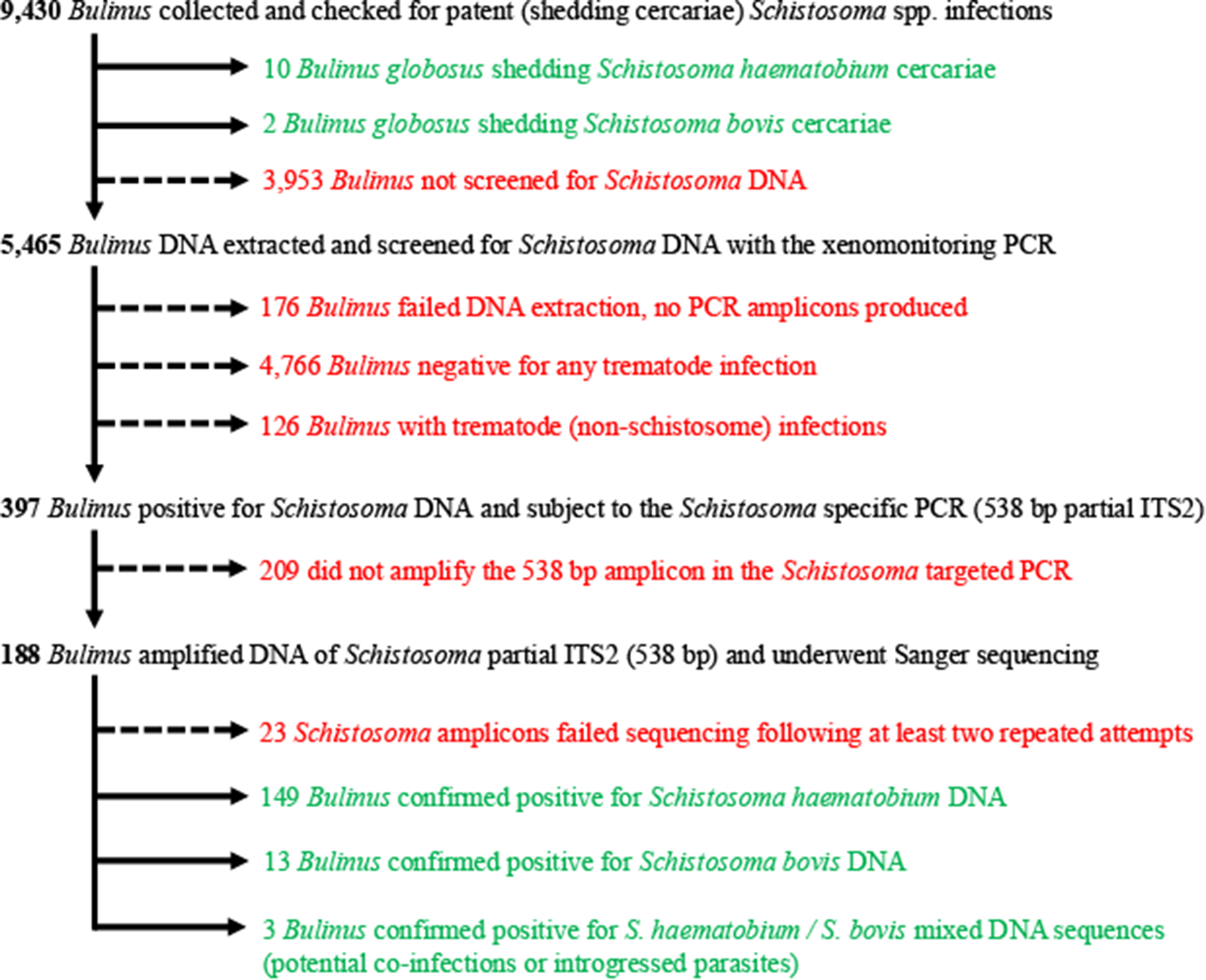
A total of 74 HWCSs were identified and surveyed in the 8 xenomonitoring shehias (Figure 1); full site information and each survey data, including ecological data, Bulinus abundance (as summarized in Supplementary Figure 2), and Schistosoma spp. identified (patent and PCR typed) from collected snails are detailed in Supplementary Table 1. In brief, B. globosus or B. nasutus were present at least once over the 4 surveys in 70% (n = 52) of the HWCSs. Out of 9430 B. globosus/B. nasutus collected from each site across the 4 xenomonitoring surveys, 12 (0.13%) patent schistosome infections were observed from B. globosus in Kinyasini (n = 10) and Kizimbani (n = 2) collected during surveys 2 and 4 (see (Pennance et al. Reference Pennance, Ame, Amour, Suleiman, Muhsin, Kabole, Ali, Archer, Allan, Emery, Rabone, Knopp, Rollinson, Cable and Webster2022a). Ten of these schistosome infections were identified as S. haematobium and 2 as S. bovis (see Pennance et al. Reference Pennance, Ame, Amour, Suleiman, Muhsin, Kabole, Ali, Archer, Allan, Emery, Rabone, Knopp, Rollinson, Cable and Webster2022a). As summarized in Figure 2, of the 9430 Bulinus collected, 5465 (58%) were randomly selected and screened using the molecular xenomonitoring assay for Schistosoma presence. Following the removal of 176 of 5289 samples giving inconclusive results (i.e. failed DNA extraction and no PCR amplicon produced), 397 (7.5%) Schistosoma-positive samples were subject to the schistosome DNA partial ITS2-targeted PCR. Snails with trematode (non-schistosome) presence represented 126 samples, and all remaining 4766 (∼90%) samples were negative for any trematode presence using these primers (Figure 2).
(A) East coast of Tanzania and the Zanzibar Archipelago (Pemba and Unguja islands). (B) Pemba (United Republic of Tanzania) showing the location of selected shehias categorized based on Schistosoma haematobium prevalence from 2012 to 2016 (Knopp et al. Reference Knopp, Ame, Person, Hattendorf, Rabone, Juma, Muhsin, Khamis, Hollenberg and Mohammed2019b) as high non-responders (dark orange), high decliners (light orange), low non-responders (dark blue) and low decliners (light blue). Location of seasonal (i.e. temporary, white circles) and permanent (i.e. never dry, green circles) freshwater bodies shown on map. (C1–C4) Freshwater bodies where Bulinus globosus and/or B. nasutus were present (black circles) or absent (white circles) or not surveyed (red circles) during malacological Survey 1 – October 2017 (C1), Survey 2 – February 2018 (C2), Survey 3 – July 2018 (C3) and Survey 4 – November 2018 (C4).

Figure 1 Long description
A) Inset map showing Pemba and Unguja island in the Zanzibar Archipelago. B) Map of Pemba displaying selected shehias categorized by prevalence from 2012 to 2016: high non-responders (dark orange), high decliners (light orange), low non-responders (dark blue) and low decliners (light blue). Seasonal water bodies are marked with white circles and permanent water bodies with green circles. C1) Survey map showing freshwater bodies where Bulinus species were present (black circles), absent (white circles), or not surveyed (red circles) in October 2017. C2) Similar survey map for February 2018. C3) Survey map for July 2018. C4) Survey map for November 2018. Each survey map indicates the distribution of Bulinus species across the island, with arrows pointing north and a scale bar indicating 20 km. The maps highlight the spatial distribution of shehia categories and freshwater bodies containing Bulinus across Pemba.
Flowchart showing the processing of the 9430 Bulinus spp. collected from Pemba across 4 malacological surveys from October 2017 to November 2018.

Figure 2 Long description
Flowchart illustrating the processing of Bulinus snails for Schistosoma DNA detection. The first section shows 9,430 Bulinus collected and checked for patent Schistosoma infections. It notes 10 Bulinus globosus shedding Schistosoma haematobium cercariae, 2 Bulinus globosus shedding Schistosoma bovis cercariae and 3,953 Bulinus not screened for Schistosoma DNA. The second section details 5,465 Bulinus DNA extracted and screened for Schistosoma DNA with xenomonitoring PCR. It includes 176 Bulinus failed DNA extraction, no PCR amplicons produced, 4,766 Bulinus negative for any trematode infection and 126 Bulinus with trematode (non-schistosome) infections. The third section shows 397 Bulinus positive for Schistosoma DNA and subject to Schistosoma specific PCR (538 bp partial ITS2). It mentions 209 did not amplify the 538 bp amplicon in the Schistosoma targeted PCR. The final section describes 188 Bulinus amplified DNA of Schistosoma partial ITS2 (538 bp) and underwent Sanger sequencing. It includes 23 Schistosoma amplicons failed sequencing following at least two repeated attempts, 149 Bulinus confirmed positive for Schistosoma haematobium DNA, 13 Bulinus confirmed positive for Schistosoma bovis DNA and 3 Bulinus confirmed positive for Schistosoma haematobium and Schistosoma bovis mixed DNA sequences, indicating potential co-infections or introgressed parasites.
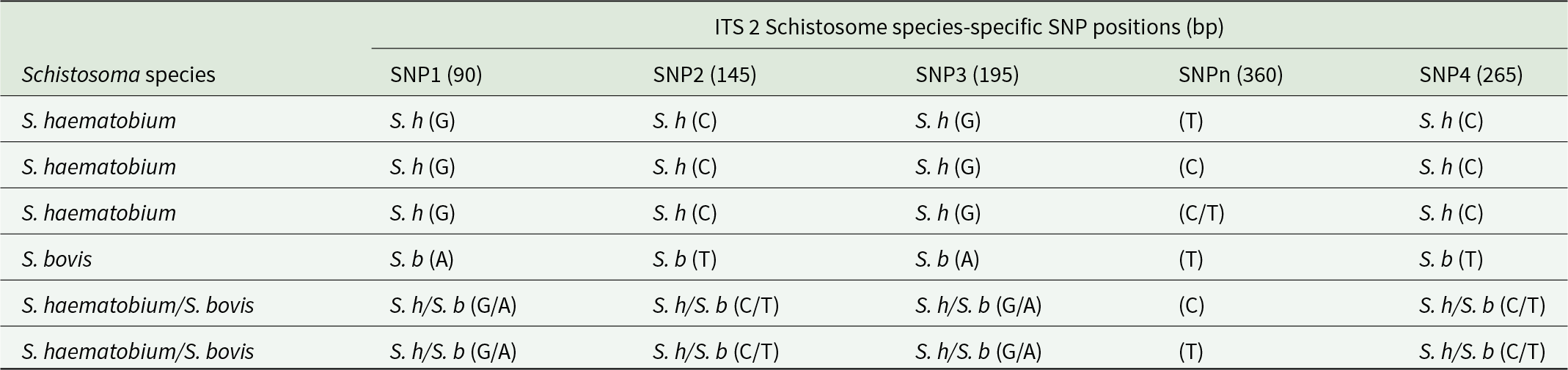
Sanger sequencing of 188 partial Schistosoma ITS2 regions (538 bp) revealed 149 S. haematobium, 13 S. bovis and 3 S. haematobium/S. bovis mixed DNA profiles, representing potential co-infections or introgressed parasites (Table 2 and Supplementary Table 2). Schistosome species identifications could not be confirmed from 23 of the amplified 188 samples that were sequenced due to failed sequencing following at least 2 repeated attempts (Figure 2). The remaining 209 of the original 397 Schistosoma positives did not amplify in the Schistosoma-targeted PCR. Comparing the 165 PCR-typed Schistosoma DNA-positive snails to the xenomonitoring assay gel electrophoresis profiles, 98 of 318 (30.8%) that amplified only the snail and schistosome band were confirmed as either S. haematobium (n = 90) or S. bovis (n = 8), whereas 64 of the 79 (81%) of the snails with snail, trematode and schistosome amplicons were confirmed as S. haematobium (n = 59) and S. bovis (n = 5).
Observed Schistosoma species-specific single nucleotide polymorphism (SNP) positions (including base position) in the ITS2 region

Table 2 Long description
Schistosoma species are compared by the nucleotide observed at five ITS2 SNP positions: 90, 145, 195, an unclassified site at 360, and 265 base pairs. Three S. haematobium entries share G at 90, C at 145, G at 195, and C at 265, while the 360 site varies as T, C, or C and T. The S. bovis entry shows A at 90, T at 145, A at 195, and T at 265, with T at the 360 site. Two entries labeled S. haematobium and S. bovis show mixed bases at the species-informative sites: G and A at 90, C and T at 145, G and A at 195, and C and T at 265. At the unclassified 360 position, the mixed entries show either C or T.
The 360 SNP is unclassified and is not recognized as a species-specific SNP.
The 538 bp ITS2 region that was successfully amplified from 165 S. haematobium and S. bovis DNA-positive snails revealed 6 different sequence profiles, including those differentiated by a fifth SNP at position 360 (see SNPn in Table 2). Double peaking was observed in 3 samples at the 4 S. haematobium and S. bovis species-specific sites, representing either DNA from co-infections of S. haematobium and S. bovis or S. haematobium × S. bovis introgressed worms being detected (Table 2).
Schistosoma haematobium DNA-positive snails
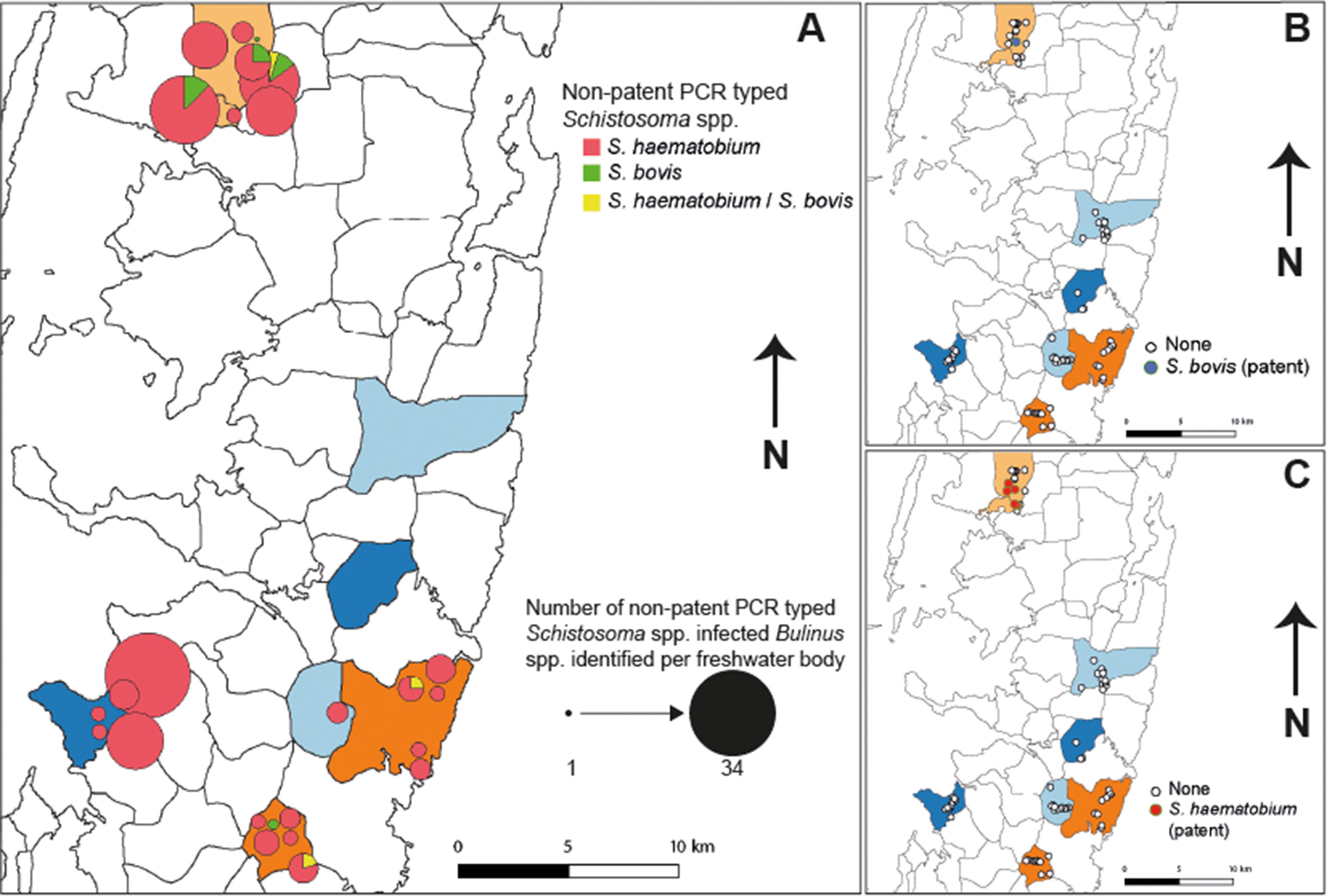
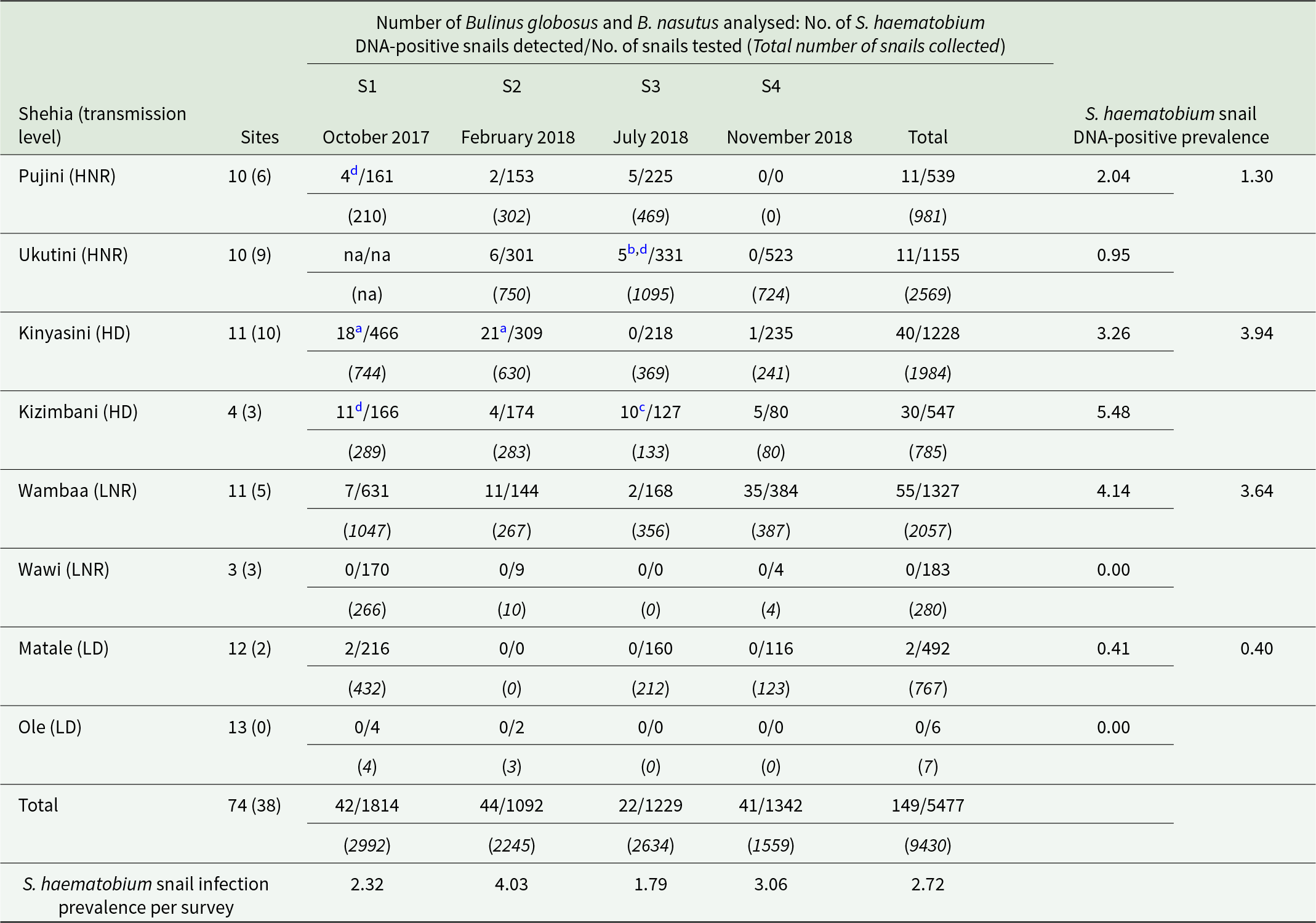
Non-patent, PCR-typed (i.e. sequencing-confirmed) S. haematobium DNA-positive snails were identified at 23 of the 52 sites where B. globosus and/or B. nasutus were collected in all shehias except for Ole and Wawi (Figure 3A and Table 3). Proportional to the number of snails extracted, the highest number of S. haematobium DNA-positive snails were observed in Kizimbani (5.48%), followed by Wambaa (4.14%), Kinyasini (3.26%) and Pujini (2.04%). Although a lower prevalence of S. haematobium DNA-positive snails was observed from sites in the low-decliner shehias (Ole, Matale) than in high-decliner shehias (Kinyasini, Kizimbani), the prevalence of S. haematobium DNA in snails in the low non-responder shehia Wambaa was unexpectedly greater than that observed in high non-responder shehias (Kinyasini, Kizimbani) (Table 3). Shehias classified as low decliners had both the least snails collected (n = 774) and the fewest positive S. haematobium PCRs (0.40%), whereas although the high non-responder shehias had the highest abundance of snails collected (n = 3550), the highest proportion of PCR-positive S. haematobium snails (3.94%) was seen in the high-decliner shehias (Table 3).
Location of selected shehias in Pemba (United Republic of Tanzania) categorized based on Schistosoma haematobium prevalence as high non-responders (dark orange), high decliners (light orange), low non-responders (dark blue) and low decliners (light blue). (A) PCR-typed Schistosoma spp. infections of Bulinus identified at freshwater bodies. Circle area is proportional to the number of PCR-typed Schistosoma spp. identified from each site across all surveys. Yellow represents snails with the presence of S. haematobium/S. bovis DNA, suggesting co-infections or introgressed schistosomes. (B) Patent S. bovis infections of B. globosus from 1 site in Kinyasini, and absence across remaining sites in Pemba. (C) Patent S. haematobium infections of B. globosus identified from 5 sites in Kinyasini (n = 3) and Kizimbani (n = 2), and absence across remaining sites in Pemba.

Figure 3 Long description
Three maps of Pemba, Tanzania, display Schistosoma spp. prevalence and PCR-typed infections. A) Shows non-patent PCR typed Schistosoma spp. infections with symbols: red for S. haematobium, green for S. bovis and yellow for S. haematobium/S. bovis. Circle size indicates the number of infected Bulinus spp. per freshwater body, ranging from 1 to 34. B) Highlights areas with S. bovis (patent) infections, marked in blue. C) Displays regions with S. haematobium (patent) infections, marked in orange. All maps include a north arrow and scale bar for reference.
PCR-typed Schistosoma haematobium DNA-positive Bulinus globosus and B. nasutus identified using the xenomonitoring assay and confirmed from sequence analysis of the 538 bp Schistosoma-specific amplicon

Table 3 Long description
Schistosoma haematobium DNA positivity in Bulinus globosus and B. nasutus is reported by shehia and survey round, alongside the number of human water-contact sites per shehia. For each shehia and survey (October 2017, February 2018, July 2018, November 2018), the table lists Schistosoma haematobium DNA-positive snails out of snails tested, plus totals and overall prevalence by survey and shehia. Overall, 149 of 5,477 snails tested were S. haematobium DNA positive. By shehia, Kizimbani shows the highest overall prevalence at 5.48 percent, followed by Wambaa at 4.14 percent and Kinyasini at 3.26 percent; Pujini is 2.04 percent and Matale is 0.41 percent, while Wawi and Ole have zero positives. Across survey rounds, overall prevalence varies, peaking in February 2018 at 4.03 percent and lowest in July 2018 at 1.79 percent, with October 2017 at 2.32 percent and November 2018 at 3.06 percent.
The number of HWCSs per shehia is given, including the number of permanent freshwater bodies shown in brackets. Shehia transmission levels are defined in Table 1 as follows: HNR, high non-responder; HD, high decliner; LNR, low non-responder; LD, low decliner.
a Three S. bovis non-patent infections were also identified.
b Five S. bovis non-patent infections were also identified.
c Two S. bovis non-patent infections were also identified.
d One snail sample showed a mixed S. haematobium/S. bovis infection (coinfection or introgressed worm).
The binomial GLMM showed no statistically significant differences in probability of S. haematobium presence in snails between the different transmission settings, although estimated marginal means did indicate higher S. haematobium presence probabilities in high-decliner shehias (1.37%) compared to low-decliner shehias (0.21%), but with substantially overlapping confidence intervals (see Supplementary File 3). A significant effect of survey was observed in the GLMM, with pairwise comparisons showing a significantly (P < 0.05) higher S. haematobium infection probability in survey 2 (February 2018, at the end of the short dry season) than Surveys 1, 3 and 4.
Schistosoma bovis DNA-positive snails
DNA of bovine S. bovis were detected in snails from Kinyasini (n = 6) (Figure 3A), where patent infections have been observed previously (Pennance et al. Reference Pennance, Ame, Amour, Suleiman, Allan, Rollinson and Webster2018). DNA of S. bovis was also detected in snails at sites in the neighbouring shehia, Kizimbani (n = 2), but also a geographically distant site, Ukutini (n = 5), located further south on the island (Figure 3A).
Snail identification and Schistosoma DNA-positive snail species
In total, 406 snails were identified as B. globosus (n = 359) and B. nasutus (n = 47) from the sites surveyed for the xenomonitoring surveys (see Pennance et al. Reference Pennance, Ame, Amour, Suleiman, Muhsin, Kabole, Ali, Archer, Allan, Emery, Rabone, Knopp, Rollinson, Cable and Webster2022a). All B. nasutus were collected from 8 separate pond sites in Pujini and co-occurred with B. globosus at 1 site (Puj11, see Pennance et al. Reference Pennance, Ame, Amour, Suleiman, Muhsin, Kabole, Ali, Archer, Allan, Emery, Rabone, Knopp, Rollinson, Cable and Webster2022a). All other sites containing Bulinus (n = 44) were identified as B. globosus (see Pennance et al. Reference Pennance, Ame, Amour, Suleiman, Muhsin, Kabole, Ali, Archer, Allan, Emery, Rabone, Knopp, Rollinson, Cable and Webster2022a).
Due to allopatric distributions of the different snail hosts, the majority of S. haematobium group DNA positives were identified from snails predicted to be B. globosus (S. haematobium = 138, S. bovis = 13, S. haematobium-bovis = 2); however, snails predicted to be B. nasutus from Pujini also had S. haematobium group DNA positives identified (S. haematobium = 10, S. haematobium-bovis = 1). One snail harbouring S. haematobium originated from the aforementioned site in Pujini where both B. globosus and B. nasutus cohabit (see Pennance et al. Reference Pennance, Ame, Amour, Suleiman, Muhsin, Kabole, Ali, Archer, Allan, Emery, Rabone, Knopp, Rollinson, Cable and Webster2022a), and it therefore cannot be assumed from the current data which species this was (B. globosus or B. nasutus).
Discussion
Here, we validate a recently developed xenomonitoring assay targeting S. haematobium and S. bovis DNA within Bulinus spp. snail vectors (Pennance et al. Reference Pennance, Archer, Lugli, Rostron, Llanwarne, Ali, Amour, Suleiman, Li, Rollinson, Cable, Knopp, Allan, Ame and Webster2020b) as an effective tool for detecting the presence of schistosomes in endemic regions in Pemba. The xenomonitoring assay was >12 times more sensitive at detecting S. haematobium presence (DNA positives) compared to traditional parasitological shedding methods, corroborating with other studies using molecular diagnostics to determine snail infections (Archer et al. Reference Archer, Cunningham, Juhász, Jones, Reed, Yeo, Mainga, Chammudzi, Kapira, Lally, Namacha, Makaula, LaCourse, Kayuni, Webster, Musaya and Stothard2024b; Andrus et al. Reference Andrus, Standley, Stothard and Wade2025). Importantly, the assay detected transmission of S. haematobium in areas where patent snail infections were not identified, including low human-infection prevalence areas where both human and snail infections using standard parasitological procedures are difficult to detect, showing the feasibility of the assay for the surveillance of potential schistosomiasis transmission in elimination settings (WHO, 2020, 2022). The greater prevalence of snails with S. haematobium DNA presence in low non-responder shehias (particularly Wambaa) when compared to the high non-responder suggests that human prevalence data determined through urine-egg microscopy may not always reflect the degree of transmission taking place in certain foci, and highlights the focal nature of transmission that makes classifying regions into broad epidemiological categories difficult. By detecting S. haematobium presence in transmission sites, it allows us to show that interventions are still needed to prevent schistosomiasis resurgence in low-prevalence areas where infected individuals are contaminating freshwater bodies containing potential snail vectors. Both B. globosus and B. nasutus were observed with S. haematobium DNA presence, and to a lesser extent S. bovis, showing that both these snail species are exposed to these schistosome species in this endemic setting. The presence of Schistosoma DNA in freshwater samples (Fornillos et al. Reference Fornillos, Sato, Tabios, Sato, Leonardo, Chigusa, Minamoto, Kikuchi, Legaspi and Fontanilla2019; Alzaylaee et al. Reference Alzaylaee, Collins, Rinaldi, Shechonge, Ngatunga, Morgan and Genner2020a) or in the snail vector signifies that waterbody contamination with miracidia is occurring. This added information should be used to inform schistosomiasis control strategies, such as behavioural interventions to educate against contamination behaviours (in this case open urination) or snail control measures, to target specific transmission areas (Knopp et al. Reference Knopp, Person, Ame, Ali, Hattendorf, Juma, Muhsin, Khamis, Mohammed and Utzinger2019a, Reference Knopp, Ame, Person, Hattendorf, Rabone, Juma, Muhsin, Khamis, Hollenberg and Mohammed2019b).
Urogenital schistosomiasis transmission through B. nasutus
From the Bulinus spp. identifications performed on a subset of samples (see Pennance et al. Reference Pennance, Ame, Amour, Suleiman, Muhsin, Kabole, Ali, Archer, Allan, Emery, Rabone, Knopp, Rollinson, Cable and Webster2022a), it can be assumed that S. haematobium DNA were detected in at least 10 B. nasutus collected in Pujini. This further confirms that this snail species is penetrated by S. haematobium in Zanzibar (Allan et al. Reference Allan, Rollinson, Smith and Dunn2009) and provides support that this vector species may play a minor role in the transmission of S. haematobium in multiple regions in Pemba (Pennance et al. Reference Pennance, Ame, Amour, Suleiman, Muhsin, Kabole, Ali, Archer, Allan, Emery, Rabone, Knopp, Rollinson, Cable and Webster2022a), especially considering that Pujini, where B. nasutus was in high abundance, is an area of high persisting human infection prevalence, suggesting sustained local transmission through B. nasutus. Although the distribution of B. globosus and B. nasutus is relatively well described for Pemba (Pennance et al. Reference Pennance, Ame, Amour, Suleiman, Muhsin, Kabole, Ali, Archer, Allan, Emery, Rabone, Knopp, Rollinson, Cable and Webster2022a), it would be beneficial to combine a Bulinus species diagnostic (Pennance et al. Reference Pennance, Ame, Ali, Amour, Suleiman, Cable and Webster2026) with this xenomonitoring methodology. This would provide species identifications for both snail vector and schistosome in parallel methods, or simultaneously as is conducted in other infectious disease monitoring tools (Tedrow et al. Reference Tedrow, Ratovonjato, Walker, Ratsimbasoa and Zimmerman2019).
Distribution of S. bovis transmission and the occurrence of co-infections with S. haematobium
The presence of S. bovis DNA within 5 B. globosus collected in Ukutini demonstrates that S. bovis transmission is likely occurring in regions outside of Kinyasini where patent infections in livestock and snails have been confirmed (Pennance et al. Reference Pennance, Ame, Amour, Suleiman, Allan, Rollinson and Webster2018, Reference Pennance, Ame, Amour, Suleiman, Cable and Webster2021). Further studies on the transmission of S. bovis in Pemba are therefore needed to elucidate the presence of bovine schistosomiasis and the risk to cattle health island-wide. Mixed ITS2 profiles of S. haematobium and S. bovis were observed within 3 snails from 3 different shehias: Kizimbani, Ukutini and Pujini. Without being able to screen individual schistosome larvae, these data are difficult to interpret since they could represent either a co-infection of S. haematobium and S. bovis or derive from an S. haematobium × S. bovis introgressed worm (Webster et al. Reference Webster, Rollinson, Stothard and Huyse2010; Platt et al. Reference Platt, McDew-White, Le Clec’h, Chevalier, Allan, Emery, Garba, Hamidou, Ame, Webster, Rollinson, Webster and Anderson2019; Pennance et al. Reference Pennance, Allan, Emery, Rabone, Cable, Garba, Hamidou, Webster, Rollinson and Webster2020a). From the 3 snails observed with mixed S. haematobium–S. bovis profiles in the current study, 2 originated from Kizimbani and Ukutini, where S. bovis DNA-positive snails were also detected, whilst the third originated from 1 site in Pujini, where only S. haematobium DNA were detected. Noteworthy is that the snail potentially co-infected with S. haematobium and S. bovis from Pujini is assumed to be B. nasutus, suggesting that this species could also play a role in S. bovis transmission on Zanzibar if infections reach patency, as is the case on mainland Africa (Brown, Reference Brown1994).
Advantages and limitations of incorporating xenomonitoring into schistosomiasis surveillance
This study also aimed to assess the feasibility of using a xenomonitoring assay in schistosomiasis surveillance and elimination following WHO recommendations (WHO, 2020). As for other xenomonitoring assays (Cook et al. Reference Cook, Xu, Msellem, Vonk, Bergström, Gosling, Al-mafazy, McElroy, Molteni and Abass2015; Garrod et al. Reference Garrod, Adams, Lingley, Saldanha, Torr and Cunningham2020), the primary advantage that the xenomonitoring assay described here offers is a more sensitive method to detect Schistosoma spp. presence in snail vectors compared to conventional methods of parasitological shedding. A second advantage is that species-level identification of the schistosomes being transmitted by the snail can be accurately deduced from sequence analysis rather than inferred from cercarial shedding patterns (Mouahid et al. Reference Mouahid, Mone, Chaib and Théron1991).
However, the cost of PCR reagents and consumables necessary for molecular xenomonitoring may be difficult to justify for regional disease control programme managers, especially if additional costs are needed for a basic laboratory set-up. Testing pooling strategies, such as those used in arthropod vectors before or after DNA extraction (Schmaedick et al. Reference Schmaedick, Koppel, Pilotte, Torres, Williams, Dobson, Lammie and Won2014; Cook et al. Reference Cook, Pilotte, Minetti, Williams and Reimer2017; Ramesh et al. Reference Ramesh, Cameron, Spence, Hoek Spaans, Melo-santos, Paiva, Guedes, Barbosa, Oliveira, Sá, Jeffries, Castanha, Oliveira, Walker, Alexander and Braga2018), to maximize the number of snails processed and minimize costs should be trialled. The processing of samples pre-PCR is also time-consuming, with a major bottleneck being the time taken to extract DNA from each snail. Following the high-throughput DNA-extraction protocol (see Supplementary File 1), it was possible for an experienced technician to perform 94 snail DNA extractions in ∼24 h, which is much shorter when compared to other more common methods (although this was not compared here), but necessitates a Qiagen Biosprint 96 workstation (Qiagen, Hilden, Germany) that is not available in many laboratories.
To circumvent the requirements for sequencing of amplicons to confirm schistosome species, diagnostic PCRs to differentiate S. haematobium and S. bovis by amplicon sizes are available (Webster et al. Reference Webster, Rollinson, Stothard and Huyse2010; Abbasi et al. Reference Abbasi, Webster, King, Rollinson and Hamburger2017; Blin et al. Reference Blin, Dametto, Agniwo, Webster, Angora, Dabo and Boissier2023) for miracidia and cercariae, although these have not been consistent, with trials on snails exposed to S. haematobium often failing to amplify (Pennance et al. Reference Pennance, Archer, Lugli, Rostron, Llanwarne, Ali, Amour, Suleiman, Li, Rollinson, Cable, Knopp, Allan, Ame and Webster2020b). In future iterations of schistosome xenomonitoring assays, alternative post-PCR analysis to differentiate species, such as those used in other disease systems, could be used (Tedrow et al. Reference Tedrow, Ratovonjato, Walker, Ratsimbasoa and Zimmerman2019). Other assay protocols, such as quantitative PCR or real-time PCR, particularly highly specific probe-based assays that inherently confirm correct DNA loci amplification without the need for DNA sequencing, may also be beneficial if access to the required equipment is possible (Kane et al. Reference Kane, Stothard, Rollinson, Leclipteux, Evraerts, Standley, Allan, Betson, Kaba, Mertens and Laurent2013; Garrod et al. Reference Garrod, Adams, Lingley, Saldanha, Torr and Cunningham2020; Alzaylaee et al. Reference Alzaylaee, Collins, Rinaldi, Shechonge, Ngatunga, Morgan and Genner2020a; Saldanha et al. Reference Saldanha, Aziku, Trima, Drapari, Garrod, Ombanya, Tirados, Betson, Mugenyi, Dunkley, Torr, Hope and Cunningham2026). Furthermore, portable nucleic acid-based diagnostic assays such as rapid polymerase amplification protocols, which can also be formulated as highly specific probe-based assays with further development, have been shown to be sensitive diagnostic tests for targeting the S. haematobium Dra1 locus in urine samples (Rosser et al. Reference Rosser, Rollinson, Forrest and Webster2015; Rostron et al. Reference Rostron, Pennance, Bakar, Rollinson, Knopp, Allan, Kabole, Ali, Ame and Webster2019), and snail xenomonitoring (Mesquita et al. Reference Mesquita, Gadd, Coelho, Cieplinski, Emery, Lugli, Simões, Fonseca, Caldeira and Webster2024). Whilst promising, all these assays are currently used for research purposes only, with further optimization and assessment needed before routine use in national disease surveillance programmes.
The future of schistosomiasis surveillance
Although improvements need to be made before the use of a S. haematobium xenomonitoring protocol for urogenital schistosomiasis surveillance on a large scale, the findings here offer good progress towards developing such a protocol. The results demonstrate that it is possible to detect S. haematobium presence by collecting only the snail vectors in both low- and high-prevalence areas, and that the probability of finding schistosome infections/presence can fluctuate seasonally, a factor that must be considered if moving towards post-elimination surveillance. Environmental DNA xenomonitoring protocols for the detection of snail and schistosome DNA within freshwater samples may also offer valuable transmission surveillance tools; however, further improvements to protocols and further demonstration of field applicability are required (Fornillos et al. Reference Fornillos, Sato, Tabios, Sato, Leonardo, Chigusa, Minamoto, Kikuchi, Legaspi and Fontanilla2019; Alzaylaee et al. Reference Alzaylaee, Collins, Rinaldi, Shechonge, Ngatunga, Morgan and Genner2020a, Reference Alzaylaee, Collins, Shechonge, Ngatunga, Morgan and Genner2020b; Mulero et al. Reference Mulero, Boissier, Allienne, Quilichini, Foata, Pointier and Rey2020; Gava et al. Reference Gava, de Carvalho, Sato, Sato, de Fava, Parreiras, de Oliveira, Teixeira, Lourenço, Dos Carvalho, Montresor, Mourão and Caldeira2025).
Supplementary material
The supplementary material for this article can be found at https://doi.org/10.1017/S0031182026102510.
Data availability statement
The datasets supporting the conclusions of this article are included within the article and its additional file.
Acknowledgements
Sanger Sequencing was provided by the Sequencing Facility at the Natural History Museum (London, UK).
Author contributions
T.P., J.C., D.R., S.K., S.M.Ali, S.M.Ame and B.L.W. conceived and designed the study. T.P., J.A., E.B.L., P.R., S.M.Ali, A.K.A., K.R.S. and S.M.Ame conducted the data gathering. T.P. performed the formal analysis and wrote the original article. All authors reviewed and edited the final manuscript.
Financial support
The study was partially funded by a Wellcome Trust Seed Award (wellcome.org) grant 207728 (awarded to B.L.W.). T.P. was funded by the Natural Environmental Research Council GW4+ DTP (nercgw4plus.ac.uk) NE/L002434/1. The funders had no role in study design, data collection and analysis, decision to publish, or preparation of the manuscript.
Competing interests
The authors declare there are no conflicts of interest.
Ethical standards
Not applicable.


 Open access

