Introduction
Development of the ovary in mouse has a unique biology since the formation of primordial follicles initiates from the late fetal period and continues during the early neonatal period (Eppig and O’Brien, Reference Eppig and O’Brien1996; Findlay et al., Reference Findlay, Hutt, Hickey and Anderson2015; Kerr et al., Reference Kerr, Duckett, Myers, Britt, Mladenovska and Findlay2006; Pepling, Reference Pepling2006). Due to this characteristic, a 1-day-old mouse ovary only contains germ cells and primordial follicles, and in turn, it can be used as a model for studying the formation and activation of primordial follicles under in vitro condition (Atrabi et al., Reference Atrabi, Akbarinejad, Khanbabaee, Dalman, Amorim, Najar-Asl, Valojerdi and Fathi2019; Eppig and O’Brien, Reference Eppig and O’Brien1996; Khodadi et al., Reference Khodadi, Atrabi, Torkzadeh, Fazli, Akbarinejad and Fathi2022; Morohaku et al., Reference Morohaku, Tanimoto, Sasaki, Kawahara-Miki, Kono, Hayashi, Hirao and Obata2016; O’Brien et al., Reference O’Brien, Pendola and Eppig2003). This animal model could assist to refine the approaches for in vitro activation of primordial follicles and development of activated follicles towards the production of MII-stage oocyte in cancer patients having experienced excision of the ovaries owing to oncotherapy (Atrabi et al., Reference Atrabi, Akbarinejad, Khanbabaee, Dalman, Amorim, Najar-Asl, Valojerdi and Fathi2019; Ford et al., Reference Ford, Beckett, Roman, McLaughlin and Sutherland2020; Sarma et al., Reference Sarma, Findlay and Hutt2019). This technique has some advantages as compared with other contemporary techniques because it does not postpone the commencement of oncotherapy as do cryopreservation of oocyte and in vitro production of the embryo and it does not potentiate the risk for re-introduction of malignant cells in the body as does transplantation of ovarian tissue (Amorim and Shikanov, Reference Amorim and Shikanov2016; Bockstaele et al., Reference Bockstaele, Tsepelidis, Dechene, Englert and Demeestere2012; Fathi et al., Reference Fathi, Rezazadeh Valojerdi, Ebrahimi, Eivazkhani, Akbarpour, Tahaei and Abtahi2017; Jeruss and Woodruff, Reference Jeruss and Woodruff2009). Nonetheless, this technique currently has some limitations since it has not been as successful in prepubertal girls as it has been in pubertal women (Luyckx et al., Reference Luyckx, Scalercio, Jadoul, Amorim, Soares, Donnez and Dolmans2013; Telfer et al., Reference Telfer, McLaughlin, Ding and Thong2008; Telfer and Zelinski, Reference Telfer and Zelinski2013). In addition, in vitro activation of primordial follicles could serve as a potential method for the preservation of endangered animal species, in which lack of mature and fertilizable oocytes is a serious restricting concern for the application of assisted reproductive techniques (ARTs) (Jewgenow and Paris, Reference Jewgenow and Paris2006; Khodadi et al., Reference Khodadi, Atrabi, Torkzadeh, Fazli, Akbarinejad and Fathi2022; Nagashima et al., Reference Nagashima, Wildt, Travis and Songsasen2019).
In this context, a number of studies have investigated the impact of various components and biological materials, including granulosa cell conditioned medium, amino acids, pyruvate, testosterone and insulin-transferrin-selenium, on the development of follicles in 1-day-old mice ovaries (Alborzi et al., Reference Alborzi, Jafari Atrabi, Akbarinejad, Khanbabaei and Fathi2020; Atrabi et al., Reference Atrabi, Akbarinejad, Khanbabaee, Dalman, Amorim, Najar-Asl, Valojerdi and Fathi2019; Atrabi et al., Reference Atrabi, Alborzi, Akbarinejad and Fathi2021; Khodadi et al., Reference Khodadi, Atrabi, Torkzadeh, Fazli, Akbarinejad and Fathi2022). More recently, the main and interaction effects of follicle-stimulating hormone (FSH) and testosterone were investigated on 1-day-old mice ovaries under in vitro conditions for 8 days, and FSH was found to increase the activation of primordial follicles, development of primary and preantral follicles and expression of some genes related to development of ovarian follicles (Torkzadeh et al., Reference Torkzadeh, Asadi, Jafari Atrabi, Eivazkhani, Khodadi, Hajiaghalou, Akbarinejad and Fathi2023). The combination of FSH and testosterone could also upregulate the expression of some factors contributing to the development of ovarian follicles; however, it did not significantly affect the morphological development of ovarian follicles based on histological analyses (Torkzadeh et al., Reference Torkzadeh, Asadi, Jafari Atrabi, Eivazkhani, Khodadi, Hajiaghalou, Akbarinejad and Fathi2023). Given that there is a time lag between molecular alterations and cellular changes (Fan et al., Reference Fan, Bialecka, Moustakas, Lam, Torrens-Juaneda, Borggreven, Trouw, Louwe, Pilgram, Mei, van der Westerlaken and Chuva de Sousa Lopes2019; Kranc et al., Reference Kranc, Brązert, Budna, Celichowski, Bryja, Nawrocki, Ożegowska, Jankowski, Chermuła, Dyszkiewicz-Konwińska, Jeseta, Pawelczyk, Bręborowicz, Rachoń, Bruska, Nowicki, Zabel and Kempisty2019; Mukherjee et al., Reference Mukherjee, Reisdorph, Guda, Pandey and Roy2012), we hypothesized that culturing for a longer period of time might lead to the manifestation of the positive effects of FSH plus testosterone combination in the morphology of ovarian follicles as well. In corroboration with this hypothesis, the extension of culture duration from 5 to 11 days has been reported to improve the growth of follicles to the preantral stage (Alborzi et al., Reference Alborzi, Jafari Atrabi, Akbarinejad, Khanbabaei and Fathi2020; Atrabi et al., Reference Atrabi, Alborzi, Akbarinejad and Fathi2021).
Accordingly, the present study was conducted to evaluate the main and interaction effects of FSH by testosterone on the development of ovarian follicles in a 1-day-old mouse ovary cultured for 12 days.
Materials and methods
Animals
Female 1-day-old Naval Medical Research Institute mice were maintained under stable conditions (temperature: 20–25°C, 40–60% humidity) in a 12 h light-dark cycle. All animal procedures were conducted in accordance with the guidelines of the Royan Ethics Committee (IR.ACECR.ROYAN.REC.1398.248). In the present study, 36 1-day-old female mice were used for histological and molecular examinations. Herein, it is worth noting that the left and right ovaries from the same mouse were used for a single experimental group to avoid any potential confounding factors concerning the side of the ovary.
Isolation of 1-day-old mice ovaries
The ovaries were isolated from newborn 1-day-old mice under a stereomicroscope (Nikon, Tokyo, Japan). Immediately after removal, ovaries were placed in α-minimum essential medium (α-MEM; Gibco, Paisley, UK) containing 10% knockout serum replacement (KSR; Gibco, Paisley, UK), supplemented with 100 IU/ml penicillin-streptomycin under mineral oil (Sigma-Aldrich, St. Louis, USA), and subsequently, the surrounding tissues were meticulously removed.
Culture of isolated ovaries and experimental design
Isolated 1-day-old mice ovaries were cultured in 96-well plates containing 300 μl of base medium containing α-MEM supplemented with 10% KSR and 100 IU/ml penicillin-streptomycin for the initial 4 days. Given that the assembly of primordial follicles continues up until post-natal day 5 (Eppig and O’Brien, Reference Eppig and O’Brien1996; Findlay et al., Reference Findlay, Hutt, Hickey and Anderson2015; Kerr et al., Reference Kerr, Duckett, Myers, Britt, Mladenovska and Findlay2006; Pepling, Reference Pepling2006) and we intended to evaluate the effect of various hormones on primordial follicles, the hormonal treatments were added to the culture media after implementation of a preliminary 4-day culture. After day 4 of culture, the ovaries were randomly allocated to four experimental groups, including (1) control group (CON; base medium), (2) FSH group (FSH; base medium plus 1.36 IU/ml FSH), (3) testosterone group (T2; base medium plus 0.1 mM testosterone) and (4) combination of FSH and testosterone group (FSH+T2; base medium plus 1.36 IU/ml FSH and 0.1 mM testosterone). Next, the ovaries were cultured for 8 additional days, and the ovarian samples were collected on day 12 for histological and molecular assessments. The ovaries allocated to various experimental groups were cultured in a single plate in each culture replicate, and each culture replicate for all experimental groups began on the same day to avoid any potential confounding factor concerning the culture plate or date for the commencement of culture. The plates were incubated at 37°C with 5% CO2, and approximately 75% of the culture medium was replaced by fresh medium every 4 days in all experimental groups.
Histological analysis
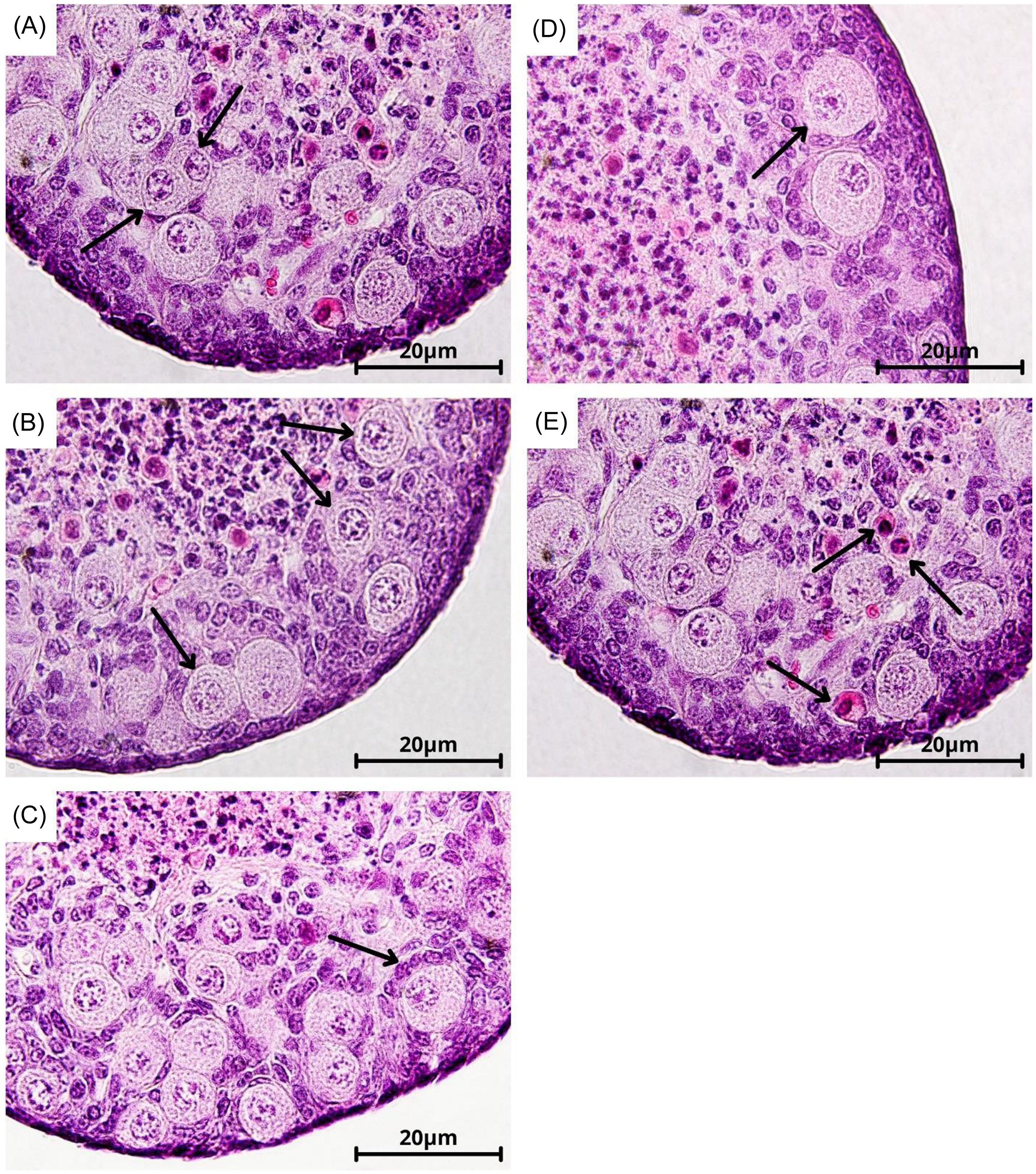
To examine follicular development in the experimental groups, the cultured ovaries (n = 10 ovaries belonging to 5 mice for each experimental group) were fixed in Bouin’s solution for 45 min and then in formalin at 4°C for at least 48 h, dehydrated with an alcohol gradient procedure, embedded in paraffin, serially sectioned at a thickness of 6 μm and mounted on glass slides. After drying at 42°C for 12 h, the sections were deparaffinized in xylene, hydrated by a gradient alcohol protocol and stained by haematoxylin and eosin. In order to avoid counting a follicle more than once, only one out of four consecutive sections was assessed under a light microscope (Nikon, Tokyo, Japan), and merely data of follicles with a clear oocyte nucleus were recorded for further analysis. The follicles were classified into primordial follicles (an oocyte surrounded by a few squamous granulosa cells; Figure 1.A), transitional follicles (an oocyte surrounded by both squamous and cuboidal granulosa cells; Figure 1.B), primary follicles (an oocyte surrounded by a single layer of cuboidal granulosa cells; Figure 1.C), preantral follicles (an oocyte surrounded by two or more layers of cuboidal granulosa cells; Figure 1.D) and atretic follicles (a degenerating oocyte with shrunken cytoplasm and deteriorated nucleus; Figure 1.E). The percentage of non-atretic follicles was considered as the proportion of intact follicles. Activation of follicles was the cumulative proportion of all activated follicles including transitional, primary and preantral follicles.
Various stages of ovarian follicles in 1-day-old mouse ovaries stained with haematoxylin and eosin staining. (A) Primordial follicles with one layer of squamous granulosa cells. (B) Transitional follicles with both squamous and cuboidal granulosa cells. (C) Primary follicles with a single layer of cuboidal granulosa cells. (D) Preantral follicles with two or more layers of cuboidal granulosa cells. (E) Atretic follicles with a degenerating oocyte. Black arrows denote the specified follicle.

Real-time PCR (RT-PCR)
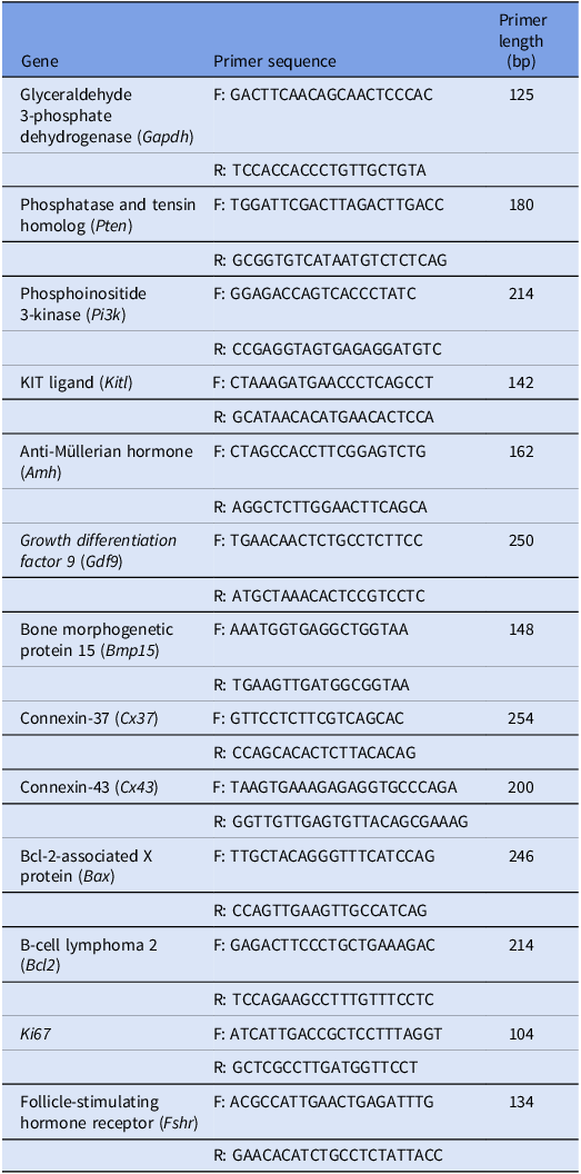
Real-time polymerase chain reaction (RT-PCR) was applied to evaluate the gene expression of factors regulating the development of ovarian follicles (Table 1). Initially, cultured ovaries in various experimental groups (n = 4 mice for each experimental group) were stored in RNA later (Qiagen, Hilden, Germany) at −80°C. Total RNA was extracted using RNeasy Plus Micro Kit (Qiagen, Hilden, Germany). Then, cDNA was synthesized from RNA using the standard oligo (dT) reverse transcription protocol considering the manufacturer’s guidelines. qRT-PCR was conducted using Power SYBR Green PCR Master Mix on an ABI Step One Plus thermocycler (Applied Biosystems, Warrington, UK). Reactions were run for 40 cycles at 95°C for 10 min, 95°C for 15 s and 61°C for 61 s, and each sample was run in duplicate. Glyceraldehyde 3-phosphate dehydrogenase (Gapdh) was used to normalize the data as a housekeeping gene. The sequence of primers used in the present study is presented in Table 1. Primer efficiency analysis was carried out by running a standard curve and calculating the slope. The primers were diluted to the concentration recommended by the kit supplier (1:10), and qPCR reactions were performed using various concentrations of the template cDNA. All the primers used in our experiment showed efficiency ranging between 90 and 110%. To confirm the success of qPCR amplifications, we conducted gel electrophoresis using the qPCR products. For this purpose, 1% agarose gel was prepared with Tris-acetate-EDTA buffer, which included a DNA ladder as a standard beside the qPCR products to assess the presence and size of DNA fragments. Electrophoresis was carried out at 150 V for 1 h. Following this, a UV transilluminator was utilized to visualize the DNA bands, which verified the success of qPCR amplifications by confirming that the bands matched the expected sizes.
Sequences of primers for real-time polymerase chain reaction test

Statistical analysis
Initially, data were tested for normal distribution using the Kolmogorov–Smirnov test (UNIVARIATE procedure). This study had a factorial experimental design, in which the main and interaction effects of two factors (i.e. FSH and testosterone) on the development of follicles and expression of genes were analyzed (two-way model). Therefore, data of histological and molecular assessments were analyzed using the generalized linear model procedure. Multiple comparisons were conducted using the LSMEANS statement. All analyses were conducted in SAS version 9.4 (SAS Institute Inc., Cary, NC, USA). Data are presented as mean ± SEM and differences were considered statistically significant at P < 0.05.
Results
Histological analysis
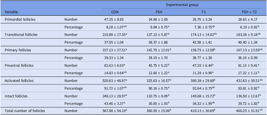
The absolute number of primordial follicles was negatively affected by the main effect of T2 (P = 0.033); however, it was not influenced by the interaction effect of FSH by T2 and the main effect of FSH (P > 0.05; Table 2). The proportion of primordial follicles was less in FSH+T2 than FSH group (P = 0.033). The proportion of primordial follicles was not affected by the main effect of FSH (P > 0.05); however, it was negatively affected by the main effect of T2 (P = 0.015; Table 2). The absolute number of transitional follicles was less in the FSH than in the CON group (P = 0.001; Table 2). The proportion of transitional follicles was not affected by the interaction effect of FSH by T2 and the main effect of FSH (P > 0.05); however, it was positively influenced by the main effect of T2 (P = 0.022; Table 2). The absolute number of primary follicles was less in the FSH and T2 groups than in the CON group (P < 0.05; Table 2). The proportion of primary follicles was not affected by the interaction effect of FSH by T2 and the main effects of FSH and T2 (P > 0.05; Table 2). The absolute number of preantral follicles was less in the FSH and T2 groups than in the CON and FSH+T2 groups (P < 0.01; Table 2). The proportion of preantral follicles was greater in the FSH+T2 group than in the FSH and T2 groups (P < 0.05). Yet neither the main effect of FSH nor the main effect of T2 affected the proportion of preantral follicles (P > 0.05; Table 2). The absolute number of activated follicles was less in the FSH and T2 groups than CON group (P < 0.05; Table 2). Activation of primordial follicles was greater in FSH+T2 than FSH group (P = 0.030). Activation of primordial follicles was not affected by the main effect of FSH (P > 0.05); however, it was positively affected by the main effect of T2 (P = 0.017; Table 2). The absolute number of intact follicles was less in the FSH, T2 and FSH+T2 groups than the CON group (P < 0.01; Table 2). The proportion of intact follicles was less in the FSH and FSH+T2 groups than the CON group (P < 0.01). The proportion of intact follicles was negatively affected by the main effect of FSH (P < 0.001); however, it was not impacted by the main effect of T2 (P > 0.05; Table 2). The total number of follicles was fewer in the FSH and T2 groups than the CON group (P < 0.05; Table 2).
The number and proportion of various stages of follicles, activated follicles and intact follicles as well as total number of follicles of the ovaries in CON, FSH, T2 and FSH + T2 groups (n = 10 ovaries belonging to 5 mice for each experimental group). Data are presented as mean ± SEM

a,bDifferent letters denote significant difference among various experimental groups (P < 0.05). Differences presented by superscripts here in this table are based on multiple comparisons by the post-hoc test LSMEANS.
Gene expression
Factors associated with development of ovarian follicles
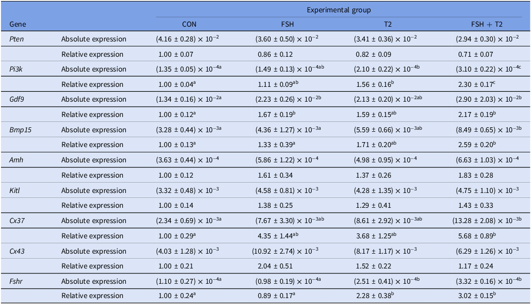
The absolute and relative expression of Pten was not affected by the interaction effect of FSH by T2 and the main effects of FSH and T2 (P > 0.05; Table 3). The absolute and relative expression of Pi3k was greater in the FSH+T2 group than in CON, FSH and T2 groups (P < 0.05); moreover, it was greater in T2 than in the CON group (P < 0.05; Table 3). The absolute and relative expression of Gdf9 was greater in the FSH and FSH+T2 groups than in the CON group (P < 0.05; Table 3). The absolute and relative expression of Bmp15 was greater in the FSH+T2 group than CON and FSH groups (P < 0.05; Table 3). The absolute and relative expression of Gdf9 and Bmp15 was positively influenced by the main effects of FSH and T2 (P < 0.05). The absolute and relative expression of Amh and Kitl was not affected by the interaction effect of FSH by T2 and the main effects of FSH and T2 (P > 0.05; Table 3). The absolute and relative expression of Cx37 was greater in FSH+T2 than in the CON group (P = 0.017; Table 3). The absolute and relative expression of Cx37 was positively influenced by the main effect of FSH (P < 0.05); moreover, the absolute expression of Cx37 was positively impacted by the main effect of T2 (P = 0.022). The absolute and relative expression of Cx43 was not affected by the interaction effect of FSH by T2 and the main effects of FSH and T2 (P > 0.05; Table 3). The absolute and relative expression of Fshr was greater in T2 and FSH+T2 groups than in CON and FSH groups (P < 0.01; Table 3). Moreover, the expression of Fshr was positively influenced by the main effect of T2 (P < 0.0001).
Absolute and relative gene expression of factors associated with development of ovarian follicles in CON, FSH, T2 and FSH + T2 groups (n = 4 mice for each experimental group). Data are presented as mean ± SEM

a,b,cDifferent letters denote significant difference among various experimental groups (P < 0.05). Differences presented by superscripts here in this table are based on multiple comparisons by the post-hoc test LSMEANS.
Factors associated with cellular proliferation and apoptosis
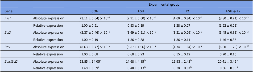
The absolute and relative expression of Ki67, Bax and Bcl2 was not affected by the interaction effect of FSH by T2 and the main effects of FSH and T2 (P > 0.05; Table 4). However, the Bax/Bcl2 gene expression ratio based on either absolute or relative expression of Bax and Bcl2, as an apoptotic index (Atrabi et al., Reference Atrabi, Akbarinejad, Khanbabaee, Dalman, Amorim, Najar-Asl, Valojerdi and Fathi2019), was less in FSH, T2 and FSH+T2 groups than CON group (P < 0.05; Table 4). Bax/Bcl2 gene expression ratio was negatively influenced by the main effects of FSH and T2 (P < 0.05).
Absolute and relative gene expression of factors associated with cellular proliferation and apoptosis in CON, FSH, T2 and FSH + T2 groups (n = 4 mice for each experimental group). Data are presented as mean ± SEM

a,bDifferent letters denote significant difference among various experimental groups (P < 0.05). Differences presented by superscripts here in this table are based on multiple comparisons by the post-hoc test LSMEANS.
Discussion
The present study was conducted to evaluate the main and interaction effects of FSH and T2 on follicular development of 1-day-old mice ovaries cultured for 12 days. To begin with, it was shown that T2 promoted the activation of primordial follicles in ovaries cultured for 12 days, which agreed with the results of previous studies in ovine (Qureshi et al., Reference Qureshi, Nussey, Bano, Musonda, Whitehead and Mason2008), porcine (Magamage et al., Reference Magamage, Zengyo, Moniruzzaman and Miyano2011) and human (Bailie et al., Reference Bailie, Maidarti, Jack, Hawthorn, Watson, Telfer and Anderson2023). Conversely, treatment with T2 in 1-day-old mice ovaries cultured for 8 days did not impact the activation of primordial follicles in our recent study (Torkzadeh et al., Reference Torkzadeh, Asadi, Jafari Atrabi, Eivazkhani, Khodadi, Hajiaghalou, Akbarinejad and Fathi2023). In this context, it is possible that the impact of T2 on 1-day-old mice ovaries required more time to materialize and its effect on ovarian follicles could be less pronounced in short-term culture systems. The positive effect of T2 on follicular activation was probably mediated through upregulation of Pi3k in the cultured ovaries because PI3K plays a key role in the activation of primordial follicles (Kim and Kurita, Reference Kim and Kurita2018; Li et al., Reference Li, Li, Zhang, Shi, Qin, Wang, Du, Ma, Chen and Cui2021a; Makker et al., Reference Makker, Goel and Mahdi2014). Likewise, T2 treatment has been reported to augment the transcription of Pi3k and activate PI3K signalling in various tissues (Li et al., Reference Li, Wang, Shi, Lu, Yu, Shi, Du and Yu2020, Reference Li, Shi, Shi and Wang2021b; White et al., Reference White, Gao, Puppa, Sato, Welle and Carson2013; Zhao et al., Reference Zhao, Liu, Wei, Jiang, Bao and Yang2016).
Unlike T2, FSH treatment did not influence the activation of primordial follicles in 1-day-old mice ovaries cultured for 12 days. By contrast, we found a positive effect of FSH on follicular activation in 1-day-old mice ovaries cultured for 8 days in our previous study (Torkzadeh et al., Reference Torkzadeh, Asadi, Jafari Atrabi, Eivazkhani, Khodadi, Hajiaghalou, Akbarinejad and Fathi2023). Comparison of the findings between the present and previous studies implicate that the positive effect of FSH may be more instantaneous than T2 but could not last for a culture system longer than 8 days. It was the same case with the effect of FSH on Pi3k expression because FSH did not influence gene expression of Pi3k in the ovaries cultured for 12 days, but FSH upregulated Pi3k in the ovaries cultured for 8 days (Torkzadeh et al., Reference Torkzadeh, Asadi, Jafari Atrabi, Eivazkhani, Khodadi, Hajiaghalou, Akbarinejad and Fathi2023). One potential explanation for such phenomena could be the dynamics in the expression of Fshr considering the length of exposure to the ligand since prolonged exposure to FSH could downregulate FSH receptor and desensitize the cells to FSH (Casarini and Crépieux, Reference Casarini and Crépieux2019; Griswold et al., Reference Griswold, Kim and Tribley2001; Simoni et al., Reference Simoni, Gromoll and Nieschlag1997). In this sense, FSH could upregulate gene expression of its receptor in the ovaries cultured for 8 days in the previous study (Torkzadeh et al., Reference Torkzadeh, Asadi, Jafari Atrabi, Eivazkhani, Khodadi, Hajiaghalou, Akbarinejad and Fathi2023); however, it failed to affect the expression of its receptor in the ovaries cultured for 12 days in the present study. Herein, it is worth noting that the neutral impact of FSH on Fshr was abrogated when FSH was used in combination with T2; furthermore, expression of Pi3k and activation of primordial follicles were the greatest in the ovaries simultaneously treated with FSH as well as T2. The upregulatory influence of T2 on FSH receptor is well-established (Fujibe et al., Reference Fujibe, Baba, Nagao, Adachi, Ikeda, Morishita, Kuno, Suzuki, Mizuuchi, Honnma, Endo and Saito2019; Laird et al., Reference Laird, Thomson, Fenwick, Mora, Franks and Hardy2017; Sen et al., Reference Sen, Prizant, Light, Biswas, Hayes, Lee, Barad, Gleicher and Hammes2014; Torkzadeh et al., Reference Torkzadeh, Asadi, Jafari Atrabi, Eivazkhani, Khodadi, Hajiaghalou, Akbarinejad and Fathi2023), yet the present study implicated that T2 can also override the desensitization of ovarian follicles to FSH due to prolonged exposure and potentiate the beneficial effects of FSH treatment on the cultured ovaries.
Neither FSH nor T2 could solely improve the transition of follicles to the preantral stage, but the combination of FSH and T2 successfully increased the proportion of preantral follicles. Enhancement in the development of activated ovarian follicles towards the preantral stage is the main target in culturing dormant primordial follicles since progression to this stage is a prerequisite before isolation of follicles for in vitro production of MII-stage oocytes, which could be used for ART purposes (Amorim and Shikanov, Reference Amorim and Shikanov2016; Khajedehi et al., Reference Khajedehi, Fathi, Akbarinejad and Gourabi2024; Khodadi et al., Reference Khodadi, Atrabi, Torkzadeh, Fazli, Akbarinejad and Fathi2022). The synergistic effect of FSH and T2 was also observed on the expression of oocyte-derived growth factors (i.e. Gdf9 and Bmp15) which was in accord with previous findings (Gilchrist et al., Reference Gilchrist, Lane and Thompson2008; Otsuka et al., Reference Otsuka, McTavish and Shimasaki2011). GDF9 contributes to not only the activation of primordial follicles but also the development of activated follicles (Gilchrist et al., Reference Gilchrist, Lane and Thompson2008; Hreinsson et al., Reference Hreinsson, Scott, Rasmussen, Swahn, Hsueh and Hovatta2002; Kedem et al., Reference Kedem, Fisch, Garor, Ben-Zaken, Gizunterman, Felz, Ben-Haroush, Kravarusic and Abir2011; Otsuka et al., Reference Otsuka, McTavish and Shimasaki2011; Vitt et al., Reference Vitt, McGee, Hayashi and Hsueh2000), and BMP15 contributes to proliferation of granulosa cells, growth of follicle and sensitization of follicle to FSH by upregulation of FSH receptor (Otsuka et al., Reference Otsuka, McTavish and Shimasaki2011; Persani et al., Reference Persani, Rossetti, Di Pasquale, Cacciatore and Fabre2014). Therefore, the positive effect of FSH+T2 on the development of follicles could be attributed to their upregulatory effects on Gdf9 and Bmp15.
Regardless, no significant effect of FSH, T2 or FSH+T2 was found on the expression of factors originating from granulosa cells (i.e. Amh and Kitl), which indicates the hormonal treatments were not as influential on the granulosa cells as they were on the oocytes, at least within a 12-day culture period. One explanation for such a phenomenon may be the fact that alterations in the oocyte precede changes in granulosa cells in newly activated follicles (Chen et al., Reference Chen, Yang, Shi, Zhang, Song and Huang2020; Zhang et al., Reference Zhang, He, Zhang, Tong, Chen, Wang, Pan and Xiao2023; Zhao et al., Reference Zhao, Feng, Zhang, Zhang, Wang, Liu, Wang, Li, Ng, Yeung, Rodriguez-Wallberg and Liu2021), and this system of in vitro culture could merely help activated follicles develop up to early steps of the preantral stage, at which limited progression in growth of granulosa cell layer occurred. In corroboration with this notion, the hormonal treatments in this study augmented gene expression of Cx37, which codes a connexin protein present at the interface between oocyte and granulosa cells and facilitates reciprocal communication between the germ cell and surrounding somatic cells (Firestone and Kapadia, Reference Firestone and Kapadia2012; Simon et al., Reference Simon, Chen and Jackson2006; Simon et al., Reference Simon, Goodenough, Li and Paul1997; Teilmann, Reference Teilmann2005). However, these hormonal treatments did not significantly impact gene expression of Cx43, which codes another protein from the connexin family localized in inter-granulosa cells gap junctions and regulates the coordinated function of granulosa cells (Firestone and Kapadia, Reference Firestone and Kapadia2012; Juneja et al., Reference Juneja, Barr, Enders and Kidder1999; Santiquet et al., Reference Santiquet, Robert and Richard2013; Teilmann, Reference Teilmann2005). Interestingly, there is evidence indicating that alterations in the expression of CX37 precede altercations in the expression of CX43 in ovarian follicles (Teilmann, Reference Teilmann2005). Nevertheless, whether the continuation of the culture of ovaries for longer than 12 days would lead to significant positive morphologic and molecular changes in granulosa cells requires further research to be addressed.
Despite the aforementioned positive effects of hormonal treatments on the development of ovarian follicles, they adversely influenced the viability of follicles in cultured ovaries. The negative effects of hormonal treatment were also evident in fewer absolute number of follicles, particularly in ovaries treated solely with FSH. In this regard, both FSH and testosterone have been reported to protect ovarian follicles from apoptosis (Shen et al., Reference Shen, Liu, Li, Teng, Zhang, Tang, Sun and Liu2014; Shi et al., Reference Shi, Guan, Yu and Liu2020; Torkzadeh et al., Reference Torkzadeh, Asadi, Jafari Atrabi, Eivazkhani, Khodadi, Hajiaghalou, Akbarinejad and Fathi2023; Yoo et al., Reference Yoo, Tanaka, Konishi, Tanabe, Taniguchi, Komura, Hayashi and Ohmichi2020). In addition, although degeneration of follicles could occur at a normal rate in the process of ovarian development (Hussein, Reference Hussein2005), the rate of degeneration in small activated follicles is believed to be commonly negligible under a normal condition (Tingen et al., Reference Tingen, Bristol-Gould, Kiesewetter, Wellington, Shea and Woodruff2009). Nevertheless, hyperactivation of primordial follicles through PI3K signalling has been observed to dramatically increase the rate of degeneration in the growing follicles (Chen et al., Reference Chen, Xia, Guan, Li and Zhang2016). As a result, one potential reason for the decreased proportion of intact follicles in ovaries treated with hormones could be the augmented rate of follicular activation and development in the corresponding ovaries rather than the direct apoptotic effect of FSH or testosterone on the ovarian follicles. As aforementioned, the effect of FSH on the progression of follicular activation and development initiates earlier than the effect of T2 on these biological processes (Torkzadeh et al., Reference Torkzadeh, Asadi, Jafari Atrabi, Eivazkhani, Khodadi, Hajiaghalou, Akbarinejad and Fathi2023), which may partly explain why the negative effect of hormonal treatments on the number of follicles was more pronounced in FSH-treated than T2-treated ovaries. Interestingly, the Bax/Bcl2 gene expression ratio as an apoptotic index was lower in all hormone-treated ovaries than in untreated ones. This finding implicates that mechanisms other than apoptosis might have been involved in the degeneration of ovarian follicles in the hormone-treated ovaries. Hence, further studies are required to unravel the mechanisms that contributed to the follicular loss following treatment with FSH and T2, and in this regard, autophagy could be one of the potential mechanisms (Bhardwaj et al., Reference Bhardwaj, Paliwal, Saraf and Sachdeva2022; Pagotto et al., Reference Pagotto, Pilotto, Mazzoldi, Nicoletto, Frezzini, Pastò and Amadori2017; Zhou et al., Reference Zhou, Peng and Mei2019).
Conclusion
In conclusion, the present study showed that treatment with the combination of FSH and T2 improved not only the activation of primordial follicles but also the growth of activated follicles towards preantral stage. This positive effect of FSH+T2 appeared to be at least partly mediated through the upregulation of Pi3k and oocyte-derived growth factors including Gdf9 and Bmp15.
Acknowledgements
The authors would like to express their gratitude to the laboratory staff at Royan Institute, Tehran, Iran for their kind assistance.
Author contributions
T.T.: data curation, methodology, investigation, project administration, writing – original draft. Z.A.: data curation, methodology, investigation, project administration, writing – original draft. M.J.A.: methodology, investigation. M.K.: methodology, investigation. F.E.: methodology, investigation. S.H.: methodology, investigation. V.A.: conceptualization, methodology, investigation, resources, supervision, formal analysis, writing – original draft, writing – review and editing. R.F.: conceptualization, methodology, investigation, resources, supervision, writing – review and editing.
Funding
The present research was supported by the University of Tehran (grant number: 30854/6/7) and Royan Institute (grant number: 98000081).
Competing interests
The authors declare no conflicts of interest.
Ethical standards
All animal procedures were conducted in accordance with the guidelines of the Royan Ethics Committee (IR.ACECR.ROYAN.REC.1398.248).