Introduction
Eggs are a popular source of nutrition as they contain several nutrients, which are essential for humans, namely, water (~75%), proteins (~12%), and lipids (~12%) (Burley and Vadehra, Reference BURLEY and VADEHRA1989; Sugino et al., Reference SUGINO, NITODA and JUNEJA1996). However, the nutrient content of eggs varies depending on breed and feeding regime. Moreover, the internal quality of eggs can change during storage and transport (Silversides and Scott, Reference SILVERSIDES and SCOTT2001). Thus, it is important to determine the chemical composition when discriminating between eggs of different quality.
For breeder eggs, the hatching rate is a critical economic factor for poultry farms. Infertile eggs or embryonic death occur in more than 10% of the eggs in hatcheries (Smith et al., Reference SMITH, LAWRENCE and HEITSCHMIDT2008). These eggs pose problems for the poultry industry because they occupy space, increase handling costs, and have the potential to harbour bacteria which could contaminate other eggs. Therefore, the early detection of non-fertile eggs and early and late embryonic death would increase the overall yield of the poultry industry.
There are many traditional methods for analysing nutrient components, quality, and embryonic death in chicken eggs, such as the Soxhlet extraction for crude fats (Song et al., Reference SONG, CHOI and OH2000), Kjeldahl method for crude protein (Lunven et al., Reference LUNVEN, DE ST MARCQ, CARNOVALE and FRATONI1973), Haugh unit (HU) method for egg quality (Haugh, Reference HAUGH1937), and candling for removing infertile eggs and dead embryos from hatching eggs. However, these methods are time consuming and unsuitable for in-line applications.
Recently, spectroscopic techniques as non-destructive, rapid and safe approaches have been successfully applied to the quantitative determination of major egg constituents (Dalle Zotte et al., Reference DALLE ZOTTE, BERZAGHI, JANSSON and ANDRIGHETTO2006; Zhao et al., Reference ZHAO, LV, JIA, CHEN, XU and QU2018). Most studies on these applications have assessed the quality of eggs and their products in terms of storage time (Xu et al., Reference XU, XU, CHEN, YAO and ZHANG2014; Coronel-Reyes et al., Reference CORONEL-REYES, RAMIREZ-MORALES, FERNANDEZ-BLANCO, RIVERO and PAZOS2018), HU (Lin et al., Reference LIN, ZHAO, SUN, CHEN and ZHOU2011; Suktanarak and Teerachaichayut, Reference SUKTANARAK and TEERACHAICHAYUT2017), air cell height (Aboonajmi and Najafabadi, Reference ABOONAJMI and NAJAFABADI2014; Aboonajmi et al., Reference ABOONAJMI, SABERI, ABBASIAN NAJAFABADI and KONDO2016), and albumen pH (Abdel-Nour et al., Reference ABDEL-NOUR, NGADI, PRASHER and KARIMI2011; Dong et al., Reference DONG, DONG, PENG and TANG2017a). The detection of infertile and non-hatchable eggs has been investigated using spectroscopic methods (Lawrence et al., Reference LAWRENCE, SMITH, WINDHAM, HEITSCHMIDT, PARK and YOON2007; Smith et al., Reference SMITH, LAWRENCE and HEITSCHMIDT2008; Liu and Ngadi, Reference LIU and NGADI2013). Furthermore, several studies have explored the feasibility of spectral analysis for the sexing of chicken eggs (Göhler et al., Reference GÖHLER, FISCHER and MEISSNER2016; Galli et al., Reference GALLI, PREUSSE, UCKERMANN, BARTELS, KRAUTWALD-JUNGHANNS, KOCH and STEINER2016; Reference GALLI, PREUSSE, UCKERMANN, BARTELS, KRAUTWALD-JUNGHANNS, KOCH and STEINER2017).
An excellent review of spectroscopic techniques expounded on their ability to determine the quality of eggs (De Ketelaere et al., Reference DE KETELAERE, BAMELIS, KEMPS, DECUYPERE and DE BAERDEMAEKER2004). This paper summarises developments published after 2004, taking some new parameters of eggs and their products into consideration.
Applications for determining chemical composition
Conventional techniques to determinate the fat, protein, and moisture content in eggs are expensive and can contaminate the samples (Lunven et al., Reference LUNVEN, DE ST MARCQ, CARNOVALE and FRATONI1973; Song et al., Reference SONG, CHOI and OH2000). Wehling et al. (Reference WEHLING, PIERCE and FRONING1988) used near-infrared reflectance (NIR) spectroscopy to measure protein and fat in spray-dried whole eggs based on three wavelengths. However, the small number of training samples again limited the applicability of this method. The use of near-infrared spectroscopy to determine the fat, moisture, and protein content in homogenised egg yolk and egg albumen (Zhao et al., Reference ZHAO, LV, JIA, CHEN, XU and QU2018) was recently reported and found that the evaluation of protein was not as accurate as that of fat but was superior to that of moisture.
Eggs are popular because of the n-3 fatty acids contained in the yolk, which can decrease the incidence of cardiovascular disease and are essential to human health. To guarantee that consumers receive appropriate amounts of these fatty acids, scientists applied hyperspectral imaging to predict the n-3 fatty acid content of eggs (Abdel-Nour and Ngadi, Reference ABDEL-NOUR and NGADI2011), achieving R 2 values of 0.89, 0.54, and 0.75 for alpha-linolenic acid (ALA), eicosapentaenoic acid (EPA), and docosahexaenoic acid (DHA), respectively. In a previous study (Dalle Zotte et al., Reference DALLE ZOTTE, BERZAGHI, JANSSON and ANDRIGHETTO2006), NIR spectroscopy was exploited to determinate ALA, EPA, and DHA contents in egg yolk, and improved R 2 values of 0.96, 0.86, and 0.87 were reported, respectively. Both studies found that ALA could be predicted most accurately. Among all studies, fat has been modelled better than protein, which supports the conclusion that the quantitative detection of fats is generally better than that of protein when evaluating the near-infrared bands (Sasic and Ozaki, Reference SASIC and OZAKI2001).
Applications for assessing egg quality
For table eggs, consumers are particularly concerned with quality and freshness. Parameters such as shell quality, freshness, and blood spots are commonly used to assess egg quality.
eggshell quality
The eggshell provides natural protection for the egg and must be strong enough to prevent cracks from occurring during the packing and transportation processes. Traditional crack detection methods mainly rely on manual methods such as visual inspection or listening for a dull, flat sound as an indicator of a crack when tapping eggs together (Lawrence et al., Reference LAWRENCE, YOON, HEITSCHMIDT, JONES and PARK2008). However, very small cracks, or micro-cracks, can go undetected by manual techniques. Narushin et al. (Reference NARUSHIN, VAN KEMPEN, WINELAND and CHRISTENSEN2004) applied infrared spectroscopy to predict eggshell quality parameters, such as shell weight, percentage, thickness, fracture force, maximal deformation and stiffness, but this method exhibited a poor predictive ability (correlation coefficients ranging from 0.29 to 0.72). In another study (Xiong et al., Reference XIONG, XU, ZHOU, YAO and CHEN2013), eggshell percentage, strength, and thickness were analysed using NIR spectroscopy with partial least-squares regression (PLSR). Recently, Dong et al. (Reference DONG, TANG, PENG and DONG2017b) evaluated eggshell thickness based on visible (VIS)/NIR spectroscopy and reported a correlation coefficient of 0.86.
Once eggs have been cracked due to poor eggshell quality, they should be identified and removed before being sent to the market. Xiong et al. (Reference XIONG, ZHU, WU and WANG2011) combined NIR and discriminant analysis (DA) to detect cracked eggs, achieving a fairly good recognition rate of 90%. Hyperspectral imaging combined with partial least squares-DA (PLS-DA) has been used to identify cracked eggs, outperforming the NIR model and achieving a classification accuracy of 97.06% (Xie and He, Reference XIE and HE2016).
egg freshness
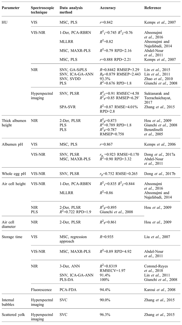
Freshness makes a major contribution to the internal quality of eggs. The traditional method of assessing egg freshness is the HU method (Haugh, Reference HAUGH1937), which requires the eggs to be broken. Recently, spectroscopic techniques have been applied to determine the freshness of eggs. A new algorithm based on support vector data description (SVDD), which overcomes the problem of unbalanced samples, has been employed to identify fresh and non-fresh eggs in terms of HU, achieving an identification rate of 93.3% (Zhao et al., Reference ZHAO, LIN, CHEN, HUANG, SUN and ZHOU2010). Three variable selection methods (spectral interval selection, genetic algorithm (GA), and successive projections algorithm (SPA)) were combined with graphically oriented local multivariate analysis to identify egg freshness based on NIR spectroscopy (Lin et al., Reference LIN, ZHAO, SUN, BI and CAI2015). Kemps et al. (Reference KEMPS, DE KETELAERE, BAMELIS, MERTENS, DECUYPERE, DE BAERDEMAEKER and SCHWÄGELE2007) combined VIS-NIR transmission spectroscopy and low-resolution proton nuclear magnetic resonance to assess egg HU, but the integration of these spectroscopic techniques did not improve prediction capability when compared with transmission spectral measurements alone. Recently, hyperspectral imaging has been used to determine the internal quality of eggs in terms of HU (Zhang et al., Reference ZHANG, PAN, TU, ZHAN and TU2015; Suktanarak and Teerachaichayut, Reference SUKTANARAK and TEERACHAICHAYUT2017), bubble formation, and scattered yolk (Zhang et al., Reference ZHANG, PAN, TU, ZHAN and TU2015). It appears that the egg HU model based on hyperspectral imaging performs better than that based on NIR or VIS-NIR spectroscopy (Table 1).
In addition to HU, Dong et al. (Reference DONG, DONG, PENG and TANG2017a) evaluated albumen pH and whole egg pH using VIS and NIR spectroscopy, reporting correlation coefficients of 0.923 and 0.752, respectively. Fourier transform near-infrared (FT-NIR) spectroscopy was applied for quantitative measurements of thick albumen (R 2 = 0.787; Berardinelli et al., Reference BERARDINELLI, GIUNCHI, GUARNIERI, PEZZI and RAGNI2005). A more robust model for the thick albumen height was obtained by data pre-processing using a Savitzky-Golay filter and second derivatives (R 2 = 0.873; Hou et al., Reference HOU, YANG, LI and XU2009); this study demonstrated the feasibility of FT-NIR for determining air cell height and air cell diameter (R 2 = 0.895 and R 2 = 0.861, respectively). Kemps et al. (Reference KEMPS, BAMELIS, DE KETELAERE, MERTENS, TONA, DECUYPERE and DE BAERDEMAEKER2006) confirmed the feasibility of VIS transmission spectroscopy for the non-destructive assessment of egg freshness.
VIS-NIR spectroscopy has been used to determine the HU and pH of the albumen, giving better determination coefficients (Abdel-Nour et al., Reference ABDEL-NOUR, NGADI, PRASHER and KARIMI2011). Furthermore, both HU and air cell height have been measured by VIS-NIR spectroscopy combined with a special regression method (Aboonajmi and Najafabadi, Reference ABOONAJMI and NAJAFABADI2014; Aboonajmi et al., Reference ABOONAJMI, SABERI, ABBASIAN NAJAFABADI and KONDO2016; see Table 1). In a study conducted by Giunchi et al. (Reference GIUNCHI, BERARDINELLI, RAGNI, FABBRI and SILAGHI2008), several freshness parameters (air cell height, thick albumen height, and HU) were assessed using FT-NIR (Table 1). They found that the HU model was worse than those for the albumen pH, thick albumen height, and air cell height. This could be due to the indirect measurement of HU based on the relationship between the weight of the intact egg and the albumen height and the dependence of the model on the reference measurement.
NIR spectroscopy has been used to assess the storage time of eggs through multivariate analysis methods such as PLS (Abdel-Nour et al., Reference ABDEL-NOUR, NGADI, PRASHER and KARIMI2011), artificial neural networks (ANNs) (Lin et al., Reference LIN, ZHAO, SUN, CHEN and ZHOU2011; Coronel-Reyes et al., Reference CORONEL-REYES, RAMIREZ-MORALES, FERNANDEZ-BLANCO, RIVERO and PAZOS2018), and PLS-DA (Giunchi et al., Reference GIUNCHI, BERARDINELLI, RAGNI, FABBRI and SILAGHI2008). In fact, PLS-DA models can classify egg samples with perfect accuracy. Fluorescence spectra were evaluated to discriminate eggs according to both their storage time and conditions (Karoui et al., Reference KAROUI, NICOLAÏ and DE BAERDEMAEKER2008). Regrettably, no fluorescence spectra have since been employed for egg assessments.
Application of spectroscopic techniques to the assessment of egg freshness.

blood and meat spots
Blood and meat spots are common defects inside eggs. The traditional method for detecting blood spots in eggs is candling, although this is labour-intensive and has variable accuracy (from 20 to 90%; Galis et al., Reference GALIS, DALE, BOUDRY and THEWIS2012). PLS-DA over a wavelength range of 500-600 nm was previously adopted to detect blood-spot eggs, achieving a discrimination rate of 96.8% (Usui et al., Reference USUI, NAKANO and MIZUTANI2005).
The above studies only considered white-shelled eggs, and blood detection accuracy in brown-shell eggs is not as high. Usui et al. (Reference USUI, NAKANO and SAITOU2006) detected blood in brown-shell eggs with a discrimination accuracy of only 85% by the traditional blood value method. The reason for the difference in accuracy is that protoporphyrin, a brown pigment in the eggshell, has similar optical properties to those of haemoglobin (De Ketelaere et al., Reference DE KETELAERE, BAMELIS, KEMPS, DECUYPERE and DE BAERDEMAEKER2004). An improved measurement system combining reflection and transmission was found to be superior to transmission-only measurements. However, for brown-shelled eggs, the detection rate remains low (De Ketelaere et al., Reference DE KETELAERE, MERTENS, KEMPS, BAMELIS, DECUYPERE and DE BAERDEMAKER2005). To overcome this problem, various methods have been developed to detect blood-spot eggs. For example, techniques using the generalised Mahalanobis distance (Usui et al., Reference USUI, NAKANO and SAITOU2006), least-squares support vector machines (Xu et al., Reference XU, XU, CHEN, YAO and ZHANG2014), binary logistic regression (Chen et al., Reference CHEN, ZHANG and XU2015), and stepwise Bayesian discriminant analysis (Zhu et al., Reference ZHU, LI, WANG, TANG, CAO and MA2017) have achieved accuracy rates of 93.3%, 91.7%, 96.9%, and 95%, respectively. It is worth mentioning the study carried out by Zhu et al. (Reference ZHU, XIE, LI, WANG and MA2015), who detected abnormal eggs, including blood-spot and meat-spot eggs, using a fusion of classifiers. The resulting model was superior to those using a single classifier or base classifier.
Application to hatching eggs
In hatching eggs, one of the main challenges is the detection of infertile and dead embryos at an early stage of incubation. Additionally, in recent years, the sexing of chickens in ovo has received increasing attention as a substitute for the culling of day-old male chicks.
fertility and embryo development
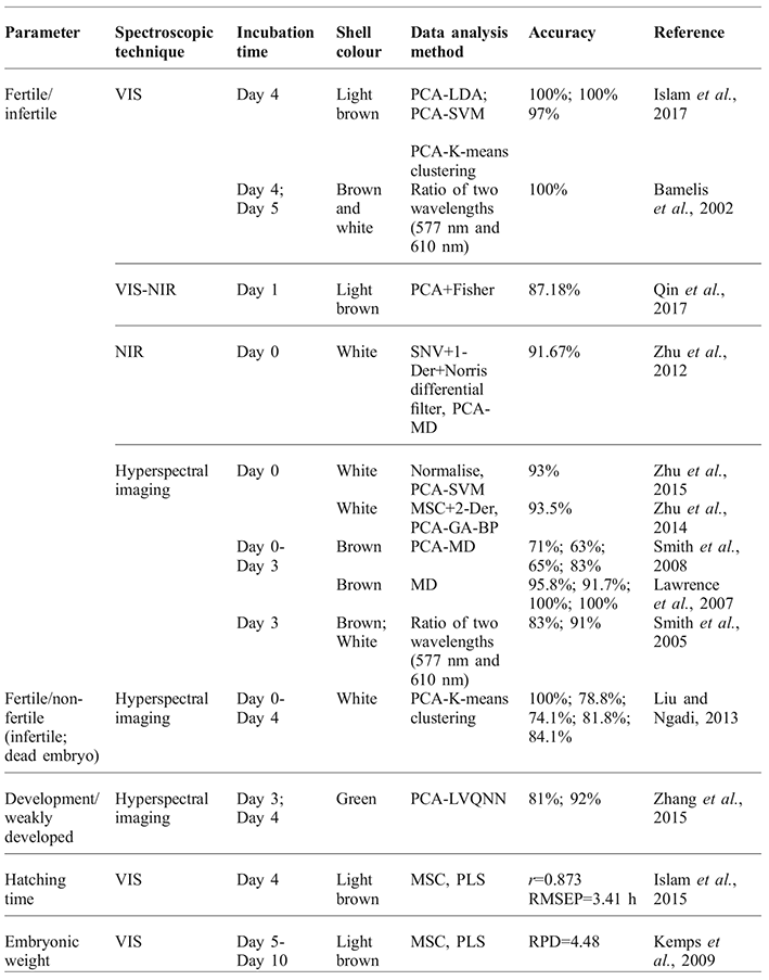
Hatchery statistics show that 8-9% of all incubated eggs are unfertilised (Das and Evans, Reference DAS and EVANS1992). Candling is traditionally used to remove infertile eggs or dead embryos from the incubator (Tian and Ma, Reference TIAN and MA2011), but this is laborious and time-consuming. The results of several spectroscopic techniques applied to the detection of fertility and embryo development are summarised in Table 2.
As early as 2002, VIS transmission spectroscopy was applied to detect embryo development in incubated eggs using the ratio of two wavelengths (577 nm and 610 nm) (Bamelis et al., Reference BAMELIS, TONA, DE BAERDEMAEKER and DECUYPERE2002). Islam et al. (Reference ISLAM, KONDO, OGAWA, FUJIURA, SUZUKI and FUJITANI2017) proposed non-destructive detection models for hatching egg fertility using VIS transmission spectroscopy. It was concluded that satisfactory classification results can only be obtained after 4-5 d of incubation through VIS transmission spectroscopy, which is very close to the candling time (Tian et al., 2011). Zhu et al. (Reference ZHU, WANG, WANG, DAI and MA2012) applied NIR spectroscopy to detect infertile eggs prior to incubation and reported an accuracy of 91.67%. Furthermore, hyperspectral imaging has been widely applied to hatching eggs (Smith et al., Reference SMITH, MAULDIN, LAWRENCE, PARK and HEITSCHMIDT2005, Reference SMITH, LAWRENCE and HEITSCHMIDT2008; Lawrence et al., Reference LAWRENCE, SMITH, WINDHAM, HEITSCHMIDT, PARK and YOON2007; Zhang et al., Reference ZHANG, PAN, TU, ZHAN and TU2015; Zhu et al., Reference ZHU, LIU, XIONG and MA2014; Reference ZHU, XIE, LI, WANG and MA2015).
For white-shell eggs, Zhu et al. (Reference ZHU, LIU, XIONG and MA2014; Reference ZHU, XIE, LI, WANG and MA2015) realised fertility detection at promising rates of 93% and 93.5% based on an SVM method and GA-propagation (GA-BP) network, respectively. The classification accuracy is generally lower for brown-shell eggs (Smith et al., Reference SMITH, MAULDIN, LAWRENCE, PARK and HEITSCHMIDT2005; Reference SMITH, LAWRENCE and HEITSCHMIDT2008). Lawrence et al. (Reference LAWRENCE, SMITH, WINDHAM, HEITSCHMIDT, PARK and YOON2007) achieved 100% accuracy at days two and three of incubation in terms of detecting the embryonic development of hatching eggs, but they used only fertile eggs and a small number of samples (N = 24).
In addition, an attempt was made to detect embryo vitality (weak embryo), although the recognition was relatively poor at 81% at day three and 92% at day four of incubation (Zhang et al., 2014). Liu and Ngadi (Reference LIU and NGADI2013) identified non-fertile eggs (infertile and dead embryo eggs) using a k-means clustering algorithm and NIR hyperspectral imaging.
As well as weak and dead embryos, there exist some slow-developing embryos in hatching eggs. Kemps et al. (Reference KEMPS, BAMELIS, MERTENS, DECUYPERE, DE BAERDEMAEKER and DE KETELAERE2010) developed a quantitative model to assess embryo weight inside the egg from five to ten days of incubation, making it possible to match the incubation length to the needs of the eggs. Similarly, to obtain a batch of homogeneous broiler eggs, scientists have predicted the chick hatching time from the day four incubation spectra (Islam et al., Reference ISLAM, KONDO, OGAWA, FUJIURA, SUZUKI, NAKAJIMA and FUJITANI2015).
Application of spectroscopic techniques to the detection of fertility and embryo development.

sexing of chickens in ovo
One-day-old male birds in hatcheries are manually sorted by feather or vent sexing (Biederman and Shiffrar, Reference BIEDERMAN and SHIFFRAR1987). The ability to recognise and remove male chicks pre-hatching would be a significant step forward in layer hatcheries. Göhler et al. (Reference GÖHLER, FISCHER and MEISSNER2016) used hyperspectral imaging for gender determination in layer lines with gender-specific down feather colour. Using totally intact hatching eggs, they obtained an accuracy of approximately 97% at 14 d of incubation. However, a study conducted by Close et al. (Reference CLOSE, BANISTER, BAUMANS, BERNOTH, BROMAGE, BUNYAN, ERHARDT, FLECKNELL, GREGORY, HACKBARTH, MORTON and WARWICK1997) demonstrated that bird embryos are capable of perceiving pain in the second half of gestation (after 10.5 d of incubation). To overcome this problem, NIR Raman spectroscopy (Galli et al., Reference GALLI, PREUSSE, UCKERMANN, BARTELS, KRAUTWALD-JUNGHANNS, KOCH and STEINER2016) and fluorescence spectroscopy (Galli et al., Reference GALLI, PREUSSE, UCKERMANN, BARTELS, KRAUTWALD-JUNGHANNS, KOCH and STEINER2017) have been applied to in ovo sexing based on differences in the composition of embryonic blood at day 3.5 of egg incubation (before the onset of pain perception), with reported accuracy rates of 90% and 93%. Nevertheless, the need for a window in the egg shell will lead to decreased hatching rates (~10%) (Galli et al., Reference GALLI, PREUSSE, UCKERMANN, BARTELS, KRAUTWALD-JUNGHANNS, KOCH and STEINER2016). A minimally invasive method was proposed by Galli et al. (Reference GALLI, PREUSSE, SCHNABEL, BARTELS, CRAMER, KRAUTWALD-JUNGHANNS, KOCH and STEINER2018) based on the same spectral technique and algorithm. In their study, the blood circulation in the extraembryonic vessels was accessed without removing the inner shell membrane.
Conclusions and future outlook
This review summarised recent advances in the quality analysis and classification of chicken eggs using several spectroscopic techniques. Hyperspectral imaging has proved to be a powerful technique for evaluating essential features, both in consumption eggs and hatching eggs. Furthermore, NIR Raman and fluorescence spectroscopy exhibit strong potential for gender determination prior to hatching. In the detection of blood-spot eggs or fertile eggs, eggshell colour proved to be a negative factor.
Numerous properties of eggs have been successfully detected by spectroscopic techniques, but there are still some barriers to overcome. First, large and representative samples are needed to obtain more robust calibrations, and simple, low-cost instruments are required to realise the online monitoring of certain properties in chicken eggs. Furthermore, in future studies, the existence or content of certain minerals in eggs may be detectable by spectroscopic techniques based on changes in physical and chemical properties caused by these minerals.
Acknowledgments
This work was supported by the Beijing Innovation Team of Modern Agricultural Industry Technical System (BAIC04-2016) and the China Agriculture Research System (CARS-41).