Introduction
Epithelial-myoepithelial carcinoma is an uncommon, low-grade carcinoma that generally occurs in the salivary glands, primarily the parotid gland, but has occasionally been observed in the trachea, larynx, hypopharynx and lacrimal gland.Reference Batsakis, Naggar and Luna1–Reference Ostrowski, Font, Halpern, Nicolitz and Barnes3
Four cases of epithelial-myoepithelial carcinoma arising in the nasal cavity have previously been reported; these were all typically low-grade malignant neoplasms.Reference Harada, Kashiwagi, Fujiura, Kusukawa and Morimatsu4–Reference Yamanegi, Uwa and Hirokawa7
In the present paper, we describe a rare case of aggressive epithelial-myoepithelial carcinoma in the nasal cavity, with high-grade histology.
Case report
A 36-year-old woman was referred to us with a nasal cavity mass.
Endoscopic evaluation revealed a 0.5 cm, irregular, protruding mass at the posterior end of the left inferior turbinate; this mass was biopsied.
A computed tomography (CT) scan was normal.
Histopathological analysis of biopsied tissue was consistent with an epithelial-myoepithelial carcinoma. Positron emission tomography (PET) CT scanning revealed no fluorodeoxyglucose uptake.
The patient underwent endoscopic excision of the tumour, extending from the posterior third of the left inferior turbinate to 1 cm anterior to the torus tubarius, and including a 1 cm surgical margin from the previous biopsy scar. The excised tissue contained no cancer cells, because of the previous biopsy.
However, on 15-month follow up, endoscopic examination revealed a mass that bled easily, located at the posterior end of the right middle turbinate. Computed tomography and magnetic resonance imaging (MRI) scans were normal. A PET CT scan revealed mild fluorodeoxyglucose uptake in the posterior portion of the right nasal cavity.
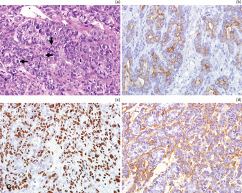
The histopathological appearance of a biopsy was consistent with epithelial-myoepithelial carcinoma. The specimen revealed a biphasic cell population of large, polygonal cells and smaller ductal cells. In some cases, the larger cells surrounded the smaller ductal cells, but more frequently the larger cells were dominant and nearly obscured the ductal cells. The tumour cells showed unusual, marked nuclear atypia, a mitotic count of ≥40/10 high-power field, and necrosis. (Mitotic figure counts of ≥2/10 high-power field are atypical in the usual form of epithelial-myoepithelial carcinoma.)
Immunohistochemical analysis revealed that the ductal tumour cells were highly reactive for cytokeratins and epithelial membrane antigen, whereas the neoplastic myoepithelial cells were positive for S-100 protein, p63 and smooth muscle actin (Figure 1).
Photomicrographs of the epithelial-myoepithelial carcinoma showing: (a) severe nuclear atypia and frequent mitoses (arrows) (H&E; ×400); (b) epithelial cells positive for cytokeratin immunostaining (×400); (c) myoepithelial cell nuclei positive for p63 immunostaining (×400); and (d) myoepithelial cells positive for cytoplasmic smooth muscle actin immunostaining (×400).

Histological and immunohistochemical findings indicated that the right nasal cavity mass was similar to the earlier left nasal cavity tumour.
The patient underwent right medial maxillectomy, and received a total of 6000 cGy post-operative radiation therapy over a six-week period.
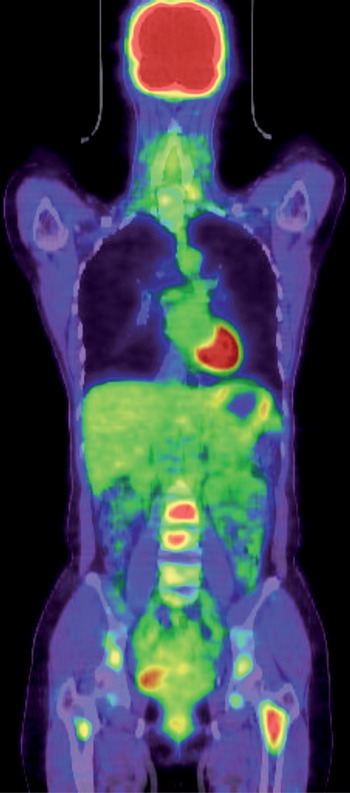
At seven-month follow up, PET CT and MRI scans revealed extensive bone metastasis in the sternum, right scapula, first rib, throughout the vertebral column, and bilaterally in the ilium bones and femurs (Figure 2).
Positron emission tomography computed tomography scan showing extensive bone metastasis in the vertebral column and bilaterally in the ilium bones and femurs.

The patient received chemotherapy plus 3000 cGy of palliative radiation therapy to the lumbar spine. However, the metastatic lesions progressed without response. At the time of writing, the patient was alive with disease.
The institutional review board of The Catholic University of Korea approved a retrospective review of the patient's medical records.
Discussion
Epithelial-myoepithelial carcinoma of intercalated duct origin is an uncommon tumour first described in 1972 by Donath et al. Reference Donath, Seifert and Schmitz8 The tumour most commonly occurs in the salivary glands, and comprises approximately 1 per cent of all salivary gland tumours.Reference Ellis, Auclair and Rosai9, Reference Lau, Goddard, Bottrill and Moffat10 Seethala et al. reported that most cases of epithelial-myoepithelial carcinoma occurred in the parotid gland (62.1 per cent), sinonasal mucoserous glands (10.3 per cent), palate (8.6 per cent) and submandibular gland (8.6 per cent).Reference Seethala, Barnes and Hunt11
The tumour is histologically characterised by well defined tubules with two cell types: an outer layer of myoepithelial cells with clear cytoplasm, which surround an inner lining of eosinophilic, cuboidal epithelial cells. The epithelial cells are positive for epithelial markers such as cytokeratins and epithelial membrane antigen, whereas the myoepithelial cells surrounding the duct-forming cells are positive for smooth muscle actin, S-100 protein, p63 and vimentin. Several types of tumours are composed of these two cell types, including epithelial-myoepithelial carcinoma, pleomorphic adenoma and adenoid cystic carcinoma.Reference Ellis and Auclair12 The cellular arrangement observed in the presented tumour excluded the possibility of an adenoid cystic carcinoma, indicating that it was either an epithelial-myoepithelial carcinoma or a pleomorphic adenoma. The World Health Organization classification of salivary gland tumours defines epithelial-myoepithelial carcinoma as a distinctive subtype of adenocarcinoma, consisting of varying proportions of two cell types, an inner layer of duct-lining cells and an outer layer of clear cells.Reference Seifert, Brocheriou, Cardesa and Eveson13
The treatment of choice for epithelial-myoepithelial carcinoma is a wide surgical excision with a clear margin, because the tumour tends to infiltrate locally.Reference Witterick, Noyek, Chapnik, Heathcote and Bedard14 However, the tumour has been reported to recur locally in the salivary glands following adequate excision, and adjuvant radiotherapy has been used to prevent local recurrence.Reference Lau, Goddard, Bottrill and Moffat15–Reference Deere, Hore, McDermott and Levine17 However, it is not clear whether radiotherapy is effective, because too few cases have been reported to enable adequate statistical analysis. Furthermore, the effect of chemotherapy on this neoplasm is uncertain.Reference Kumagai, Suzuki, Matsuura, Takahashi, Hashimoto, Suzuki and Tezuka16
Salivary gland epithelial-myoepithelial carcinoma is typically a low-grade malignant neoplasm characterised by slow, local, infiltrating growth and possible recurrence. In rare cases, the tumour can give rise to remote metastases that are eventually lethal.Reference Collina, Gale, Visona, Betts, Cenacchi and Eusebi18–Reference Alos, Carrillo, Ramos, Baez, Mallofre and Fernandez20
Four cases of epithelial-myoepithelial carcinoma arising in the nasal cavity have previously been reported. As all of these tumours were typically low-grade malignant neoplasms with mild atypia, they were surgically excised, and none recurred.Reference Harada, Kashiwagi, Fujiura, Kusukawa and Morimatsu4–Reference Yamanegi, Uwa and Hirokawa7
• Epithelial-myoepithelial carcinoma is uncommon, low-grade and generally occurs in salivary glands
• Nasal cavity cases are very rarely reported, and are typically low-grade malignancies
• A nasal cavity case with high-grade histology and highly aggressive behaviour is reported
However, in the present case, the epithelial-myoepithelial carcinoma recurred on the contralateral side 15 months after the primary surgery, and distant metastasis were discovered seven months after the second surgical procedure. On histopathological analysis, the tumour cells showed unusual, marked nuclear atypia, a mitotic count of ≥40/10 high-power field, and necrosis. Mitotic figure counts of ≥2/10 high-power field are atypical in the usual type of epithelial-myoepithelial carcinoma. The highly aggressive behaviour of this tumour probably correlates with the observed high histological grade, anaplasia, high mitotic index and necrosis.
Conclusion
This paper describes a very rare case of an aggressive epithelial-myoepithelial carcinoma in the nasal cavity, with high-grade histology.