Blood cultures are considered the gold standard for detecting bloodstream infections; they facilitate prompt and directed antimicrobial therapy for patients with sepsis. Reference Weinstein and Doern1–Reference Dawson4 However, false-positive blood culture results can lead to inappropriate clinical evaluation and treatment, leading to unnecessary patient risk. Reference Hall and Lyman2,Reference Bates, Goldman and Lee3,Reference Alahmadi, Aldeyab and McElnay5–Reference Gander, Byrd, DeCrescenzo, Hirany, Bowen and Baughman7 Blood culture contamination with skin microflora is believed to be the primary cause of false-positive blood culture results; however, needle contamination and collector contamination have also been implicated. Reference Hall and Lyman2,Reference Bell, Bogar, Plante, Rasmussen and Winters8,Reference Bowen, Coleman and Cunningham9 Reported institutional blood-culture contamination rates vary significantly, from 0.6% to 10%, and the Clinical Laboratory Standards Institute recommends that institutions strive to achieve a contamination rate <3%. Reference Hall and Lyman2,Reference Dawson4 Efforts to reduce blood-culture contamination include the use of dedicated phlebotomists, the use of diversion devices, and ensuring proper sterile technique when collecting cultures. Reference Hall and Lyman2,Reference Dawson4,Reference Gander, Byrd, DeCrescenzo, Hirany, Bowen and Baughman7–Reference Zimmerman, Assous, Yinnon and Wiener-Well15
Reported risk factors associated with blood-culture contamination include poor collection method, staff competency, increased patient age, presence of comorbidities, and patient illness severity. Reference Hall and Lyman2–Reference Alahmadi, Aldeyab and McElnay5,Reference Chih-Jan, Chi-Jung and Hsiang-Chin16,Reference Qamruddin, Khanna and Orr17 However, most of the relevant studies are relatively small, are performed over short periods, or focus on provider-specific risk factors rather than patient-specific risk factors. Additionally, with the introduction of the Centers for Medicare and Medicaid Services sepsis core measure (SEP-1), 18–21 the practice of “code sepsis” in emergency departments to expedite blood culture collection is increasing. Although this intervention likely improves time to antibiotic administration, it may compromise sterile technique, which worsens contamination rates. Since the introduction of code sepsis at our institution, emergency-department blood-culture contamination rates have increased to >6%.
Small studies, with short-term follow-up, have shown that patients with false-positive blood cultures receive more aggressive work-ups and treatment and that they experience increased lengths of stay (LOS), hospital costs, patient morbidity rates, and mortality rates. Reference Hall and Lyman2,Reference Bates, Goldman and Lee3,Reference Alahmadi, Aldeyab and McElnay5–Reference Gander, Byrd, DeCrescenzo, Hirany, Bowen and Baughman7 To better assess patient-specific risk factors and clinical outcomes associated with blood-culture contamination, we performed a large, retrospective, case-control study among hospitalized patients following blood cultures collected in our emergency department over a 4-year period.
Methods
Study design
A single-center, retrospective, case-control study was performed at a 509-bed academic hospital in Little Rock, Arkansas. Electronic medical records of adult inpatients with blood culture collected in the emergency department between May 2014 and March 2018 were reviewed. The blood culture first performed per admission was defined as a blood-culture episode. The study was determined to be non–human-subject research by our institutional review board (IRB no. 228078).
Patient selection
Patients with positive blood culture revealing microbial contaminants were considered cases and patients with negative blood culture were considered controls. Patients with positive blood culture not meeting microbial contaminant definition (true positive) were excluded from all analyses. Patients were also excluded if they were <18 years old, were discharged directly from the emergency department without inpatient admission, or if the blood culture was collected outside the emergency department. Blood-culture collections on inpatient units were excluded to better evaluate the clinical impact of blood-culture contamination at the time of admission on the entire hospitalization. Cultures collected from sources other than blood were excluded.
Blood culture collection
Standardized institutional blood-culture collection guidelines recommend the collection of 2 sets of blood samples from 2 peripheral sites, 15 minutes apart, following antiseptic sterilization. Dedicated phlebotomy teams were not present in the emergency department; thus, blood culture collection was performed primarily by registered nurses (>97%). During the study period, aseptic blood culture collection technique training was performed upon hire to the emergency department without routine assessment or return demonstration.
Determination of contaminant cultures
Microbial contaminants were defined according to the laboratory guidelines (ie, 1 of 2 sets positive for an organism rarely pathogenic, such as coagulase negative staphylococci, Micrococcus, Corynebacterium, Bacillus spp nonanthracis, or viridans streptococci) adopted from previously published recommendations. Reference Weinstein and Doern1 If only 1 set of blood cultures was obtained, positive cultures were considered true positives.
Data collection
Patient demographics, comorbid conditions, vital signs on presentation, laboratory data, and clinical data were extracted from the electronic medical record on all eligible patients during the study period. Comorbid conditions included congestive heart failure (CHF), chronic obstructive pulmonary disease (COPD), diabetes mellitus (DM), hypertension (HTN), liver disease, peripheral vascular disease (PVD), chronic kidney disease (CKD), paralysis, drug abuse, and presence of a malignancy. The identity and position of staff responsible for collecting each blood culture were also extracted from the electronic medical record for each eligible blood culture.
Determination of sepsis status
Systemic inflammatory response syndrome (SIRS) criteria included temperature ≥38°C or <36°C, heart rate >90 per minute, respirations >20 per minute, and/or white blood cell count >12,000 or <4,000 cells/μL occurring within the first 2 hours of presentation. Sepsis was defined by having met 2 or more SIRS criteria because this implied clinical concern for infection given that all included patients had blood culture obtained. Sepsis plus shock was defined as meeting sepsis criteria and also having hypotension within the first 2 hours of presentation, defined as a systolic pressure <100 mmHg.
Code sepsis definition
Upon code sepsis activation, several individuals are called to respond urgently to the bedside to obtain patient work-up and administer treatments. Details of code sepsis activation are provided in the Supplementary Material (online). Code sepsis was introduced into the emergency department in March 2015 and could be activated by a nurse of physician when 2 or more SIRS criteria were met and the patient was either hypotensive or a physician determined that activation was appropriate.
Risk factor analysis
Patient-specific data including age, sex, race, body mass index (BMI), comorbid conditions, activation of code sepsis, and sepsis status were compared between cases and controls. Risk factors identified as significant (P < .05) were included in multivariate analysis. Age and BMI were assessed as continuous variables. A separate provider-specific analysis calculating the contamination rate for individual staff was performed. Providers with <10 blood culture samples collected during the study period were not included in the provider-specific analysis.
Clinical outcome analysis
The primary outcome was hospital length of stay (LOS). Secondary outcomes included length of antimicrobial therapy (LOT) defined as the number of days that a patient received any systemic antimicrobial agent, total days of antimicrobial therapy (DOT) defined as the aggregate sum of individual antimicrobial days of therapy, infectious disease (ID) consultation, transthoracic echocardiograms (TTEs), transesophageal echocardiograms (TEEs), total hospital charges, vancomycin utilization, acute kidney injury (AKI) defined as an increase in serum creatinine >0.3 mg/dL over a 48-hour period or >1.5× increase over a 7-day period, hospice referral, and in-hospital mortality. Adjusted analyses were performed to control for age, race, BMI, comorbidities, and presence of sepsis on presentation.
Statistical analysis
Continuous variables were analyzed using the Student t test, and categorical variables were assessed using the χ Reference Hall and Lyman2 test or the Fisher exact test. An adjusted analysis to assess risk factors associated with contaminated blood culture was performed using logistic regression for dichotomous outcomes, linear regression for assessing the effect of contaminated blood culture on LOS, and generalized linear model with γ log link to estimate the effect of contaminated blood culture on inpatient costs. A P value < .05 was considered statistically significant, and all analyses used 2-tailed tests. All analyses were performed using SAS version 9.3 software (SAS Institute, Cary, NC).
Results
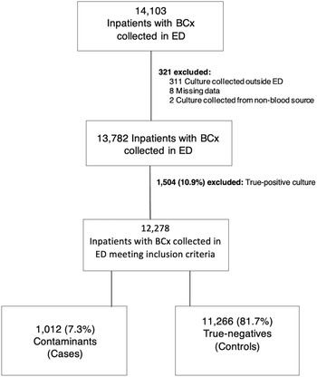
Of the 14,103 blood-culture episodes that occurred during the study period, 13,782 were reviewed based on culture results. Moreover, 1,504 (10.9%) true-positive blood cultures were excluded, leaving 1,012 (7.3%) contaminanted blood cultures (ie, cases) and 11,266 (81.7%) negative blood cultures (ie, controls) (Fig. 1). Peripheral blood culture collection was common (98%), and collection from central access was rare (2%).
Study flow chart. Note. BCx, blood culture; ED, emergency department.

Risk-factor analysis results
Baseline patient demographic and clinical data are outlined in Table 1. In univariate analysis, the following patient-specific factors were associated with blood-culture contamination: increasing age, black race, increasing BMI, presence of specific comorbidities, activation of code sepsis, and sepsis plus shock on presentation. Multivariate adjusted analysis revealed that increasing age, black race, increasing BMI, COPD, paralysis, and sepsis plus shock on presentation were independent risk factors for blood-culture contamination, whereas metastatic cancer was protective (Table 2). Characteristics were similar when blood cultures were obtained by collectors with high and low contamination rates (Supplementary Table 1 online).
Patient Characteristics and Univariate Risk Factor Analysis for Blood Culture Contamination

Note. BCx, blood culture; BMI, body mass index; CHF, congestive heart failure; COPD, chronic obstructive pulmonary disease; DM, diabetes mellitus; HTN, hypertension; PVD, peripheral vascular disease; CKD, chronic kidney disease; SIRS, systemic inflammatory response syndrome. Results shown in bold are statistically significant.
a Shock refers to systolic blood pressure <100 mmHg.
Multivariate Analysis of Risk Factors for Blood Culture Contamination

Note. BMI, body mass index; CHF, congestive heart failure; COPD, chronic obstructive pulmonary disease; DM, diabetes mellitus; OR, odds ratio; CI, confidence interval. Results shown in bold are statistically significant.
a Adjusted for age, race, BMI, comorbidities, and sepsis on presentation.
b Shock refers to systolic blood pressure <100 mmHg.
Clinical outcome analysis results
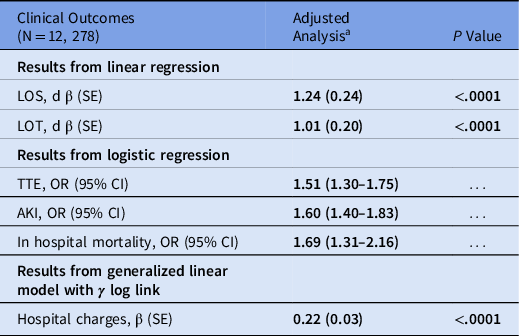
Clinical outcomes of patients with blood-culture contamination are shown in Tables 3 and 4, with univariate and multivariate analyses, respectively. After adjusting for age, race, BMI, comorbidities, and sepsis status, patients with blood-culture contamination had a higher LOS as compared to patients without contaminated blood culture (unadjusted: 7.9 days vs 6.6 days; adjusted: β = 1.24 ± 0.24; P < .0001). Similarly, patients with contaminated blood culture had higher antibiotic LOT (unadjusted: 6.2 days vs 5.2 days; adjusted: β = 1.01 ± 0.20; P < .001), hospital charges (unadjusted: $36,008 vs $28,875; adjusted: β = 0.22 ± 0.03; P < .0001), rate of AKI (unadjusted: 36.7% vs 26.3%; aOR, 1.60; 95% CI, 1.40–1.83), frequency of TTE orders (unadjusted: 27.4% vs 19.2%; aOR, 1.51; 95% CI, 1.30–1.75), and in-hospital mortality (unadjusted: 8.0% vs 4.6%; aOR, 1.69; 95% CI, 1.31–2.16) compared to patients without contaminated blood culture. In univariate analysis, vancomycin was ordered more frequently (81% vs 65%; P < .0001) and administered longer (mean DOT, 3.5 days vs 2.5 days; P < .0001) in patients with contaminated blood cultures, though individual drug use was not included in the multivariate model (Table 3).
Unadjusted Clinical Outcomes Associated With Blood Culture Contamination

Note. LOS, length of stay; LOT, length of treatment; DOT, days of treatment; ID, infectious diseases; TTE, transthoracic echocardiogram; TEE, transesophageal echogardiogram; PK, pharmacokinetic; AKI, acute kidney injury. Results shown in bold are statistically significant.
Multivariate Analysis of Clinical Outcomes Associated With Blood Culture contamination

Note. LOS, length of stay; LOT, length of treatment; TTE, transthoracic echocardiogram; AKI, acute kidney injury; OR, odds ratio; CI, confidence interval; β, beta coefficient; SE, standard error. Results shown in bold are statistically significant.
a Adjusted for age, race, BMI, comorbidities, and sepsis on presentation.
Analysis of individual collector contamination rates
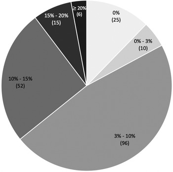
Results are presented in Figure 2. During the study period, 204 individual providers collected ≥10 blood cultures, with a mean of 32.2 blood-culture collections per collector. Also, 35 collectors (17.2%) maintained a contamination rate <3%, and 25 of these collectors had no contamination episodes. However, 73 collectors (35.8%) had an individual contamination rate >10%. The mean collector contamination rate was 8.2%.
Distribution of individual contamination rates among 204 collectors from the University of Arkansas for Medical Sciences Emergency Department during the study period. Data are from collectors with 10 or more collections.

Discussion
Across various studies, contaminated blood cultures have shown evidence of severe negative outcomes among patients: increased costs, inappropriate antibiotic usage, increased length of stay, and adverse outcomes. Reference Hall and Lyman2–Reference Souvenir, Anderson and Palpant6,Reference Self, Mickanin and Grijalva22–Reference van der, Yuri and Miller24 Our study is unique in that, to our knowledge, we have one of the largest data sets contributing to this growing volume of data, including 12,278 blood culture episodes with 1,012 contaminants over a 4-year study period. Our findings add to the body of evidence with sizeable power given the sample. Furthermore, we have provided evidence for risk factors predisposing patients to contamination while also highlighting the devastating clinical outcomes for patients with contaminated cultures. Additionally, our observations regarding collector contamination rates reveal a wide range of individual levels of efficacy in sterile technique during blood culture collection.
The patients most at risk for contamination were of older age, black race, higher BMI, and had comorbidities such as CHF, COPD, and paralysis. Black patients were disproportionately at increased risk for blood-culture contamination (aOR, 1.32; 95% CI, 1.15–1.51), whereas white patients demonstrated a protective trend. Our data suggest that patients considered “difficult sticks” for venipuncture had higher rates of contamination, which is consistent with common risk factors previously reported in the literature such as older age, presence of comorbidities, and severity of illness. Reference Bates, Goldman and Lee3,Reference Chih-Jan, Chi-Jung and Hsiang-Chin16 These differences in risk are likely due to the difficulty of the venipuncture: the darker skin of black patients makes isolating peripheral veins more difficult visually; obese individuals’ increased subepidermal fat masks peripheral veins; older individuals typically have more tenuous, collapsed, or fragile veins; and patients presenting with shock or severe sepsis have collapsed veins due to low blood pressure, which can create a sense of urgency that could contribute to poor technique or shortcuts in culture collection. When observing their blood culture collections, other researchers have noted that staff exhibit poor hand hygiene, poor antisepsis, or repalpate the veins without adequate sterilization, despite the presence of institutional standards. Reference Bowen, Coleman and Cunningham9,Reference Hall, Domenico, Self and Hain12 These practices do not seem unusual when considering how the difficulty of venipuncture differs from patient to patient.
We investigated the impact of code sepsis activation on blood-culture contamination rates. Upon code sepsis activation, several individuals are called to respond urgently to the bedside to obtain patient work-up and to administer treatments, which could promote poor blood-culture collection technique. In our study, activation of code sepsis led to more frequent blood-culture contamination (42.9% vs 34.8%), though this finding was not statistically significant in multivariate analysis (aOR, 1.16; 95% CI, 0.98–1.36). However, code sepsis was only in practice during a portion of our study, and this finding should be investigated further. It is not surprising that patients with sepsis plus shock have higher contamination rates, but the significance level of this finding is concerning because there is a high likelihood that these individuals have a true bacteremia contributing to their presentation. Their increased risk of contamination predisposes them to inadequate work-up and treatment, wasting valuable time and resources for their treatment, as shown in the literature and by our clinical outcomes.
The primary outcome of hospital LOS was significantly longer in patients with contaminated blood cultures than those with negative blood cultures (7.9 days vs 6.6 days; β = 1.24; P < .0001) after controlling for age, race, BMI, and presence of sepsis on presentation. This 24% increase in LOS is a significant finding; increased LOS places patients at increased risk of adverse events and adds to hospital costs. This trend is supported in the literature. Some have shown similar marginal increases, such as in Gander et al Reference Gander, Byrd, DeCrescenzo, Hirany, Bowen and Baughman7 who noted a 1-day increased median LOS (5 days vs 4 days). Others have suggested more dramatic increased LOS, such as Alahmadi et al, Reference Alahmadi, Aldeyab and McElnay5 who noted a 5.4-day average difference in LOS between cases and controls. Reference Hall and Lyman2–Reference Dawson4
Patients with contaminated blood cultures also had increased total antimicrobial exposure (6.2 days vs 5.2 days; P < .0001), more frequent vancomycin orders (81.3% vs 64.9%; P < .0001), and longer vancomycin DOT (3.5 days vs 2.5 days; P < .0001) compared to controls. The 25% vancomycin prescription increase and 40% DOT increase were significantly higher compared to control patients. These data support the findings reported in other studies, such as van der Heijden et al, Reference van der, Yuri and Miller24 who extrapolated nearly 293 extra vancomycin orders due to contaminated cultures, and Souvenir et al, Reference Souvenir, Anderson and Palpant6 who warned of the reflexive use of vancomycin in cases of coagulase negative staphylococci contamination contributing to increased costs and antibiotic misuse. Antibiotic resistance has become an ever-growing concern, particularly with the emerging threat of vancomycin-resistant Enterococcus. Reference Hall and Lyman2,Reference Dawson4,Reference Souvenir, Anderson and Palpant6 Unnecessary use of antibiotics, such as vancomycin, predispose patients to adverse effects such as AKI and increased patient and hospital costs, reinforced both by the literature and the data presented herein. Reference Bates, Goldman and Lee3,Reference Dawson4,Reference Souvenir, Anderson and Palpant6,Reference van der, Yuri and Miller24 In our study, acute kidney injury (36.7% vs 26.3%; aOR, 1.60; 95% CI, 1.40–1.83) was more frequent in patients with contaminated cultures likely due to increased vancomycin exposure.
Strikingly, our study revealed an increase in rate of in-hospital mortality (8.0% vs 4.6%; aOR, 1.69; 95% CI, 1.31–2.16) in patients with blood-culture contamination. Although this association is hard to prove retrospectively, it was statistically significant in multivariate analysis adjusting for age, race, BMI, and sepsis status. The association between increase in LOS, antibiotic exposure, AKI rate, and procedures (including echocardiogram) with blood-culture contamination in our study likely leads to a more complicated hospitalization, especially in patients with the noted risks for contamination. This important finding stresses the need to mitigate unnecessary blood-culture contamination.
Lastly, in an unadjusted analysis, patients with blood-culture contamination in our study had a higher average hospital charge of an additional $7,132 compared to patients with a true-negative blood culture ($36,007 vs $28,874; P < .0001). After adjusting for age, race, BMI, and sepsis status, the inpatient costs were 24% higher in those with a contaminated blood culture than those without a contaminated culture (β = 0.22 ± 0.03; P < .0001). Our finding is similar to the $7,500 increased hospital costs reported by Alahmadi et al. Reference Alahmadi, Aldeyab and McElnay5
Our findings contribute to a volume of evidence that blood-culture contamination is a serious hurdle for adequate, efficient, and safe healthcare for patients. Various efforts have been studied to address this problem. Dedicated phlebotomy teams are a common suggestion that has strong evidence for efficacy in reducing not only contamination rates but also overall hospital costs. Reference Hall and Lyman2,Reference Dawson4,Reference Gander, Byrd, DeCrescenzo, Hirany, Bowen and Baughman7,Reference Garcia, Spitzer and Beaudry10,Reference Garcia, Spitzer, Kranz and Barnes11,Reference Self, Talbot, Paul, Collins and Ward23 Diversion devices, where blood is isolated into a separate chamber between venipuncture and collection of the specimens have also shown promise in reducing contamination rates, particularly in a study by Bell et al Reference Bell, Bogar, Plante, Rasmussen and Winters8 that showed an 82.8% reduction in contamination and projected $641,792 in savings over the course of their 7-month study. Reference Rupp, Cavalieri, Marolf and Lyden14 Some have even reported changes as simple as educating proper technique, providing a report card, and instituting institutional standards for blood-culture collection showing significant reductions in their overall contamination rates. Reference Bowen, Coleman and Cunningham9–Reference Ramirez, Gordón and Cortes13,Reference Zimmerman, Assous, Yinnon and Wiener-Well15 These findings are of particular interest to our institution given our findings of wide variation in blood-culture contamination rates between individual collectors. The results of our study were provided to a multidisciplinary quality improvement team consisting of nurses, physicians, pathologists, microbiologists, and others to address collector rates >10%, leading to enhanced education and feedback for individual collectors.
Our study has several limitations. First, this study was a retrospective, case-control study, and we were unable to standardize or validate every finding. Our data collecton was limited to the data recorded in the electronic medical record. This aspect of our study also makes drawing direct conclusions from the data difficult because we were unable to confirm our suspicions regarding culture technique. However, the volume of data analyzed adds power to the data despite this limitation. Second, inpatient units were excluded, and evaluating a single department of a single institution lessens the generalizability of our conclusions. However, this study is the largest date on blood-culture contamination risk-factor analysis and clinical outcome analysis. Additionally, our data appear to correlate well with other single-institution emergency-department studies in the literature. A third limitation is the lack of a true gold standard for determining contaminant blood culture from true bacteremia, which makes the study of this phenomenon difficult. Although similar studies have relied on manual chart review and clinical determination of “contaminant” versus “true bacteremia,” our study relied on the guidelines in place within the microbiology laboratory. In some cases, clinical bacteremias may have been categorized as contaminants and some true contaminants may have been categorized as true bacteremias. However, our process of identifying contaminants followed the national standards proposed by the Clinical Laboratory Standards Institute and was consistent throughout the study, removing subjectivity. Additionally, the evaluation of code sepsis was limited to years 2–4 of the study because this practice was not yet in place during year 1. To account for this, analysis of this variable only included patients during the period when code sepsis was in practice. Finally, our results showed that patients with severely ill presentations had increased risks of contaminated blood culture and, subsequently, patients with contaminated blood cultures had worse outcomes. Severity of illness at presentation may contribute to the worse outcomes noted in this study. However, these outcomes remained statistically significant, even when controlling for sepsis status, age, BMI, and race.
In conclusion, blood-culture contamination remains an ever-present problem in routine medical practice. This large risk factor and clinical outcome study provides important data that can be used by institutions to support changes in practice. Mitigating unnecessary blood-culture contamination will require novel interventions and culture change to help reduce the negative burden that this essential medical test carries.
Acknowledgments
We give special thanks to Dr Robert McGehee, Dr Brett Bailey, Dr Randy Maddox, and Ms Kim Gates for support during this study. Data for the study were provided by the Arkansas Clinical Data Repository (AR-CDR) maintained by the Department of Biomedical Informatics in the the College of Medicine at the University of Arkansas for Medical Sciences (UAMS). The AR-CDR is approved to operate as an enterprise data resource supporting research across the UAMS. Data in the AR-CDR were obtained from the UAMS electronic medical record, tumor registry, billing, and cancer genomic data and compromises encounters since May 1, 2014.
Financial support
Justin Klucher received 2 grants during this investigation: $3,000 from the UAMS College of Medicine and Translational Research Institute and an additional $4,000 from the Infectious Disease Society of America as a recipient of the Grants for Emerging Researchers and Mentorship.
Conflicts of interest
All authors report no conflicts of interest relevant to this article.
Supplementary material
To view supplementary material for this article, please visit https://doi.org/10.1017/ice.2021.111