Introduction
The clinical electroencephalograph (EEG) is a completely noninvasive and relatively inexpensive test of brain function that is readily and widely available, even in out-of-the-way medical centers. Despite decades of publications attesting to the role of the clinical EEG in diagnosing and managing psychiatric disorders, the procedure remains highly underutilized in the practice of psychiatry. The understanding of its use is central for neuropsychiatry. Given that the value of any diagnostic test lies in the balance between its cost and degree of risk to patients (including inconvenience) and the clinical importance of the information it yields, we will marshal evidence in this review that the cost/benefit ratio of obtaining EEG testing (particularly when properly guided) tips heavily in favor of the test. We will also speculate on the reasons why the clinical EEG is underutilized in psychiatry.
The visually inspected EEG (vEEG), frequently referred to as a standard EEG, can detect various forms of abnormalities, each with its own clinical significance. In brief, vEEG-detected abnormalities can be paroxysmal, meaning the abnormality appears and disappears, or is stationary (i.e., is present throughout a certain state of vigilance, usually wakefulness). For decades, psychiatrists thought, but were never sure, that paroxysmal EEG discharges without overt, conventionally defined seizures (i.e., isolated epileptiform discharges [IEDs]) may have such behavioral consequences as emotional lability, irritability, or temper dyscontrol.Reference Shelley, Trimble and Boutros 1 This remains the case today. The acronym IED is employed throughout this manuscript to denote the occurrence of epileptiform discharges in the complete absence of clinically diagnosable seizures. This is a distinction from interictal discharges in epileptic individuals. While there are no studies directly comparing the two phenomena, interictal discharges are capable of getting propagated and leading to seizure activity while IEDs are not. This difference may not be meaningful, but until studies are conducted, we prefer to be specific as regards to which phenomenon we are alluding. The most important unanswered question remains the value of detecting IED activity in a nonepileptic psychiatric patient in predicting favorable responses to anticonvulsant treatment.Reference Boutros, Kirollos, Pogarell and Gallinat 2
Slowing of the EEG is also seriously underinvestigated in psychiatric patients. The extent of medical follow-up of abnormal screening EEGs secured from psychiatric patients, particularly those reporting slow-wave abnormalities as a single finding, varies widely.Reference Struve and Boutros 3 The practicing neuropsychiatrist must be thoroughly familiar with what the EEG can detect in general, what the clinical implications are, and also the shortcomings of vEEG testing. The clinician must also be able to make a judgment, based on the clinical report, as to whether or not the test performed was adequate to the question asked.
The vEEG can be profitably complemented by computer quantification of the EEG (QEEG) signal.Reference Coburn, Lauterbach, Boutros, Black, Arciniegas and Coffey 4 This added dimension is now readily available, as all new EEG systems are digital. QEEG could soon become the standard EEG method applied in psychiatry due to its rather huge promise.Reference Coburn, Lauterbach, Boutros, Black, Arciniegas and Coffey 4 – Reference Grossi, Olivieri and Buscema 7
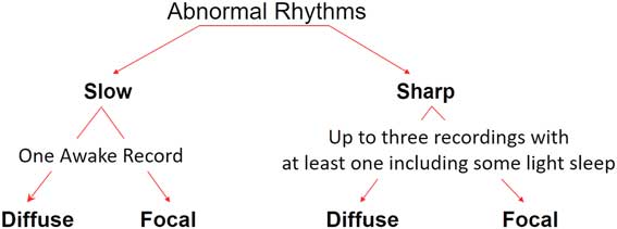
Figure 1 depicts the two major forms of EEG abnormalities (paroxysmal and slowing). The diagram stresses the fact that, if the expected abnormality is slowing of the EEG (focal or generalized), then a single recording during full wakefulness is usually sufficient. On the other hand, when the suspected abnormality is likely to be paroxysmal in nature, where it is not infrequent that a single recording can be falsely negative, repeat recording (perhaps with sleep deprivation or more intense activation procedures) may be necessary. It should be stated here that the optimal number or duration of recordings for maximal yield has not been established for any psychiatric condition.
The two major forms of EEG abnormalities (paroxysmal and slowing).

Role of Epileptiform Activity in Psychiatric Conditions
Episodic psychiatric symptoms are not uncommon and include panic attacks, repeated violent acts (particularly when seemingly unmotivated), dissociative episodes, some cases of autism spectrum disorders, as well as some forms of rapid cycling mood disorders.Reference Boutros, Bowyer, Wang, Urfy and Loeb 8 A number of single-case reports have also suggested that some cases of borderline personality disorder may in fact be epilepsy spectrum disorder (ESD) (see Boutros et al.Reference Boutros, Torello and McGlashan 9 for a review). Some evidence has accumulated over the years that at least in a subset of patients exhibiting these symptoms there may be evidence for the presence of focal cortical/subcortical hyperexcitability,Reference Boutros, Galloway, Ghosh, Gjini and Bowyer 10 the same form of abnormality underlying most forms of epilepsy.Reference Elisevich, Shukla and Moran 11 In these cases, the condition could be conceptualized as an ESD with significant treatment implications.Reference Boutros, Bowyer, Wang, Urfy and Loeb 8 There is currently no clear demarcation of this category of symptoms or their prevalence. How these symptoms occur, what would be an appropriate workup, and possible treatments are all answerable research questions.Reference Boutros, Bowyer, Wang, Urfy and Loeb 8
The ESD concept does remain controversial. It does not imply that ESDs are epileptic disorders, though they may share some manifestations like episodic clinical manifestations and responsiveness to some antiepilepsy drugs (AEDs), particularly those with mood-stabilizing properties.Reference Boutros, Kirollos, Pogarell and Gallinat 2 In order to diagnose such a condition as epilepsy, the behavioral episodes need to be a representation of one of the recognized seizure manifestations.Reference Engel 12 Hence, the proposition that the IEDs may be contributing physiologically to the observed behavior but in fact not causing seizures may be a more appropriate designation and that an ESD designation would be more applicable. The detection of an IED in a nonepileptic psychiatric patient with episodic or treatment-unresponsive symptoms may be important for a full biopsychosocial conceptualization of the patient’s condition.
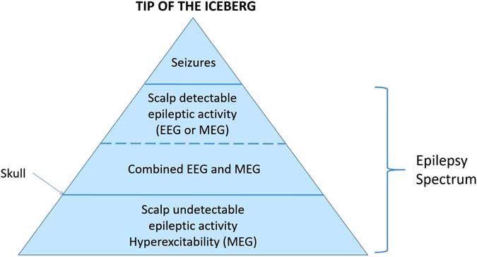
Figure 2 depicts the concept of ESD. As depicted, epilepsy sits on the top of a pyramid of decreasing degrees of cortical excitability. The wide bottom of the pyramid is the “hypothesized” large populations of psychiatric patients exhibiting paroxysmal behaviors who are neither epileptic nor exhibit frank paroxysmal activity on their EEGs or magnetoencephalograms (MEGs) and who may be harboring regions of focal hyperexcitability. EEG alone or in combination with magentoencephalography (see below) can help in detecting cases of ESD in the second or third tier of Figure 2 but not as of yet the cases within in the wide bottom tier of the pyramid. It has been known for many years that subcortical EEG changes occur in the absence of cortical abnormalities.
The concept of ESD. Clinical epilepsy sits on top of a pyramid of decreasing degrees of cortical excitability. The wide bottom of the pyramid is the “hypothesized” large populations of psychiatric patients exhibiting paroxysmal behaviors who may be harboring regions of focal hyperexcitability.

Panic disorder (PD) is a special condition where the clinical manifestations of the attacks can in fact be a reflection of epileptic activity (like in fear auras) or ESD, a not infrequent challenging clinical differential diagnostic issue.Reference Boutros, Gjini, Moran, Chugani and Bowyer 13 A number of reports have been published suggesting that in some treatment-refractory PD patients AEDs may be useful.Reference Boutros, Gallinat and Pogarell 14 Currently, use of AEDs in PD patients is either reserved for patients with demonstrable epileptic activity or as a last resort when nothing else has worked.Reference Toni, Cassano and Perugi 15 , Reference Hoffman and Mathew 16 In fact, Hoffman and MathewReference Hoffman and Mathew 16 did not list AEDs among possible treatment alternatives for PD.
A similar situation exists in individuals with repeated violent episodes. As early as the mid-1940s, it was recognized that criminals had a higher prevalence of EEG abnormalities. Among psychiatric populations, the group of “psychopaths” had the largest incidence of either borderline or frank abnormalities, which consisted mainly of diffuse background slowing in unmedicated patients and/or paroxysmal activity.Reference Hill and Watterson 17 These authors concluded that the more aggressive the patient, the more likely that the EEG would be abnormal.Reference Hill and Watterson 17 Despite the passing of seven decades, this work remains undisputed and seminal. As is strongly argued in the present review, these different EEG abnormalities signify widely different neuropathologies and should never be lumped together under “abnormal EEGs.” The sparse subsequent research further supports and elaborates on the above observations, suggesting that habitual aggressors exhibit a significant increase in EEG abnormalities as compared to nonhabitual aggressors,Reference Williams 18 with more frontal region abnormalities but more diffuse and epileptic activity in the nonhabitual aggressors. Furthermore, 70% of patients who have committed violent offences against strangers tended to have bilateral paroxysmal EEG discharges.Reference Howard 19
Autistic Spectrum Disorders
A recent review of vEEG abnormalities in autistic spectrum disorders (ASDs) (in the absence of epilepsy) reveals the well-documented high rate of deviations in this population.Reference Boutros, Lajiness-O’Neill, Zillgitt, Richard and Bowyer 20 The most notable observation is that, despite the strong available evidence for significant EEG abnormalities in this population, EEG is hardly ever utilized to work up these individuals, and, even more importantly, there has been absolutely no research in this area in more than two decades.
Whether or not the presence of IEDs predicts a positive response of psychiatric symptoms to anticonvulsants is a wide open research question. A stronger correlation (or larger effect size) between the presence of IEDs and good clinical response to AEDs would be strong evidence that the detected IEDs are in fact contributing to the clinical features being treated. The reverse cannot be said to constitute strong evidence against the hypothesis. Nonetheless, if studies show both a strong link between the presence of IEDs and good clinical response to AEDs as well as a strong link between the absence of IEDs and a lack of clinical response to AEDs, then one can propose that current EEG technology is adequate and that the false negative rate is not a significant factor (see below).
Studies attempting to answer this question in ASD children (this being a good population in which to try this, as IEDs are relatively common, thus obviating the need to screen a very large number of patients) have provided some encouraging data. Gillberg and SchaumannReference Gillberg and Schaumann 21 described two cases of infantile autism without clinical seizures where EEG abnormalities were not discovered until relatively late in the course of the psychiatric disorder. Anticonvulsant medications led to the complete disappearance of psychotic symptoms and to simultaneous disappearance of the pathological EEG changes. Hollander and his groupReference Hollander, Dolgoff-Kaspar, Cartwright, Rawitt and Novotny 22 conducted a retrospective pilot study to determine whether divalproex sodium is effective in treating the core dimensions and associated features of autism. They included 14 patients with either autism, Asperger’s syndrome, or pervasive developmental disorder. Subjects were included irrespective of having had a history of seizures or EEG abnormalities. Some 10 of the 14 patients who completed the trial (71%) were rated as responsive to treatment (mean dose=768 mg/day, range=125–2500 mg/day). Improvement was noted in the core symptoms of autism as well as in the associated features of affective instability, impulsivity, and aggression. All patients with abnormal EEGs were rated as responders. Chez and colleaguesReference Chez, Chang, Krasne, Coughlan, Kominsky and Schwartz 23 used valproic acid to treat 176 ASD patients with epileptic discharges. In follow-up EEGs (10 months later, on average), 46.6% had normalized, and an additional 17% showed improvement. None had worsened. The authors concluded that a more proactive approach to evaluation and treatment is justified, as some of the abnormalities may be reversible. These studies need to be set alongside the fact that epilepsy develops in some cases of ASD during the teenage or early adult years, changing the management and care needs of such patients considerably. A more recent studyReference Swatzyna, Tarnow, Turner, Roark, MacInerney and Kozlowski 24 strongly supported the contribution of vEEG to the practice of personalized medicine in ASD children.
Cognitive Problems
Frye et al.Reference Frye, Butler, Strickland, Castillo and Papanicolaou 25 retrospectively reviewed the charts of 22 children with atypical cognitive development who did not respond to standard educational therapy and demonstrated discharges on EEG. Only 9% of the sample had a history of seizures, but the majority of children had multifocal discharges on their EEGs. Of the 20 patients subsequently treated with antiepileptic medications, 70% demonstrated definite improvement by the next clinic visit. This study suggested that children with EEG discharges and developmental cognitive disorders demonstrate an identifiable pattern of symptomatology and discharges on EEG that respond well to AED treatment. It also demonstrated that monitoring in an epilepsy-monitoring unit is significantly superior to routine outpatient vEEGs in detecting paroxysmal discharges. Larsson et al.Reference Larsson, Bakke and Bjørnæs 26 provided evidence from a placebo-controlled double-blind crossover study that antiepileptic drugs (in this case, levetiracetam) can significantly decrease the frequency of epileptic discharges in these individuals irrespective of whether or not they had manifest epilepsy.
The above studies support the need for a more proactive approach to evaluation and suggest that treatment is justified as some of the physiological abnormalities may be reversible, a conclusion supported by the work of Pressler et al.Reference Pressler, Robinson, Wilson and Binnie 27 who showed that treatment of interictal epileptiform discharges can improve behavior in epileptic children with behavioral problems. Despite the above reports, a review by TharpReference Tharp 28 concluded that there is no justification for use of anticonvulsants when epilepsy cannot be clinically diagnosed. A confident answer to this question relies almost solely on the ability to reliably detect paroxysmal activity, even when rare, small-in-amplitude, or emanating from deeper brain structures that are behaviorally salient.
The behavioral effects of IEDs appear to be region-dependent, as hippocampal spikes are associated with cognitive disruption,Reference Kleen, Scott, Holmes and Lenck-Santini 29 and spikes during neonatal development impair reference memory and long-term potentiation in rodents as adults.Reference Khan, Zhao, Miller and Holmes 30 IEDs emanating from the amygdalar region would be expected to correlate with fear manifestations (as in panic attacks).Reference Gallinat and Hegerl 31
Slowing of the EEG
As stated above, the extent of medical follow-up of abnormal EEGs of psychiatric patients, particularly those reporting slow-wave abnormalities (both focal or generalized) as the sole finding, varies widely. In a retrospective review of screening EEGs for psychiatric inpatients in a state hospital, 103 consecutive cases of abnormal EEGs with generalized, focal, or paroxysmal slowing as the only EEG finding were identified. Despite suggestions for medical follow-up, less than half (44.6%) of the patients received subsequent study. However, 74.2% of the patients considered to be without organic pathophysiology at the time of the EEG had such positive organic findings on medical follow-up.Reference Struve and Boutros 3 Prominent among these findings were unsuspected brain atrophy and ventricular enlargement, neoplasms or vascular abnormalities, epilepsy, and unsuspected toxicity.Reference Struve and Boutros 3 A focal slow-wave abnormality should be alarming to the clinician, as it could signify a focal lesion. The nature and consequences of the focal slowing must be verified in order to perform a proper biopsychosocial assessment and devise proper development of an informed management and/or rehabilitation plan.
Delirium is the most common clinical syndrome that leads to consulting by a psychiatrist in general hospitals.Reference Brown 32 The fifth edition of the Diagnostic and Statistical Manual of Mental Disorders (DSM–5) recognizes three possible clinical presentations: hyperactive, hypoactive, and mixed (being the most common presentation). Hypokinetic delirium is the most likely to be missed and perhaps where an EEG would be most useful. 33 Hypokinetic delirium also frequently occurs in psychiatric patients due to a metabolic encephalopathy developing in the course of treatment with neuroleptics, particularly when anticonvulsants are used in combination with various typical and atypical neuroleptics.Reference Tonkonogy and Geller 34 All the diagnostic symptoms of delirium can be witnessed in acute psychosis.Reference Tonkonogy and Geller 34 In clinical states where the accurate assessment of sensorium is not possible, differentiation between delirium and acute psychosis may be impossible based on clinical evaluation only. Engel and RomanoReference Engel and Romano 35 emphasized that EEG was by far the most sensitive and reliable indicator of delirium. The concurrent altered mental state and the slowing of background EEG activity provided solid evidence for delirium. As Engel and Romano showed, follow-up of slow-wave activity reveals changes that mirror the clinical state. This can be invaluable, especially when there are diagnostic challenges about the diagnosis of delirium and concerns that the mental state may be deteriorating. Finally, The DSM–5 recognizes an “attenuated delirium syndrome.” The role of EEG in detecting this syndrome is yet to be defined.
Furthermore, patients who are unable or unwilling to participate in clinical evaluation for the assessment of their mental status may pose a significant challenge to clinicians. The role of the EEG in evaluating such patients has not been fully evaluated. Javanbakht et al.Reference Javanbakht, Amirsadri, Arfken, Dewald and Boutros 36 included two groups of psychiatric emergency room patients—15 patients with difficult-to-assess mental status (DAMS) and 15 patients oriented to time, place, and person who were seen in the same psychiatric emergency room—and 10 healthy controls. An abbreviated 16-channel EEG (AbEEG, allowing for determination of the dominant background rhythm) recording was performed for all subjects. Four subjects in the DAMS and none in the other patient and healthy control groups exhibited background rhythms strongly suggestive of a delirium. The preliminary data suggest that clinical examination of a DAMS person in the psychiatric emergency room may benefit from EEG utilization. These limited data also show that EEGs can be obtained in an acute psychiatric setting.
Shortcomings of the vEEG
A critical issue is the ability of a scalp EEG to detect IEDs. Current guidelines for an outpatient EEG recording vary widely from as short as 15 to as long as 120 minutes.Reference Cascino, Trennerry and So 37 Of major importance is the need to attain stages I and II sleep when the purpose of the test is to identify IEDs. Sleep deprivation has been shown to increase the yield for paroxysmal activity over and above assuring the recording of EEG during sleep.Reference Leach, Stephan, Salveta and Brodie 38 Most EEG studies in psychiatric populations where IEDs are sought are usually comprised of a single recording, with a lack of IEDs simply equated with the absence of IEDs. In fact, only 29–56% of bona fide epileptic patients will have an IED on a single recording of 30-minute duration.Reference Goodin and Aminoff 39 By repeating the EEGs up to six times, the yield can reach 82%.Reference Marsan and Zivin 40 Losey and Uber-ZakReference Losey and Uber-Zak 41 examined the time to first IED in extended recordings from patients suspected of having epilepsy. They reported that 47% of all IEDs occurred later than the 20-minute duration currently officially recommended. At least 11% of their records did not yield an IED until after 40 minutes, which is the basis for recommending that the minimal recording duration in psychiatric patients, when IEDs are suspected, should be no shorter than 60 minutes. This recommendation is supported by Chochoi and colleagues,Reference Chochoi, Tyvaert, Derambure and Szurhaj 42 who provided additional evidence that longer recordings (beyond the required 20 minutes) were necessary to detect paroxysmal activities. In case of a negative study, a repeat following an all-night sleep deprivation is recommended with a longer duration of 2 to 3 hours.Reference Losey and Uber-Zak 41 Improving technology so that smaller IEDs at the cortex can be detected at the scalp has not occurred. In the absence of pressure applied from practicing psychiatrists and neuropsychiatrists, such efforts will not be forthcoming.
The disadvantages of visual analysis of the EEG include significant questions about reliability. This problem is compounded by the lack of widely accepted normal standards developed specifically for psychiatric populations.Reference Boutros, Mirolo and Struve 43 The overwhelming majority of studies delineating the boundaries of normality of EEG have lacked control for most psychiatric disorders, and most conspicuously for personality disorders and substance abuse.Reference Boutros, Mirolo and Struve 43 Finally, EEG abnormalities commonly do not reflect specific pathological processes. For example, diffuse slowing of the EEG can result from a wide range of pathologic processes (metabolic or degenerative) that are capable of causing encephalopathy, and slowing can be viewed as a “final common pathway” of EEG cerebral dysfunction. The EEG, nonetheless, can be useful in evaluating the degree of severity of the ongoing pathologic process. The EEG is thus a good positive test, as the presence of IEDs would constitute strong evidence for focal hyperexcitability but is not a good negative test (i.e., it cannot be used as strong grounds to rule out the presence of focal hyperexcitability).
Future Directions
Controversial sharp waves
The so-called “controversial sharp waves or spikes” deserve specific mention (for a more expanded discussion, please refer to Hughes and WilsonReference Hughes and Wilson 44 or HughesReference Hughes 45 for examples of these patterns). Five EEG patterns were consistently observed to be more prevalent in psychiatric populations than either healthy or nonpsychiatric patient control populations.Reference Boutros 46 Despite the increased prevalence in psychiatric patients, defining the patterns’ exact neurobiological basis and clinical correlates has proven to be an elusive goal. It should be noted that none of the following patterns predictably correlate with an epileptic disorder. Recent advances in the ability to localize the neural source generators of activity recorded on the scalp can be illuminating once the clinical correlates of these controversial patterns are elucidated. Further probing into the clinical correlates of these EEG patterns can be facilitated by an increased flow in psychiatric EEG recordings and the availability of advanced localization and prolonged monitoring facilities.
We list the names of these waveforms without discussion due to space limitations, but the reader can find more expanded discussions in BoutrosReference Boutros 46 : the 14- and 6-Hz positive spikes,Reference DeLong, Rosenberger, Hildreth and Silver 47 the 6-second spike and wave discharges,Reference Boutros, Hughes and Weiler 48 the small sharp spikes (SSS),Reference Hughes and Gruener 49 the rhythmic midtemporal discharges,Reference Boutros, Hughes and Weiler 48 , Reference Anderson and Vanderspek 50 and the Wicket spikes.Reference Krauss, Abdallah, Lesser, Thompson and Niedermeyer 51 Of particular interest is the noted association between the presence of the SSS and affective disorders.Reference Small 52 , Reference Small, Milstein and Medlock 53 Even more intriguing is the possible association between SSS and suicidality.Reference Struve, Saraf, Arko, Klein and Becka 54 For a more comprehensive review of available literature related to these controversial waveforms, the interested reader is referred to Boutros.Reference Boutros 46 At a minimum, the EEGer providing the interpretation must be trained in detecting these phenomena and must report them when present. It must be left up to the referring clinician to decide if they want to take any action based on their own knowledge of the phenomenon.
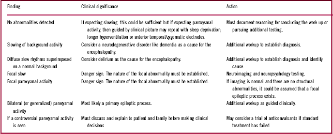
Table 1 lists the EEG abnormalities frequently seen in psychiatric patients, their potential clinical significance, and the further actions that may be necessary. It goes without saying that, while retaining a high degree of suspicion, clinicians should have a reason for ordering the EEG and must document any results and any subsequent action based on those results.
EEG abnormalities frequently seen in psychiatric patients, clinical significance, and further action

Why is the vEEG underutilized in psychiatry?
It is likely that the diversion between psychiatry (moving away from being brain-based and evidence-based) and neurology (rejecting any notion without strong evidential support) has caused the field of clinical electroencephalography to move away from psychiatry and find a secure home in neurology departments.Reference Boutros 55 , Reference Boutros 56 In addition, the following factors are likely to have contributed to the current underutilization of EEG in psychiatric practices. Perhaps most important is the lack of exposure of trainees (i.e., psychiatry residents) to clinical EEG during training. The complete unavailability of clinical neurophysiology fellowships for psychiatrists (at least in the United States) further decreases the interest of psychiatry trainees to learn about this technique. The lack of studies addressing the issue of clinical correlates and the therapeutic implications of each type of EEG abnormality in the various psychiatric populations further places a significant distance between daily practice and the EEG laboratory. This knowledge gap and the paucity of clinical use of EEG is one that should be addressed in any neuropsychiatric training program.
The Development of Newer Technologies Allowing Better Detection of IEDs
Among the newer technologies, magnetoencephalography holds special promise for better detection of epileptic-like activity, particularly from deeper structures. Recently, it became apparent that the coregistration of electrical and magnetic data yields complementary information. Furthermore, recent advances in the field of epilepsy surgery have led to identification of regions of abnormally elevated focal coherence as indicative of regions of hyperexcitability.Reference Elisevich, Shukla and Moran 11 A recently developed and tested new methodology—coherence imaging–to assess neural coherence within neural tissue (as contrasted to coherence assessed at surface electrode recording locations)Reference Bowyer 57 promises to be able to identify brain regions of focal hyperexcitability that may be contributing to episodic behavior and qualifying as an ESD (the wide bottom of the pyramid in Figure 2). The proposition that increased focal coherence may be a reflection of hyperexcitability and perhaps reflective of an ESD was recently tested in a limited dataset of panic disorder patients.Reference Boutros, Galloway, Ghosh, Gjini and Bowyer 10 A readily testable hypothesis is whether or not the presence of focal hyper-coherence (as detected by MEG) predicts a favorable clinical response to AEDs, particularly if the focus is located within behaviorally salient brain regions (e.g., frontal or limbic structures).
Conclusions
Although the existing EEG literature is replete with reports of abnormalities in association with neuropsychiatric disorders, only a few generalizations can be made about particular EEG patterns and disorders. The strong (and relatively straightforward) correlation between EEG abnormalities and epilepsy has overshadowed the more complex relationship between EEG abnormalities and psychiatric disorders. The increased recognition, especially with inpatient EEG monitoring of patients with difficult-to-manage seizure disorders, that nonepileptic seizures are much more frequent than recognized previously needs to be addressed. Many have been diagnosed as having epilepsy on the basis of an abnormal EEG, which corresponds to some of the wave patterns discussed above, a diagnosis that was not only wrong, but dangerous. Many get sucked into an epilepsy environment, taking anticonvulsants in increasing doses, and the underlying psychopathology worsens, as does the frequency of their attacks. Moreover, the prevailing concept of “not treating the EEG” led to further deemphasizing of such EEG deviations. A well-established fact is that the incidence of EEG abnormalities, both slow-wave abnormalities and epileptiform discharges, are higher in psychiatric populations as compared to control groups.Reference Shelley, Trimble and Boutros 1 The implications and understanding of the IEDs have continued to evolve with research into the genetic and metabolic bases of these events,Reference Noebels 58 intracranial electrophysiology, and the relationship of epileptiform discharges (EDs) with neuropsychological functions.Reference Aarts, Binnie, Smit and Wilkins 59 Accumulating data thus has begun to shift the older concept of the interictal EDs (in epileptic patients) away from that of electrophysiological events that are not associated with cognitive or behavioral consequences to events that are the phenotypic expression of a variety of cellular disorders.Reference Fisch 60
We thus conclude by strongly suggesting that the presence of IEDs in nonepileptic individuals with psychiatric symptoms should be treated as a variant/covariant in electrophysiological studies. The same cannot be as positively stated for the absence of IEDs. Nonetheless, if adequate EEG workups were performed (including awake and asleep EEGs, ambulatory EEG or MEG) with negative results, including an individual in a no-IED group would be justifiable by today’s available technology.
Finally, it is not known whether currently available seizure medications are effective in normalizing hyperexcitable brain tissue that has not yet become capable of inducing seizures. Scattered reports suggest that a few of these medications may have some efficacy in this regard, but further research is needed to examine these efficacies, particularly in newly diagnosed ASD patients.Reference Boutros, Lajiness-O’Neill, Zillgitt, Richard and Bowyer 20 The use of some anticonvulsants in the management of mood instability and bipolar disorders reinforces the overlap between electrophysiological variables, anticonvulsant medications, and regulation of mood.
As all of the above-raised issues in this review are significantly more pertinent to psychiatry, and especially neuropsychiatry, as compared to neurology, it seems reasonable to recommend that clinical electrophysiology laboratories be established in every academic psychiatry department.Reference Pogarell, Hegerl and Boutros 61 This change would allow for teaching trainees, providing a clinical service closer to where the patients actually reside (particularly if housed in an out-of-the way state psychiatric facility).Reference Boutros 62
Disclosures
Dr. Boutros hereby declares that he has no conflicts of interest to disclose.