




No CrossRef data available.
Article contents
Trichinella papuae and Trichinella zimbabwensis induce infection in experimentally infected varans, caimans, pythons and turtles
Published online by Cambridge University Press: 03 March 2004
Abstract
The discovery of Trichinella zimbabwensis in farm crocodiles of Zimbabwe has opened up a new frontier in the epidemiology of the Trichinella genus. The objective of the present study was to investigate the infectivity of encapsulated species (T. spiralis, T. nativa, T. britovi, T. murrelli and T. nelsoni) and non-encapsulated species (T. pseudospiralis, T. papuae and T. zimbabwensis) in caimans (Caiman crocodilus), varans (Varanus exanthematicus), pythons (Python molurus bivittatus) and turtles (Pelomedusa subrufa) raised at their natural temperature range (26–32 °C). Mice and chickens were used as controls. At 6 days post-infection (p.i.), adult worms were detected in the small intestine of reptiles infected with T. papuae and T. zimbabwensis, of chickens infected with T. pseudospiralis and of mice infected with all encapsulated and non-encapsulated species. At 60 days p.i., T. papuae and T. zimbabwensis adult worms were collected from the intestine of varans and caimans and larvae from muscles of the four reptile species, T. pseudospiralis larvae from muscles of chickens, and larvae of all Trichinella species from mouse muscles. The highest reproductive capacity index of both T. papuae and T. zimbabwensis was observed in varans. The results show that T. papuae and T. zimbabwensis are able to complete their entire life-cycle in both poikilothermic and homoiothermic animals.
Keywords
Information
- Type
- Research Article
- Information
- Copyright
- 2004 Cambridge University Press
INTRODUCTION
For 145 years following their discovery (Owen, 1835), nematodes of the Trichinella genus were considered to infect only mammals (Campbell, 1988). In 1980, a non-encapsulated species, Trichinella pseudospiralis, was found to also infect birds (Shaikenov, 1980). In 1995, non-encapsulated larvae belonging to a new species, Trichinella zimbabwensis, were discovered in farm crocodiles (Crocodylus niloticus) of Zimbabwe (Pozio et al. 2002), representing the first report of naturally infected reptiles. Although attempts have been made to infect reptiles with all 5 encapsulated Trichinella species (T. spiralis, T. nativa, T. britovi, T. murrelli and T. nelsoni) and with T. pseudospiralis, all of these attempts failed when the animals were bred at their natural temperature range (i.e. 29–30 °C) (Kapel et al. 1998), whereas in snakes, lizards and turtles, the life-cycle of T. spiralis was successfully reproduced when the animals were bred at 37 °C (i.e. the body temperature of mammals) (Pozio, 2001).
The objectives of the present study were to evaluate the infectivity of all Trichinella species, the development of the nurse cell and the critical point of development in experimentally infected reptiles belonging to the orders Loricata, Squamata and Chelonide.
MATERIALS AND METHODS
Experimental animals
Tropical carnivorous reptiles were used, specifically: 15 spectacled caimans (Caiman crocodilus, synonymous of Caiman sclerops L., 1758), a species originating from Central and South America, age 6–8 months, weight 350–550 g; 15 savannah monitors (referred to as ‘varans’) (Varanus exanthematicus Bosc, 1792), a species originating from equatorial Africa, age: 2–3 years, weight 300–450 g; 40 Burmese pythons (Python molurus bivittatus Kuhl, 1820), a species originating from Southeast Asia, age 6–8 months, weight 350–850 g; and 40 African helmeted turtles (Pelomedusa subrufa Lacépède, 1788), a species originating from equatorial Africa, age about 2 years, weight 50–100 g. All animals were born and raised in captivity. Caimans and pythons were kept 1 to a terrarium, whereas varans and turtles were kept 2 to a terrarium. The terrariums for caimans and turtles consisted of 2/3 water and 1/3 dry substratum. The terrariums for pythons and varans had a water tank and a substratum consisting of small pieces of cluster-pine bark. All animals were kept under a day/night rhythm of 12/12 h. Pythons and turtles were kept at 28–30 °C during the day and at 26–28 °C at night; caimans and varans were kept at 30–32 °C during the day and at 28–30 °C at night (the temperature refers to the air). During the study, the body temperature of reptiles was measured every 2 weeks. Since the source of heat was not inside the terrarium, the body temperature was, in all cases, quite similar to the air temperature of the terrarium. Pythons and varans were fed laboratory mice; turtles were fed fish or meat; and caimans were fed fish. The frequency of feeding depended on the specific species. Forty Swiss CD1 female mice weighing 25 g and 32 two-week-old chickens weighing 80 g were used as controls. Animals were housed and treated according to the European directive 8/609 EEC.
Experimental infections
Eight Trichinella reference strains were used to infect the animals. Of these 5 were encapsulated species: T. spiralis (code ISS3), T. nativa (code ISS10), T. britovi (code ISS2), T. murrelli (code ISS35) and T. nelsoni (code ISS29); and 3 were non-encapsulated species: T. pseudospiralis (code ISS13), T. papuae (code ISS572) and T. zimbabwensis (code ISS1029).
Larvae were collected from skinned and eviscerated mouse carcasses by artificial digestion, following a protocol slightly modified from Pozio (1987). The digestive fluid consisted of 0·5% hydrochloric acid, 0·5% pepsin 1[ratio ]10000, and PBS at 40 °C. Mouse carcasses were cut into small pieces and minced for 30 s in a blender with the digestive fluid at a concentration of 1[ratio ]40 (w/vol). The mixture was then incubated at 39–40 °C, stirring constantly, for either 15 min (for non-encapsulated species) or for 30 min (for encapsulated species). Muscle larvae were then allowed to sediment at 39–40 °C for 15 min, and the supernatant was discarded. The muscle larvae were washed 4 times with warm PBS and counted 3 times under a dissection microscope at a magnification of 20–40×.
The reptiles and chickens were inoculated per os with 3000 larvae/animal. A 2 ml syringe connected to a silicone stomach tube was used for varans, pythons, and caimans, whereas, for turtles and chickens, the syringe was connected to a gavage needle. For varans, pythons, and caimans, a small aluminium bar with a hole in the middle was used to keep the animal's jaws open and prevent the stomach tube from being crushed. The control mice were inoculated per os with 500 larvae/animal using a 2 ml syringe connected to a gavage needle. To ensure that all of the larvae entered the stomach, the stomach tube was flushed with water following larva inoculation. The animals of each species were divided into groups of 5. The turtles, pythons, chickens, and mice were inoculated per os with the 8 Trichinella species; the varans and caimans were inoculated per os with T. papuae, T. zimbabwensis and T. nelsoni. For each group of 5 animals, 2 specimens were killed 6 days post-infection (p.i.); the other 3 specimens were killed 60 days p.i.
Worm collection from the intestine
The reptiles were killed by hypothermia, whereas the mice and chickens were killed by CO2 inhalation to collect intestinal worms at 6 and 60 days p.i. To facilitate the recovery of adult worms from the intestine, the reptiles were not fed during the week prior to their killing and the chickens and mice were not fed for 1 day prior to sacrifice. The intestine was collected, opened longitudinally, and incubated in PBS at 37 °C for 2 h, following a previously published protocol (Blair, 1983). Worms were searched for in the sediment under a microscope at a magnification of 20–40×; they were counted 3 times and then stored in 70% ethyl alcohol.
Muscle larvae collection
At 60 days p.i., the reptiles, chickens and mice were skinned and eviscerated. For the turtles, pythons and mice, the entire carcass was digested separately for each animal, whereas for the chickens, varans and caimans, muscles were collected from the bones, which were also scraped with a scalpel to collect the largest possible amount of muscle to allow for the identification of predilection muscles. For these latter animals, to determine the preferential muscles, digestion was conducted separately for single muscles or muscle groups, according to the protocol reported above. For all animals, the larvae were collected, counted 3 times, and stored in 70% ethyl alcohol for the morphological and molecular studies. The reproductive capacity index (RCI) (i.e. the number of larvae collected from muscles divided by the number of larvae given per os) was evaluated for each animal. In total, 100 larvae of each of the Trichinella species which had succeeded in developing in varans and caimans were given per os to Swiss CD1 mice immunosuppressed with cyclophosphamide (4 mg/mouse) at 0 and 4 days p.i., to evaluate their infectivity. At 40 days p.i., these mice were killed, skinned, and eviscerated; the entire carcass was digested and the larvae were counted.
Worm morphology and the ultrastructure of the nurse-cell larva complex
To assess the influence of the host on worm morphology, the total length and width at the midbody was measured for 20 adult worms (10 females and 10 males) collected from the small intestine of varans, caimans and mice at 6 days p.i. and for 20 muscle larvae collected from varans, caimans and mice at 60 days p.i. A descriptive univariate statistical analysis (mean and standard deviation) and Student's t-test (two-tailed) were used to evaluate the statistical differences in the size of muscle larvae and adult worms.
For the histological and ultrastructural analyses of the nurse cell-larva complex, small pieces of intercostal muscle were collected from varans, caimans, and mice at 60 days p.i. The muscle samples were fixed in 0·1 M cacodylate buffer (pH 7·2) containing 2·5% glutaraldehyde for 4 h at 4 °C. The samples were then washed in the same buffer and post-fixed in 1% OsO4 in cacodylate buffer for 1·5 h at 4 °C. All samples were dehydrated in ethanol and embedded in Epon 812 for sectioning. For light-microscopy, semi-thin sections (0·5 μm) were stained with 0·5% toluidine blue and examined under a Zeiss photomicroscope III. Thin sections (80 μm), stained with uranyl acetate and lead citrate, were examined under a Zeiss EM900 transmission electron microscope (TEM).
Molecular identification of adult worms and larvae
To confirm that the adult worms and larvae that were collected from the animals belonged to the same species used for experimental infection, a multiplex PCR analysis (Zarlenga et al. 1999) was used, according to a previously published protocol (Pozio & La Rosa, 2003).
RESULTS
Detection of intestinal worms
At 6 days p.i., in mice, we detected live adult worms (i.e. both male worms and females with developed embryos in the uterus) belonging to all 8 Trichinella species, whereas in chickens, only T. pseudospiralis worms were found (Table 1). For all reptile species, only live worms of T. papuae and T. zimbabwensis, probably at the adult stage (i.e. no well developed embryo detected in the uterus), were detected. In turtles and pythons, a few dead L1 larvae belonging to T. spiralis, T. murrelli and T. nelsoni were detected (Table 1).

At 60 days p.i., live adult worms (including females with embryos in the uterus) were detected only in varans and caimans. A mean of 138 T. papuae worms (range 98–187) and 127 T. zimbabwensis worms (range: 62–165) were detected in varans; a mean of 85 T. papuae worms (range 39–124) and 79 T. zimbabwensis worms (range 18–131) were detected in caimans.
Detection of larvae in muscles
At 60 days p.i., larvae of all 8 Trichinella species were detected in mice, with an RCI ranging from 1·2 for T. papuae to 80·5 for T. spiralis; larvae were also detected in chickens infected with T. pseudospiralis and in all reptile species infected with T. papuae and T. zimbabwensis (Table 2). In reptiles, the RCI was higher for T. papuae than for T. zimbabwensis. The RCI was highest for varans (41·4 for T. papuae) and lowest for turtles (0·05 for T. zimbabwensis) (Table 2). In none of the reptile species were larva of any encapsulated species or of the non-encapsulated species T. pseudospiralis detected.
Table 2. Average larvae per gram (lpg), standard deviation (S.D.) and reproductive capacity index (RCI) in the muscles of three specimens of each host species (for chickens only two specimens were examined) 60 days post-infection (For turtles, pythons and mice, lpg refers to the weight of the entire skinned and eviscerated carcass; for varans, caimans and chickens, lpg refers to the weight of most of the striated muscles.)

Preferential muscles were studied in varans and caimans. In both animals, the tongue was the most infected muscle, followed by muscles of the anterior legs, of the posterior legs, muscles of the tail, intercostal muscles, and muscles of the spinal column (Table 3).

For neither T. papuae nor T. zimbabwensis were significant differences in the size of the intestinal worms observed when comparing varans, caimans, and mice 6 days p.i. The muscle larvae of T. papuae and T. zimbabwensis detected in mice 60 days p.i. were significantly smaller than those detected in varans and caimans at 60 days p.i. (Table 4).

The degenerative patterns observed in the muscles of caimans and varans were very similar when comparing T. papuae with T. zimbabwensis. At 60 days p.i., in caimans, larvae were enclosed in muscle fibres, which were disarranged in transversal sections, as revealed by light microscopy. Neither a nurse cell-like structure nor a fibrous envelope was observed around the larva-muscle complex (Fig. 1A). The TEM micrographs revealed that the structure of the muscle was altered: the typical fibrous structure had disappeared and several vacuoles (i.e. degenerate mitochondria) were present (Fig. 1B and C). As a consequence of the sarcomer dissociation, clusters of highly degenerated mitochondria were coalesced around the larva (Fig. 1D). The degenerative events in the muscle cells of infected varans were quite similar to those observed in caimans. In varans, light microscopy revealed larvae enclosed in a disarranged fibrous structure (Fig. 2A). Neither a nurse cell-like structure nor inflammatory cell infiltrates were observed. The TEM micrographs showed an interruption in the continuity of the fibrous architecture: clusters of dissociated muscle fibres and vacuoles (i.e. degenerate mitochondria) were present (Fig. 2B and C). In the muscle, where sarcomers were totally desegregated, a large number of mitochondria were coalesced (Fig. 2D). In some mitochondria, the matrix was partially conserved, whereas in others it was totally vacuolated (Fig. 2D).

Fig. 1. Larvae of Trichinella zimbabwensis in muscles of a caiman 60 days post-infection. (A) Longitudinal semi-thin section showing an intracellular larva (l) enclosed in a disarranged muscle fibre (mf). Neither a nurse-cell-like structure nor fibrous bundles surrounding the larva-muscle complex are present. (B) TEM micrograph showing the loss of the contractile texture of the muscle fibre (mf) and the presence of vacuolated mitochondria (arrows). l, Larva. (C) Higher magnification of the infected muscle cell. Vacuolated mitochondria (asterisks) and residual bodies (arrows) are present. l, Larva. (D) Clusters of highly degenerated mitochondria (m) surrounding the larva (l).

Fig. 2. Larvae of Trichinella zimbabwensis in muscles of a varan 60 days post-infection. (A) In the semi-thin section the larva (l) is enclosed in the disarranged muscle fibre (mf). mc, Normal muscle cell. (B) TEM micrograph showing the discontinuity of the muscle fibre (mf) architecture. Several vacuolated mitochondria (arrows) are present. l, Larva. (C) Higher magnification showing remnants of the muscle fibre (mf) and clusters of degenerate and vacuolated mitochondria (arrows). l, Larva. (D) In the region where the sarcomers are totally lacking, clusters of mitochondria (m) surround the larva (l). Note that the matrix of some mitochondria is beginning to become vacuolated (arrows).
In mice infected with T. papuae and T. zimbabwensis, radical changes had occurred in the microarchitecture of the muscle fibre around the muscle larvae at 60 days p.i. Specifically, the muscle larvae appeared to be enclosed in a fibrous structure delimited by an irregular coat. As expected for non-encapsulated species, the collagen capsule that is typical to encapsulated species was not present, and no infiltration of inflammatory cells was observed around the infected muscle fibres (Fig. 3A). The TEM micrographs showed a nurse-cell in which myofilaments were totally disarranged and interspersed with clusters of vacuolated mitochondria, small vesicles, tubules containing electron-dense material and lamellar bodies (Fig. 3B). In some samples, the texture of the muscle cell around the intracellular larvae appeared to be partially conserved (Fig. 3C). In the outer zone of the nurse-cell, a thin layer of collagen fibres was observed (Fig. 3C). In some degenerate muscle cells, clusters of collagen fibres were scattered in the nurse-cell (Fig. 3D).

Fig. 3. TEM micrographs of larvae of Trichinella zimbabwensis in muscles of mice 60 days post-infection. (A) The muscle fibres that surround the larva (l) appear partially disarranged. Remnants of myofilaments (arrows) are interspersed with clusters of mitochondria (m). At the periphery of the collagen fibres (f), the texture of the muscle cell (mc) appears to be partially conserved. (B) The fibre cells are totally modified and the contractile elements are replaced by cisternae (c) of the endoplasmic reticulum. The mitochondria (m) appear to be swollen and vacuolated and the matrix has totally disappeared. (C) Nurse-cell-like structure (nc) showing a hypertrophic nucleus (n) with the nucleolus (arrow head) in the transcriptional phase. Note the clusters of small mitochondria (asterisks) and the rough endoplasmic reticulum (arrows). l, Larva. (D) Details of the nurse-cell-like structure showing clusters of small mitochondria (m) and the endoplasmic reticulum (arrows).
The multiplex PCR analysis of adult worms and muscle larvae collected from the different host species confirmed that the parasites belonged to the Trichinella species used to infect them. At 40 days p.i., CD1 mice inoculated with larvae of T. papuae and T. zimbabwensis collected from the muscles of the four reptile species were positive (RCI=8·4 for T. papuae and RCI=25·3 for T. zimbabwensis in immunosuppressed mice), indicating that the larvae present in reptile muscles at 60 days p.i. were infective.
Clinical signs
In reptiles, it is very difficult to evaluate the clinical signs of disease, although it is known that the trophic activity and the moulting process are good indicators of health. In our study, the varans stopped eating for about 2 weeks, beginning at around 2 weeks after infection, which could correspond to the invasive phase of the larvae in muscles. None of the other reptiles showed any changes in trophic activity. In none of the animals were decreases in agility observed, not even in varans, which harboured up to 3000 larvae per g of muscle.
DISCUSSION
The present study shows that both T. papuae and T. zimbabwensis establish as infective muscle larvae in reptiles. This biological characteristic is consistent with the biochemical and molecular findings of these two Trichinella species and with their capacity to cross between them and to produce a viable but not reproductive F1 generation in a murine model (Pozio et al. 2002).
In a previous study, a nurse cell-like structure with a thin envelope of collagen fibres at the periphery of the muscle-larva complex had been observed in the muscles of a crocodile experimentally infected with T. zimbabwensis 18 months earlier (Pozio et al. 2002). In the present study, a period of 60 days appeared to be insufficient for the complete development of the nurse cell-larva complex in caimans and varans, whereas this time-period was sufficient in mice. These results suggest that the infective larvae of T. papuae and T. zimbabwensis produce the same degenerative patterns in the muscle cells of both cold-blooded and warm-blooded animals yet that the rapidity at which the changes in the muscle-fibre architecture occur is strongly influenced by the physiology of the vertebrate group.
In caimans and varans, at 60 days p.i., infective L1 larvae of T. papuae and T. zimbabwensis were detected in muscle cells, yet the TEM micrographs showed that these cells had not completed their development into a nurse cell. These findings suggest that the development of newborn larvae into infective L1 larvae occurs independently of the changes in the structure of the muscle cell and that this development does not require the formation of the nurse cell. By contrast, these events appear to be closely correlated in homoiothermic animals (Despommier, 1998).
The finding that the muscle larvae of both T. papuae and T. zimbabwensis were smaller in mice than in reptiles at the same times post-infection suggests that larva size is an adaptive character related to the host species. Perhaps the greater size of the larvae in reptiles is related to the slower development of the nurse cell in these poikilothermic animals, which would allow more time for the larvae to grow.
The lack of clinical signs in varans and caimans, together with the high levels of infection, is consistent with the hypothesis that these animals act as potential reservoirs of T. papuae and T. zimbabwensis. Reptiles belonging to the Varanidae and Crocodilidae families are carnivores with scavenger behaviour, which is the most important biological and ecological character of animals known to act as Trichinella reservoirs (e.g. fox, racoon dog, wolf, bear and hyena) (Campbell, 1988). In light of these findings, reptile meat should not be excluded as a potential source of human trichinellosis. The very low number of T. papuae and T. zimbabwensis larvae detected in pythons and turtles, in spite of a very high infecting dose, seems to suggest that these species do not play an important role in the epidemiology of Trichinella. This is consistent with the trophic spectrum of pythons and turtles, which probably does not include Trichinella carriers. Nonetheless, that the worm burden in turtles and pythons was low could have been related to the temperature at which these animals were raised (26–30 °C), which is 2 degrees lower than the temperature used for varans and caimans (28–32 °C).
Experimental infections of reptiles with T. spiralis have been attempted several times, yet the success of these experiments depended on the temperature at which the animals were raised. Specifically, Texas horned lizards (Phrynosoma cornutum) were not successfully infected at their activity temperature (20–28 °C), yet viable larvae were recovered from the muscles of these lizards when the animals had been kept at 37 °C for 1 month (Jordan, 1964). Similarly, the European turtle (Emys orbicularis) (Guevara Pozo & Contreras-Pena, 1966), the Caucasian Agama (Agama caucasica) (Asatrian, Movsessian & Gevorkian, 2000) and the long-nosed viper (Vipera ammodytes) (Cristea & Perian, 1999) were successfully infected with an encapsulated species of Trichinella (probably T. spiralis) only when kept at 37 °C. Encapsulated species (T. spiralis, T. nativa, T. britovi, T. murrelli and T. nelsoni) and one non-encapsulated species (T. pseudospiralis) have been tested in caimans raised at 29 °C without success (Kapel et al. 1998). Among other cold-blooded vertebrates, attempts to infect 5 species of fresh water fish, raised at 18–22 °C, with T. britovi failed (Moretti et al. 1997).
These data, together with molecular (Zarlenga et al. 2004) and biochemical data (La Rosa, Marucci & Pozio, 2004), suggest that temperature plays a role in the speciation mechanism of Trichinella. Specifically, the non-encapsulated species T. papuae and T. zimbabwensis can develop at temperatures ranging from 26 °C to 40 °C, both in cold- and warm-blooded animals (i.e. tropical reptiles and mammals); the non-encapsulated species T. pseudospiralis can develop at temperatures from 37 °C to 42·5 °C (i.e., in homoiothermic animals, specifically, mammals and birds); and all of the encapsulated species develop at 37–40 °C (i.e. only in mammals) (Table 5). The presence in the genus Trichinella of different species which infect reptiles and mammals or birds and mammals or only mammals strongly suggests that this nematode group is ancient and that it evolved with the evolution of reptiles into mammals and birds (i.e. from poikilothermic to homoiothermic vertebrates).

Finally, to the best of our knowledge, T. papuae and T. zimbabwensis are the only two parasites known to complete their entire life-cycle independently of whether the host is homoiothermic or poikilothermic. This seems to indicate that these two Trichinella species are capable of activating different physiological mechanisms, according to the specific vertebrate class.
We are grateful to Mr Claudio Bagnoli for his assistance in raising the reptiles. This work was in part funded by the project ‘TRICHIPORSE’ of the European Union, Contract QLK1-CT-2001-01156.
References
REFERENCES
ASATRIAN,
A.,
MOVSESSIAN,
S. &
GEVORKIAN,
A.
(2000).
Experimental infection of some reptiles with T. spiralis and T. pseudospiralis. Abstract Book, p. 154, The 10th International Conference on Trichinellosis, 20–24 August, Fontainebleau, France.
BLAIR, L. S.
(1983).
Laboratory Techniques. In Trichinella and Trichinosis (ed. Campbell, W. C.), pp. 563–570.
Plenum Press,
New York and London.CrossRef
CAMPBELL, C. W.
(1988).
Trichinosis revisited, another look at modes of transmission.
Parasitology Today 4, 83–86.CrossRefGoogle Scholar
CRISTEA, G. &
PERIAN, A.
(1999).
Experimental infestation with Trichinella larvae in vipers.
Revista Romana de Parasitologia 9, 45.Google Scholar
DESPOMMIER, D. D.
(1998).
How does Trichinella spiralis make itself at home?
Parasitology Today 14, 318–323.Google Scholar
GUEVARA POZO, D. &
CONTRERAS-PENA, J. A.
(1966).
Trichinella spiralis (Owen 1835), experiencias de infestacion en diversos vertebratos.
Revista Iberica de Parasitologia 26, 239–288.Google Scholar
JORDAN, C. A.
(1964).
Experimentally trichinized Texas horned lizards.
Journal of Parasitology 50, 24.Google Scholar
KAPEL, C. M.,
WEBSTER, P.,
BJORN, H.,
MURRELL, K. D. &
NANSEN, P.
(1998).
Evaluation of the infectivity of Trichinella spp. for reptiles (Caiman sclerops).
International Journal for Parasitology 28, 1935–1937.CrossRefGoogle Scholar
LA ROSA, G.,
MARUCCI, G. &
POZIO, E.
(2004).
Biochemical analysis of encapsulated and non-encapsulated species of Trichinella (Nematoda, Trichinellidae) from cold- and warm-blooded animals reveals a high genetic divergence in the genus.
Parasitology Research (in press).Google Scholar
MORETTI, A.,
PIERGILI FIORETTI, D.,
PASQUALI, P.,
MECHELLI, L.,
ROSSODIVITA, M. E. &
POLIDORI, G. A.
(1997).
Experimental infection of fish with Trichinella britovi: biological evaluations. In
Trichinellosis
(ed. Ortega Pierres, G.,
Gamble, R.,
Van Knapen, F. &
Wakelin, D.), pp. 135–142.
Centro de Investigacion y Estudios Avanzados del Instituto Politecnico Nacional, Mexico, D.F. Mexico.
OWEN, R.
(1835).
Description of a microscopic entozoon infesting the muscles of the human body.
Transactions of the Zoological Society of London 1, 315–323.Google Scholar
POZIO,
E. (1987).
Isoenzymatic typing of 23 Trichinella isolates. Tropical Medicine and Parasitology 38, 111–116.Google Scholar
POZIO, E.
(2001).
New patterns of Trichinella infections.
Veterinary Parasitology 98, 133–148.CrossRefGoogle Scholar
POZIO, E.,
FOGGIN, C. M.,
MARUCCI, G.,
LA ROSA, G.,
SACCHI, L.,
CORONA, S.,
ROSSI, P. &
MUKARATIRWA, S.
(2002).
Trichinella zimbabwensis n.sp. (Nematoda), a new non-encapsulated species from crocodiles (Crocodylus niloticus) in Zimbabwe also infecting mammals.
International Journal for Parasitology 32, 1787–1799.Google Scholar
POZIO, E. &
LA ROSA, G.
(2003).
PCR-derived methods for the identification of Trichinella parasites from animal and human samples.
Methods in Molecular Biology 216, 299–309.Google Scholar
POZIO, E.,
OWEN, I. L.,
LA ROSA, G.,
SACCHI, L.,
ROSSI, P. &
CORONA, S.
(1999).
Trichinella papuae n.sp. (Nematoda), a new non-encapsulated species from domestic and sylvatic swine of Papua New Guinea.
International Journal for Parasitology 29, 1825–1839.Google Scholar
SHAIKENOV, B. S.
(1980).
Spontaneous infection of birds with Trichinella pseudospiralis Garkavi, 1972.
Folia Parasitolica 27, 227–230.Google Scholar
ZARLENGA, D. S.,
CHUTE, M. B.,
MARTIN, A. &
KAPEL, M. O.
(1999).
A single multiplex PCR for unequivocal differentiation of six distinct genotypes of Trichinella and three geographical genotypes of Trichinella pseudospiralis.
International Journal for Parasitology 29, 1859–1867.CrossRefGoogle Scholar
ZARLENGA, D. S.,
LA ROSA, G.,
POZIO, E. &
ROSENTHAL, B.
(2004).
Identification and classification within the genus Trichinella, with special emphasis on non-encapsulated species.
Veterinary Parasitology (in press).Google Scholar

Table 1. Number of adult worms of Trichinella collected from the small intestine of two specimens for each host species 6 days post-infection
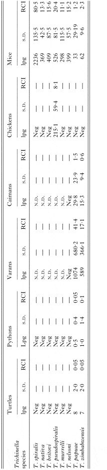
Table 2. Average larvae per gram (lpg), standard deviation (S.D.) and reproductive capacity index (RCI) in the muscles of three specimens of each host species (for chickens only two specimens were examined) 60 days post-infection

Table 3. Average number of Trichinella larvae per g of muscle in three varans and three caimans infected with T. papuae or T. zimbabwensis

Table 4. Morphological features and standard deviation (S.D.) of Trichinella papuae and Trichinella zimbabwensis larvae (L1) collected from muscles of mice, varans and caimans (all measurements are in μm) 60 days post-infection

Fig. 1. Larvae of Trichinella zimbabwensis in muscles of a caiman 60 days post-infection. (A) Longitudinal semi-thin section showing an intracellular larva (l) enclosed in a disarranged muscle fibre (mf). Neither a nurse-cell-like structure nor fibrous bundles surrounding the larva-muscle complex are present. (B) TEM micrograph showing the loss of the contractile texture of the muscle fibre (mf) and the presence of vacuolated mitochondria (arrows). l, Larva. (C) Higher magnification of the infected muscle cell. Vacuolated mitochondria (asterisks) and residual bodies (arrows) are present. l, Larva. (D) Clusters of highly degenerated mitochondria (m) surrounding the larva (l).
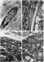
Fig. 2. Larvae of Trichinella zimbabwensis in muscles of a varan 60 days post-infection. (A) In the semi-thin section the larva (l) is enclosed in the disarranged muscle fibre (mf). mc, Normal muscle cell. (B) TEM micrograph showing the discontinuity of the muscle fibre (mf) architecture. Several vacuolated mitochondria (arrows) are present. l, Larva. (C) Higher magnification showing remnants of the muscle fibre (mf) and clusters of degenerate and vacuolated mitochondria (arrows). l, Larva. (D) In the region where the sarcomers are totally lacking, clusters of mitochondria (m) surround the larva (l). Note that the matrix of some mitochondria is beginning to become vacuolated (arrows).
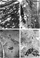
Fig. 3. TEM micrographs of larvae of Trichinella zimbabwensis in muscles of mice 60 days post-infection. (A) The muscle fibres that surround the larva (l) appear partially disarranged. Remnants of myofilaments (arrows) are interspersed with clusters of mitochondria (m). At the periphery of the collagen fibres (f), the texture of the muscle cell (mc) appears to be partially conserved. (B) The fibre cells are totally modified and the contractile elements are replaced by cisternae (c) of the endoplasmic reticulum. The mitochondria (m) appear to be swollen and vacuolated and the matrix has totally disappeared. (C) Nurse-cell-like structure (nc) showing a hypertrophic nucleus (n) with the nucleolus (arrow head) in the transcriptional phase. Note the clusters of small mitochondria (asterisks) and the rough endoplasmic reticulum (arrows). l, Larva. (D) Details of the nurse-cell-like structure showing clusters of small mitochondria (m) and the endoplasmic reticulum (arrows).
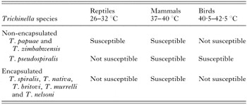
Table 5. Susceptibility of reptiles, mammals and birds to the eight Trichinella species and relationship between Trichinella species and the temperature range of the host
 You have
Access
- 62
- Cited by