Introduction
Unlike mammals, laying hens have a unique bone turnover synchronised with a daily egg laying cycle, indicating that rapid remodelling occurs in laying hen bones. Because of rapid bone turnover and extensive calcium mobilisation from bones for eggshell formation, bone fractures and osteoporosis in laying hens during the egg production are a concern in the poultry industry (Wilson and Thorp, Reference WILSON and THORP1998; Riczu et al., Reference RICZU, SAUNDERS-BLADES, YNGVESSON, ROBINSON and KORVER2004; Kim et al., Reference KIM, DONALSON, HERRERA, KUBENA, NISBET and RICKE2005 and 2007). Furthermore, bone related problems are directly related to economic parameters, such as egg production and eggshell quality. Thus, understanding bone metabolism in laying hens is important to maintain efficient production and improve animal welfare and minimise health issues. In this review, concepts and methods to understand bone metabolism in laying hens will be discussed.
Three types of laying hen bone
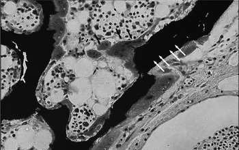
Avian females have a unique bone metabolism related to egg formation. Unlike mammals, female avian species have three distinctive regions of bones: cortical, cancellous, and medullary bones (van de Velde et al., Reference VAN DE VELDE, VERMEIDEN and BLOOT1985; Fisher and Schraer, Reference FISHER and SCHRAER1982; Kim et al., Reference KIM, DONALSON, STALLON, BLOOMFIEOLD, KUBENA, NISBET and RICKE2007). Cortical bone is a compact structural bone, which is tightly packed and highly organised. Cancellous bone, often synonymously called trabecular bone, is a three-dimensional lattice-shaped honeycomb architecture located in the end of bones (Odgaard, Reference ODGAARD1997). In contrast to cortical and cancellous bones, medullary bone is a special kind of woven bone lying in the marrow cavities of laying hens (van de Velde et al., Reference VAN DE VELDE, VERMEIDEN and BLOOT1985). A histological section of medullary bone of the single comb white Leghorn hen is shown in Figure 1. Medullary bone is a highly labile secondary bone which develops in sexually mature female avian females and is formed in response to oestrogen and androgen in the blood (Fisher and Schraer, Reference FISHER and SCHRAER1982).
Medullary bone of Single comb White Leghorn hen. White arrows are medullary bone. Black arrows are osteoclasts.

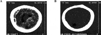
Elevated activities of osteoblasts and osteoclasts are found in medullary bone. Medullary bone matrix has higher proteoglycan content compared to cortical bone (Candlish and Holt, Reference CANDLISH and HOLT1971). Keratan sulphate is considered the primary glycosaminoglycan in medullary bone whereas chondroitin sulphate is the major glycosaminoglycan in cortical bone (Fisher and Schraer, Reference FISHER and SCHRAER1982). The bone density images of tibia from a normal hen and a nine-day feed withdrawal hen using peripheral quantitative computed tomography (pQCT) are shown in Figure 2.
The peripheral quantitative computed tomography (pQCT) images of normal hens (A) and 9-day feed withdrawal hens (B) (Kim et al., Reference KIM, DONALSON, STALLON, BLOOMFIEOLD, KUBENA, NISBET and RICKE2007).

Kim et al. (Reference KIM, DONALSON, STALLON, BLOOMFIEOLD, KUBENA, NISBET and RICKE2007) reported that tibiae of a normal hen contains dense medullary bone while medullary bone of a feed-withdrawal hen is absent, indicating that medullary bone is a labile component of bone and susceptible for bone resorption in laying hens. This bone is turned over according to a daily egg cycle and contributes 35 to 40% of the calcium for eggshell formation (Mueller et al., Reference MUELLER, SCHRAER and SCHRAER1964; Candlish, Reference CANDLISH1971; Buss and Guyer, Reference BUSS and GUYER1984). Furthermore, several studies have suggested that medullary bone may play a significant role in the development of osteoporosis in laying hens, which is one of the major animal welfare and economic issues (Wilson and Thorp, Reference WILSON and THORP1998; Wilson et al., Reference WILSON, SOLOMON and THORP1998; Cransberg et al., Reference CRANSBERG, PARKINSON, WILSON and THORP2001). There is an inverse relationship between the amount of medullary bone and cortical bone wherein the quantity of medullary bone increases at the cost of cortical bone during the egg production cycle (Wilson and Thorp, Reference WILSON and THORP1998; Whitehead and Fleming, Reference WHITEHEAD and FLEMING2000; Cransberg et al., Reference CRANSBERG, PARKINSON, WILSON and THORP2001). Thus, structural bone loss and the development of osteoporosis in laying hens are associated with the modelling and remodelling of medullary bone, which serves as a primary calcium source for eggshell formation (Wilson and Thorp, Reference WILSON and THORP1998; Cransberg et al., Reference CRANSBERG, PARKINSON, WILSON and THORP2001).
Bone resorption by osteoclast
Osteoclasts are bone-resorbing cells, which digest the inorganic and organic bone matrix by their secreted protons and proteinases, and subsequently translate the released ionized calcium from the bone into basolateral surface through the cytoplasm by endocytosis. These are large, multinucleated cells formed by the fusion of hematopoietic mononuclear precursors. Their developed cytoplasm is rich in mitochondria and lysosomal bone-digestive enzymes such as cathepsins and metalloproteinases. The characteristic features of the osteoclasts are that tartrate-resistant acid phosphatase (TRAP) activity is detected as a specific marker and that osteoclasts extend the fine projections of the plasma membranes, termed ruffled borders, at the apical surface (toward the bone surface). The ruffled borders are the specific areas where osteoclasts secrete protons and proteinases, and absorb the bone-released calcium and phosphorus. Ruffled borders are surrounded by a clear zone and the microenvironment of bone resorption is strictly maintained.
Medullary bone osteoclasts alternately cease and accelerate the bone resorption during the egg-laying cycle, and supply calcium for eggshell formation (Miller, Reference MILLER1992; Dacke et al., Reference DACKE, ARKLE, COOK, WORMSTONE, JONES, ZAIDI and BASCAL1993, Dacke, Reference DACKE2000; Sugiyama and Kusuhara, Reference SUGIYAMA and KUSUHARA2001). While the osteoclast population does not change during the 24-hour egg-laying cycle in chickens and quails, morphometric and ultrastructural studies demonstrate considerable changes in osteoclast structure that correlate with the changing calcium requirement during the egg-laying cycle (Miller, Reference MILLER1977, Reference MILLER1981; van de Velde et al., Reference VAN DE VELDE, VERMEIDEN, TOUW and VELDHUIJZEN1984b; Sugiyama and Kusuhara Reference SUGIYAMA and KUSUHARA1993, Reference SUGIYAMA and KUSUHARA1994a). When an egg is in the infundibulum, isthmus or magnum of the oviduct, osteoclasts lose the ruffled borders and bone resorption ceases. However, when an egg is in the shell gland of the oviduct and the egg begins to be calcified, osteoclasts develop ruffled borders and bone is resorbed for eggshell formation.
In soft-eggshell producing hens, the medullary bone osteoclasts lack ruffled borders, and do not supply the calcium for eggshell formation. For these hens, medullary bone matrix is highly calcified with irregular lamellar structures, as observed in cortical bone (Yoshiko et al., Reference YOSHIKO, KUSUHARA and ISHIDA1987, Reference YOSHIKO, KUSUHARA and ISHIDA1988). Therefore, the population and activity of osteoclasts represent the calcium supply for eggshell formation from medullary bone, and is widely used as a criterion of calcium metabolism of egg-laying hens (Fleming et al., Reference FLEMING, MCCORMACK, MCTEIR and WHITEHEAD2006; Fleming, Reference FLEMING2008).
In general, the modulation of osteoclast activities is mainly regulated by both the parathyroid hormone (PTH) and calcitonin (CTN) calcium-regulating hormones. PTH is an 88-amino acid polypeptide in its native form, and is secreted by chief cells of parathyroid glands in response to low plasma calcium levels. Van de Velde et al. (Reference VAN DE VELDE, LOVERIDGE and VERMEIDEN1984a) measured plasma PTH bioactivity during the chicken egg-laying cycle by a cytochemical bioassay. This is elevated during the period of eggshell calcification after which it falls to a low level. CTN is a polypeptide hormone of 32 amino acids, and CTN secretion is regulated primarily by rising plasma calcium levels leading to an increased secretion from the C cells of the ultimobranchial glands. In egg-laying quail, plasma CTN concentrations are at their highest after oviposition and fall as eggshell calcification proceeds, rising at the end of calcification (Dacke et al., Reference DACKE, BOELKINS, SMITH and KENNY1972; Dacke, Reference DACKE2000). Taken together, it is presumed that CTN would stop the osteoclastic bone resorption of medullary bone when an egg is in the magnum of the oviduct, and PTH would stimulate osteoclastic bone resorption for eggshell formation during the period when an egg is in the shell gland of the oviduct. In support of this presumption, the injection of PTH into hens during the period when an eggshell is not calcified causes the development of ruffled borders of osteoclasts, thus increasing plasma calcium levels (Miller, Reference MILLER1978). PTH in organ culture systems of chicken medullary bone stimulates the development of ruffled borders and the acid phosphatase activity of osteoclasts, and, conversely, CTN inhibits them (Sugiyama et al., Reference SUGIYAMA, OHASHI and KUSUHARA1993: Sugiyama and Kusuhara, Reference SUGIYAMA and KUSUHARA1994b).
Relationship between bone quality and egg production
Bone quality is closely related with egg production and eggshell quality. Osteoporosis in laying hens compromises egg production and shell quality. Cransberg et al. (Reference CRANSBERG, PARKINSON, WILSON and THORP2001) evaluated the relationship between body weight, egg production and skeletal abnormalities characteristics of osteoporosis at 45 weeks of age. The authors observed that laying hens showing severe osteoporosis experienced a body weight loss and an 18% decrease in egg production compared to healthy hens. Average body weight decline in laying hens was correlated with a 15 to 20% loss of skeletal calcium reserves and with the induction of osteoporosis. Because bone contributes substantial amounts of calcium in each eggshell, bone properties of laying hens would likely exhibit a negative correlation with eggshell qualities. Bishop et al. (Reference BISHOP, FLEMING, McCORMACK, FLOCK and WHITEHEAD2000) have characterised the hens selected for resistance or susceptibility to osteoporosis based on a bone index. The hens susceptible to osteoporosis had significantly higher eggshell thickness and better eggshell quality but lower bone quality compared to the hens selected for resistance to osteoporosis. This result suggests that the hens susceptible to osteoporosis have more active bone resorption to supply eggshell calcium. Kim et al. (Reference KIM, DONALSON, HERRERA, KUBENA, NISBET and RICKE2005) indicated that eggshell qualities were negatively correlated with bone qualities. Shell weight exhibited negative correlations with dry bone weight (-0.388; P < 0.01), ash weight (-0.305; P < 0.05), and percent ash (-0.450; P < 0.001). Percent shell also had negative correlations with bone, fresh weight (-0.467; P < 0.001), dry weight (-0.539; P < 0.001), fat-free dry weight (-0.354; P < 0.01), ash weight (-0.363; P < 0.01), and percent ash (-0.450; P < 0.001). Shell thickness was negatively correlated with fresh bone weight (-0.348; P < 0.05), dry bone weight (-0.452; P < 0.001), and ash weight (-0.281; P < 0.05).
Kim et al. (Reference KIM, FORD, MITCHELL, ELKIN and LEACH2004) have evaluated the bones of the restricted ovulator (RO) hens using histological analysis and dual-energy x-ray absorptiometry (DEXA). This was of particular interest because the RO hens could be a useful model for studying avian bone metabolism related to eggshell formation and egg production. The RO hens are generally unable to ovulate because of a point mutation in the oocyte VLDL receptor gene (Bujo et al., Reference BUJO, YAMAMOTO, HAYASHI, HERMANN, NIMPF and SCHNEIDER1995) which expresses a protein product that mediates the uptake of yolk precursors from the circulation (Nimpf and Schneider, Reference NIMPF and SCHNEIDER1991). Therefore, RO hens do not have the cyclic calcium metabolism associated with eggshell formation. There is a substantial increase in cortical bone thickness and the amount of medullary bone deposited in the bones from the RO hens when compared to the wild-type (WT). The intense toluidine blue staining of the medullary bone of both phenotypes is a reflection of the higher glycosaminoglycan content of this bone. Kim et al. (Reference KIM, FORD, MITCHELL, ELKIN and LEACH2004) also found that the RO hens had significantly higher humerus, femur, and tibia mineral contents and densities compared to the WT hens. These results support the concept that eggshell formation increases calcium mobilisation from bones and reduces bone quality in laying hens.
Methods to assess bone density, geometry, strength and cellular activities
Various methodologies are commonly available to quantify bone status in poultry. Bone ash fraction (g/g dry weight), bone mineral content (BMC) and bone mineral density (BMD; BMC normalised to some defined area), and bone mechanical properties are often used as indicators of bone mass and bone strength. Histological methods are required if it is necessary to assess bone cell activity on specific bone surfaces and compartments, and this can be critical in determining whether alterations in bone formation and/or bone resorption are the mechanisms underlying altered bone mass or strength.
bone ashing
Bone ashing literally yields, by burning off all organic material, the total mineral content of a given known dry weight of bone. Ash weight is a fundamental measure of bone mineral content, expressed most often as percentage fat-free dry weight. As such it is direct verification of the degree of mineralisation of the bone tissue itself independent of all porosities. This is its primary advantage over all the bone densitometry techniques discussed in the following section because the bone mineral density using bone densitometry techniques includes soft tissue (e.g. marrow) volumes as well as the multiple minute porosities in cortical bone in the volume measurement that provides the denominator for calculating bone mineral density. However, one can use the bone mineral content data (grams of mineral in the sample) derived from densitometry techniques to provide information similar to ash weight, but reproducibility will be negatively affected whenever any region of interest is selected that is less than the whole bone area.
Bone ash is strongly predictive of bone mineral density and of bone breaking strength, with correlation coefficients (r) of 0.92 (Zhang and Coon, Reference ZHANG and COON1997) and 0.77 (Hester et al., Reference HESTER, SCHREIWEIS, ORBAN, MAZZUCO, KOPKA, LEDUR M.C. and MOODY2004), respectively. The procedure is relatively simple, requiring only an ashing oven and a sensitive balance, and no special technical skills. However, bone ash cannot distinguish between cortical, cancellous, or medullary bone compartments, which may be important to study objectives.
densitometric techniques
Digitised fluoroscopy, dual energy X-ray absorptiometry (DEXA), and peripheral quantitative computed tomography (pQCT) are commonly used techniques in assessing bone mineral content (BMC) and bone mineral density (BMD). The first, digitised fluoroscopy, is an adaptation of radiographic absorptiometry in which the bird's bone is co-exposed with an aluminium step wedge to a single low-dose X-ray; the video output is captured from the image intensification system in fluoroscopy mode. The relevant quantitative data (radiographic density in mm Alumium equivalent) can be derived afterwards using NIH-Image software (Fleming et al., Reference FLEMING, MCCORMACK and WHITEHEAD2000). The primary advantages of this procedure are its relative low cost, the low radiation exposure involved, speed of data acquisition, and greater accessibility, since digitised fluoroscopy utilises equipment usually available in veterinary radiology departments and software available in the public domain. Digitised fluoroscopy can be performed relatively quickly on unanaesthetised animals (Fleming et al., Reference FLEMING, MCCORMACK and WHITEHEAD2000), and might be the method of choice if screening of large populations of birds is required. However, digitised fluoroscopy has a relatively low sensitivity and resolution, cannot correct for large variations in overlying soft tissue volume, nor separate out cortical, cancellous, and medullary bone compartments.
Dual energy x-ray absorptiometry, more commonly known by the acronym DEXA, is the gold standard for clinical use in diagnosing osteoporosis in human patients. Most absorptiometer manufacturers offer software that is marketed for use in small animals. DEXA can be used to assess both body composition (distinguishing lean and fat mass from bone mass) (Mitchell et al., Reference MITCHELL, ROSEBROUGH and CONWAY1997) and bone mineral content/bone mineral density (BMC/BMD) in small animals and, with some ingenuity, can be performed on unanaesthetised animals (Hester et al., Reference HESTER, SCHREIWEIS, ORBAN, MAZZUCO, KOPKA, LEDUR M.C. and MOODY2004). The use of photons of two different energy levels allows for the technique to be used on bone surrounded by large amounts of soft tissue, a key limitation of the earlier technique of single photon absorptiometry. The user defines a two-dimensional bone area of interest on the scan image. The total bone mineral detected is normalised to this planar area, and BMD expressed as mg/cm2; some researchers refer to this as ‘areal’ BMD, to distinguish it from true volume-based density measures. By dividing scanned bones during analysis into smaller regions of interest, one can assess mixed bone sites (e.g. metaphyses) versus cortical bone sites (mid-shaft regions of long bones). For laying hens, a region of interest can be specified (e.g. diaphysis of the humerus) where large fluctuations in BMD reflect gains and losses of medullary bone, such as those observed during induced moult (Hester et al., Reference HESTER, SCHREIWEIS, ORBAN, MAZZUCO, KOPKA, LEDUR M.C. and MOODY2004). The radiation exposure involved is minimal, and whole bones can be imaged; both of these factors represent distinct advantages over computed tomography (CT) techniques. Although full-size DEXA scanners are rather expensive, smaller versions designed to measure BMD in the human wrist are an appropriate size for many birds (pDEXA, Norland Medical Systems, Fort Atkinson, WI). To achieve acceptable reproducibility for research purposes, it is critical to standardise positioning of the bird's limb, because DEXA cannot correct for varying bone thickness within the designated region of interest. This latter challenge also limits the use of DEXA in rapidly growing animals, as absolute bone size will be dramatically different (Carter et al., Reference CARTER, BOUXSEIN and MARCUS1992). The resolution is also much lower than that achieved with computed tomography.
For more precise and detailed information about compartment-specific BMD and cross-sectional geometry, quantitative computed tomography scans offer tremendous improvements over DEXA. In particular, peripheral quantitative computed tomography (pQCT) has achieved more widespread use in small animal research over the last ten years, and minimises radiation exposure of vulnerable organs in the animal's core by delimiting the relatively exposure to limb bones (hence, the ‘peripheral’ in pQCT); the radiation dose is less than 0.1 mSv/hour (Gasser, Reference GASSER2003). During scans, an X-ray source and coupled detectors rotate around the animal's limb, collecting data in two dimensions, which converts to three dimensions after accounting for scan slice thickness (0.5 to 1.0 mm on most models). Although an entire bone can be scanned, this is time-consuming and prolongs anaesthesia time if collected in vivo; more often, the user will specify regions of interest and scan one to five consecutive slices. Resolution depends on the machine model and user choices, but can be as low as 70 microns. When scanning a mixed bone site (cortical plus cancellous or medullary bone), it is advisable to average results over two to four consecutive slices to minimise variability due to positioning error.
Absolute bone mineral content or density for the whole bone or for a region of interest specified by the user can also be measured. The resulting BMD values reflect a true volumetric value (vBMD, in mg/cm3); in addition, a wide variety of cross-sectional geometric variables can be computed. The most frequently used will be cross-sectional area and cross-sectional (polar) moment of inertia. Both of these have a critical impact on a bone's mechanical properties and resistance to fracture, and are required if one wishes to calculate material properties after conducting mechanical testing of bone specimens once harvested from the animal. Further, cross-sectional areas and BMC/vBMD variables can be computed separately for the cortical and cancellous (or medullary) bone compartments within the same scan slice. This may be of critical importance to answering experimental hypotheses about medullary bone in laying hens (Kim et al., Reference KIM, FORD, MITCHELL, ELKIN and LEACH2004, Reference KIM, DONALSON, MITCHELL, KUBENA, NISBET and RICKE2006; Mazzuco and Hester, Reference MAZZUCO and HESTER2005). For example, recent data demonstrate a selective reduction in BMD of medullary and metaphyseal cancellous bone during a nine-day moulting period, whereas there were no significant differences in cortical bone density over the same period (Kim et al., Reference KIM, DONALSON, STALLON, BLOOMFIEOLD, KUBENA, NISBET and RICKE2007).
mechanical testing of bone
Most of the aforementioned imaging techniques provide data on bone quantity (BMC, cross-sectional area) or bone quality (BMD) with the ultimate purpose of providing inferences about a key function of bone: its strength and resistance to fracture. When animals are euthanized at the end of an experiment, bones can be harvested for testing of various mechanical properties that subsequently provide the most convincing evidence about alterations in bone strength with the experimental intervention or animal husbandry practice in question. Performing these tests require specialized equipment (e.g. Instron or MTS systems) that can impose loads of known magnitudes at specific rates, interfaced with dedicated computers and software. These devices are standard equipment in any engineering school or testing facility; in addition, desk-top models for testing small specimens are available.
Long bones are typically subjected to three- or four-point bending or torsional tests, whereas vertebral bone or cylindrical specimens sampled from a long bone are tested in compression between two parallel platens. Most often these loads are applied ‘to failure’ (i.e. until the bone fractures). Hence, these are destructive tests but one can still determine ash weight of the samples if all bone fragments are retrieved. Data derived from load-displacement curves when testing whole bones are used to describe structural properties, most frequently, ultimate or fracture force and stiffness. The latter derives from the slope of the load versus displacement curve well before fracture occurs and is important to the bone's functional interaction with muscle in locomotion. If ultimate force and stiffness are normalised to the bone's size and cross-sectional geometry, the resulting variables (most commonly, ultimate stress and elastic modulus) then describe the bone tissue material (or intrinsic) properties independent of bone size. This is most critical when comparing animals of different ages or genders, where variations in bone size may mask important differences in the mechanical properties of the bone tissue itself. Even though larger bones are almost always stronger, they may have poorer material properties, as we previously demonstrated in aging rats (Bloomfield et al., Reference BLOOMFIELD, HOGAN and DELP2002). For more complete information, refer to a comprehensive tutorial by Turner and Burr (Reference TURNER and BURR1993).
histomorphometry
Histological analyses of bone micro-architecture and bone cell activity can provide valuable information in order to evaluate bone growth and development in avian models. Although micro-computed tomography scans can provide an assessment of cancellous bone microarchitecture and volume, this expensive technique may not be widely available. Under-mineralised bone specimens can be embedded in hard plastic resins, sectioned at 4- 8 microns, and then stained for mineralised bone (e.g. von Kossa or Goldner's); this is usually followed by a counterstain to highlight cells. If cancellous bone microarchitecture is of interest, the most commonly measured variables are bone volume per specified tissue volume (%BV/TV) and trabecular number, thickness, and trabecular spacing. With increasing %BV/TV, one usually observes increases in trabecular number and thickness and a corresponding decline in trabecular spacing; the opposite case applies with loss of cancellous bone. These analyses can be applied, of course, to medullary bone in avian models as well as to cancellous bone in the vertebrae or long bone metaphyses; both represent metabolically active bone that changes rapidly with endocrine or nutritional interventions (Wilson et al., Reference WILSON, SOLOMON and THORP1998; Kim et al., Reference KIM, FORD, MITCHELL, ELKIN and LEACH2004).
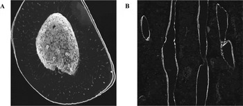
Histomorphometry remains the only method by which bone formation rate can be assessed in a site- and envelope-specific manner. Urine or blood biomarkers of bone resorption and formation are useful in indicating systemic (i.e. whole skeleton) alterations in osteoclast and osteoblast activity, but provide no information as to which skeletal site(s) are impacted. Fluorochrome labels (e.g. calcein, oxytetracycline) are administered by injection to the experimental animal on two separate days before sacrifice. We are unaware of any evidence that fluorochrome labelling is toxic or impacts on normal function; it does not affect body weight or feed consumption in laying hens (Hudson et al., Reference HUDSON, BRITTON, ROWLAND and BURHR1993). These fluorochromes bind to circulating calcium for 24-48 hours after the injection; any bone surface undergoing mineralisation during this period will capture the labelled calcium and exhibit a bright label when examined under epifluorescent light with the appropriate filters (Figure 3).
Fluorochrome labels in bones for measuring bone formation rate.

Our laboratory has confirmed that intraperitoneal injections of 5 to 10 mg calcein/kg body weight in mature poultry yield clearly visible labels. At sites capturing a label at both injection times, parallel double labels will be observed, permitting the assessment of inter-label widths. It should be noted that this labelling procedure is of lesser utility in very young animals, e.g. broiler chicks, as matrix formation and mineralisation are developing so rapidly that labelling yields a diffuse broad band. The average inter-label width over a specific bone envelope (periosteal, endocortical, or cancellous/medullary) defines mineral apposition rate (MAR, microns/day). MAR reflects the average activity of osteoblasts at focal sites with double labels, whereas percentage mineralising surface (calculated from the length of single- and double-labelled surfaces within a region of interest or on a particular bone envelope) indicates the number of osteoblast teams active during the labelling period. Bone formation rate is then a product of MAR and percentage mineralising surface and is specific to the bone surface and anatomical site measured (Parfitt, Reference PARFITT1983). Histomorphometric analysis can also be used to quantify bone growth and development with measurements of longitudinal growth and growth plate morphology. Measuring inter-label width of the calcifying front at the growth plate yields a longitudinal growth rate; in addition, growth plate area and height, hypertrophic chondrocyte zone height, and proliferative and hypertrophic chondrocyte number can be described (Ohashi et al., Reference OHASHI, ROBLING, BURR and TURNER2002). These measures are particularly critical when evaluating the impact of an intervention or husbandry practice on growth and development of young poultry.
Conclusions
Since skeletal problems in laying hens are important economic, welfare, and health issues for the poultry industry, better understanding of bone metabolism in laying hens is important to enhance productivity and improve animal welfare. In order to achieve this goal, the selection of proper methodologies is critical. Several methods, such as bone ashing, densitometric techniques, mechanical testing, or histomorphometry, can be used for measuring bone status in laying hens. However, we should understand that each method has its own advantages and disadvantages. It is necessary to adapt and develop new methodologies for evaluating bone quality in order to continuously improve our knowledge of bone metabolism in laying hens.
Acknowledgement
The authors would like to thank Dr. Bill Guenter in University of Manitoba for helpful suggestions and comments. This Review was supported by Manitoba Egg Farmer, Manitoba Agri-Food Research and Development Initiative (ARDI) and Natural Science and Engineering Council (NSERC)-Discovery.