Introduction
Although genomewide transcriptome studies are readily available for many tissues and cell types, functional annotation of the entire mammalian genome remains a daunting challenge. Several consortium-based genomewide knockout and mutagenesis projects are well underway, with the goal of generating mutant alleles for every gene in the mouse (Austin et al., Reference Austin, Battey and Bradley2004; Guan et al., Reference Guan, Ye, Yang and Gao2010). While these resources are immensely valuable and continue to produce data through large-scale phenotype screening efforts, usually they will not functionally assess genes during development other than to document lethality when an essential gene is abrogated. We have undertaken an RNAi-based screen during mouse preimplantation, aimed at identification of genes involved in epigenetic and developmental events during the first cell lineage decisions in vivo. During the initial phase of this screen we have identified many genes required for blastocyst formation. In an effort towards functional genome annotation, here we characterize four genes that are each essential during the morula to blastocyst transition in the mouse. This report documents the preimplantation requirement of Splicing Factor 3b, subunit 1 (Sf3b1 also called SAP155 and Sf3B155); 0610009D07Rik (hereafter called by the human orthologue gene name, Sf3b14, but also called HSPC175, Ht006, P14, SAP14, Sf3b14a)); Ribosomal RNA Processing 7 homolog A (S. cerevisiae, Rrp7a); and Ribosomal Protein L7-like 1 (Rpl7l1).
Sf3b1 has been shown in vitro to be an integral member of both the major and minor spliceosome and binds to both sides of the pre-mRNA branch site (Das et al., Reference Das, Xia, Palandjian, Gozani, Chyung and Reed1999; Gozani et al., Reference Gozani, Potashkin and Reed1998; Wang et al., Reference Wang, Chua, Seghezzi, Lees, Gozani and Reed1998). Sf3b1 regulates ceramide Bcl-x alternative splicing through binding to ceramide-responsive RNA cis-element 1 in pre-mRNA, preventing splicing and resulting in exon inclusion (Massiello et al., Reference Massiello, Roesser and Chalfant2006). Mice homozygous for a knockout allele of Sap155 die at the 16–32-cell stage and, interestingly, heterozygotes show homeotic transformations that are rescued in an Mll null background (Isono et al., Reference Isono, Mizutani-Koseki, Komori, Schmidt-Zachmann and Koseki2005). U2snrp/Sf3b1 have been shown to interact with polycomb group genes ZFP144 and RNF2 (Isono et al., Reference Isono, Mizutani-Koseki, Komori, Schmidt-Zachmann and Koseki2005), possibly explaining the homeotic transformations due to shifts in Hox gene expression boundaries. Although morula-stage lethality was noted by Isono et al. (Reference Isono, Mizutani-Koseki, Komori, Schmidt-Zachmann and Koseki2005) limited characterization of the preimplantation phenotype was performed.
While genomewide expression analyses suggest that Rpl7l1 transcripts are increased in the early gastrula organizer and during pancreas differentiation (Hoffman et al., Reference Hoffman, Zavaglia, Witzsche, Ruiz de Algara, Beach, Hoodless, Jones, Marra and Helgason2008; Tamplin et al., Reference Tamplin, Kinzel, Cox, Bell, Rossant and Lickert2008), there are currently no published reports specifically on Rpl7l1 function. Similarly, the Rrp7a and Sf3b14 genes have been annotated in cDNA sequencing projects (Kawai et al., Reference Kawai, Shinagawa and Shibata2001; Okazaki et al., Reference Okazaki, Furuno and Kasukawa2002), but remain functionally undocumented.
Here, we report the preimplantation expression patterns of Sf3b14, Sf3b1, Rpl7l1, and Rrp7a and show that each is required for the morula to blastocyst transition. In the absence of each protein, cell fate decisions are initiated, which suggest cell lethal phenotypes rather than specific developmental requirements of these factors. We also report that zygotic transcription does initiate, but reduced mRNA and protein levels of several genes occurs in the absence of Rpl7l1 and Rrp7a and suggest critical functions in mRNA translation and/or ribosome function during mammalian development. We also present data that indicate an overall decrease in multi-exon transcript levels in the absence of Sf3b14 and Sf3b1, confirming a role in RNA splicing for these genes in vivo.
Materials and methods
Embryo production and collection
B6D2F1 females (Jackson Laboratories Stock 100006) received an intra-peritoneal injection of pregnant mare serum gonadotropin (PMSG; 5 IU/animal), followed by stimulation with human chorionic gonadotropin (hCG; 5IU/animal) 48 h later. Mice were mated with B6D2F1 males. The following morning, females were removed from the stud cages and examined for the presence of a vaginal plug. If a plug was present, embryos (0.5 dpc) were collected via dissection of the ampulla in M2 medium followed by 5 min in 0.1% hyaluronidase (Sigma). All animal studies were approved by the Animal Care and Use Committee, University of Massachusetts.
Microinjection and embryo culture
To create the microinjection needle (injector), borosilicate capillary tubes (Sutter Instruments) were pulled with a Sutter p-87 glass puller (Sutter Instruments), creating a fine tapered point. The needle tip was cut to make a 1–2 μM diameter opening and the needle was then bent to 15o using a microforge (DeFronbrun). This needle was back filled with mercury and rinsed with 10% PVP (Sigma) prior to use. The embryo-holding glass needle (holder) was created using the same borosilicate capillary tubes and pulled as above, cut at 85 μM, fire polished and bent to 15°. Microinjection was performed using a piezo impact-drive injector (PrimeTech) mounted on a Diaphot inverted microscope (Nikon) with Hoffman Modulation Contrast objectives and condenser. Micromanipulation of embryos was performed using TransferMan NK 2 (Eppendorf) in M2 medium (Chemicon) with 0.01% PVP (Sigma). Each embryo was injected with approximately 5 pl dsRNA. Following microinjection, embryos were transferred into microdrops of M2 medium for 10 mi at 37°C. Embryos were transferred to and cultured in 35 mm Petri dishes (Falcon) in 30–50μl drops of potassium simplex optimised medium (KSOM) (Chemicon) under 3 ml light mineral oil (Fisher Scientific) in 5% CO2, 5% O2 and 90% N2 at 37°C in a humidified incubator.
dsRNA production
For T7-RNA polymerase-mediated double-stranded RNA (dsRNA) production, Sf3b14, Sf3b1, Rpl7l1, Rrp7a and green fluorescent protein (GFP)-specific polymerase chain reaction (PCR) primers were designed using 40-base-pair oligos that contained the T7 binding sequences followed by gene specific sequences as follows: GFP TAATACGACTCACTATAGGGCACATGAAGCAGCACGACTT and TAATACGACTCACTATAGGGTGCTCAGGTAGTGGTTGTCG; Rpl7l1 TAATACGACTCACTATAGGGGCTTTGGAGGTGCCTGATAA and TAATACGACTCACTATAGGGGGAGGAAAGGGCACAGAAAT; Rrp7a TAATACGACTCACTATAGGGCACCTTGTGAAGAGCGGAAT and TAATACGACTCACTATAGGGTCTGTGAAGGCCAAATCCTC; Sf3b1 TAATACGACTCACTATAGGGTGTGGAGTTGGCAAACAAAG and TAATACGACTCACTATAGGGCATACAGCACCACACCCAAG; Sf3b14 TAATACGACTCACTATAGGGCGAAGGCGATCAGGTTTAGA and TAATACGACTCACTATAGGGGGGAGGATCTGTGTTGATGC. PCR products were amplified from genomic DNA and purified. dsRNAs were created using the MEGAscript T7 in vitro transcription kit (Ambion) followed by DNase treatment (Roche), purification with NucAway Spin Columns (Ambion) and then phenol/chloroform extracted. dsRNAs were re-suspended in nuclease-free water (Integrated DNA Technologies) and diluted to 1 μg/μl for microinjection. dsRNA was stored at −80°C until use.
RNA extraction and quantitative PCR
RNA extraction was performed with Roche High Pure RNA Isolation Kit (Roche). cDNA was synthesized with both random hexamers and oligo-dT primers as described previously (Griffith et al., Reference Griffith, Trask, Hiller, Walentuk, Pawlak, Tremblay and Mager2011). Real-time reverse transcription (RT)-PCR was performed using 0.75 embryonic equivalents with Taqman probe-based gene expression assays from Applied Biosystems as follows: 2X Quanta PerfeCTa Supermix Low ROX, 20X Vic-labelled ActB (#4352341E) or GapD (#4352339E), 20X Gene Expression Assays (Oct4 Mm00658129_gH; Cdx2 Mm01212280_m1; Nanog Mm01617761_g1; Sf3b1 Mm00473100_m1; Sf3b14 Mm01310843_m1; Rpl7l1 Mm00786031_s1; Rrp7a Mm00780551_s1; Peg3 Mm00493299_s1; Snrpn Mm02391920_g1) and water to a total of 20 μl. Reactions were run in triplicate on a Stratagene Mx3005p real-time PCR machine with a thermal profile of: 1 cycle of 50°C for 30 s, 1 cycle of 95°C for 2 min, then 40 cycles of 15 s at 95°C and 30 s at 60°C.
Fixation, embedding and sectioning
Preimplantation embryos were embedded in a pellet of NIH3T3 cells and prepared for histology by fixation in 4% paraformaldehyde (PFA) for 2 h at room temp or overnight at 4°C. Embryos were dehydrated through a series of ethanol washes; 15 min each in 25%, 50%, 75% ethanol diluted in phosphate-buffered saline (PBS), followed by two 100% ethanol washes. Embryo/cell pellets were embedded and sectioned as described previously (Griffith et al., Reference Griffith, Trask, Hiller, Walentuk, Pawlak, Tremblay and Mager2011).
Immunohistochemistry
Sections were deparaffinized with three 5-min xylene washes and rehydrated with three 5-min washes in 100% ethanol, followed by successive 1-min washes in 90%, 80%, 70% ethanol and then water. Antigen retrieval was performed by boiling for 11 min in 0.01 M Tris base pH 10.0 with 0.05% Tween 20. After slides cooled to room temperature they were washed twice in phosphate-buffered saline/Tween 20 (PBT) for 2 min and blocked with 0.5% milk in PBT for 2 h at room temperature in a humidified chamber. Primary antibody was applied in 0.05% milk/PBT overnight at 4°C in a humid chamber. Three 15-min PBT washes preceded a 1-h secondary treatment at 1:500 in 0.05% milk/PBT in a humid chamber at room temperature. Slides were washed in PBT for 15 min twice and then in PBS for 15 min. Nuclei were stained with 4′,6-diamidino-2-phenylindole (DAPI; Roche or Molecular Probes) in PBS (1:10,000) for 10 min and then rinsed with PBS. Slides were sealed and coverslipped with Prolong Gold (Invitrogen). Primary antibodies were used at the following concentrations: Phospho-p53 9284 Cell Signaling 1:200; Sf3b14 AP48076 AbGent 1:200; Sf3b1(SAP155) ab39578 abcam 1:200; Pol II phosphoS2 ab5095 abcam 1:200; Rpl7l1 16707–1-AP Proteintech Group 1:200; and Rrp7a sc-86109 Santa Cruz 1:200. AlexaFluor 488 Donkey anti-rabbit A21206 (molecular probes) was used as secondary antibody at 1:500.
Whole mount embryo immunofluorescence
Whole mount preimplantation embryo immunofluorescence (IF) analysis was performed as previously described (Maserati et al., Reference Maserati, Walentuk, Dai, Holston, Adams and Mager2011). Primary antibodies were used at the following dilutions: Oct4 sc-5279 Santa Cruz 1:200, Nanog RCAB0002P-F Cosmo Bio Co 1:200, Cdx2 AM392–5M BioGenex 1:1000. Secondary antibodies from Molecular Probes were used at 1:500: AlexaFluor 546 Donkey anti-goat A11056, AlexaFluor 488 Donkey anti-rabbit A21206, AlexaFluor 488 Donkey anti-mouse A21202 and AlexaFluor 546 Donkey anti-mouse A10036.
Imaging
Digital images of whole mount embryos and sectioned embryos were taken with a Nikon Eclipse TE2000-S inverted fluorescence microscope and QImaging Retiga Exi Fast 1394 camera with NIS-Elements BR Software.
Results and Discussion
Sf3b14, Sf3b1, Rpl7l1, and Rrp7a are required for blastocyst development
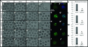
Single cell zygotes were microinjected with double-stranded RNA (dsRNA) that had been designed specifically to functionally knockdown (KD) each gene. While dsGFP-injected control embryos developed normally to the blastocyst stage (Fig. 1 A), injection of dsSf3b14 (Fig. 1 B, K), dsSf3b1 (Fig. 1 C, L), dsRpl7l1 (Fig. 1 D, M) and dsRrp7a (Fig. 1 E, N) resulted in nearly complete failure of blastocyst formation. Interestingly, embryos developed from 1-cell to morula without any observed morphological or developmental differences, including formation of compacted morula at 72 hours post fertilization (hpf). Despite the normal appearance at 72 hpf, KD of each of these four genes resulted in visibly degenerating embryos at 96 hpf, a time when dsGFP control embryos had developed to the blastocyst stage (Fig. 1 A–E).
Sf3b14, Sf3b1, Rpl7l1 and Rrp7a are required for blastocyst formation. Evaluation of developmental progression at 24 (2-cell stage), 48 (8-cell stage), 72 (morula stage) and 96 (blastocyst stage) hours post fertilization (hpf), following microinjection of double-stranded RNAs designed to target Sf3b14 (A), Sf3b1 (B), Rpl7l1 (C) and Rrp7a (D). Note the morphologically normal progression until the compacting morula stage but failure to develop into blastocysts in each case. Consistent with apparent death of knockdown embryos at 96 hpf, p53 is activated in many blastomeres indicating apoptosis (compare (F) with (G–J)). Quantification of blastocyst formation (K–N). Error bars represent standard error of the mean (SEM). Asterisks indicate t-test P-values <0.01. The number of microinjected embryos is indicated for each group in charts (K–N).

Given the rapid and complete failure of blastocyst formation, we first asked if functional KD of these critical factors resulted in increased apoptosis. Although Trp53 (p53) protein is present in blastomeres of the normal preimplantation embryo, it is present in its inactive form. Immunofluorescence detection with an antibody specific to active p53 (phosphorylated at serine 15) indicated that apoptosis had been initiated in many (but not all) blastomeres in dsSf3b14, dsSf3b1, dsRpl7l1 and dsRrp7a embryos (compare Fig. 1 F with Fig. 1 G–J).
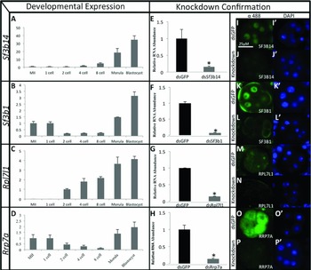
As KD embryos failed to develop past the morula to blastocyst transition, we next sought to determine the normal expression pattern of each gene and validate gene-specific KD. As shown in Fig. 2 A, Sf3b14 transcripts were barely detectable in ovulated oocytes metaphase II (MII), 1-cell, 2-cell and 4-cell embryos, with a rapid rise in gene expression occurring between the 8-cell, morula and blastocyst stages. Sf3b1 showed a similar expression profile, although appreciable maternal transcripts could be detected in the oocyte and zygote (Fig. 2 B). Rpl7l1 was not detected in MII or zygote stages, and expression levels increased steadily from the 2-cell through to the blastocyst stage (Fig. 2 C). Rrp7a expression closely resembled that of Sf3b1, being present in the oocyte, reduced during cleavage stages, and then increasing dramatically during the morula and blastocyst stages. The dynamic expression profiles of these gene was one of the criteria for inclusion in our screen – the peak of expression in morula/blastocyst implies functional importance at that developmental window, and matches the observed blastocyst failure phenotypes observed.
Wild-type expression patterns and knockdown (KD) validation of Sf3b14, Sf3b1, Rpl7l1 and Rrp7a. RT-qPCR results show dynamic expression of each gene during preimplantation development (A–D). Quantitative assessment of KD efficiency resulting from gene-specific dsRNA injection shows robust loss of mRNA for each targeted transcript (E–H). Detection of endogenous proteins by immunofluorescence (IF) in control and KD embryos at the 8-cell stage show nuclear localization of Sf3b14 (I), Sf3b1 (K), nucleolar localization of Rpl7l1 (M) and cytoplasmic localization of Rrp7a (O). Knockdown embryos show drastic reduction/absence of protein (J, L, N and P). Error bars represent standard error of the mean (SEM). Asterisks indicate t-test P-values <0.01.

Microinjection of dsSf3b14, dsSf3b1, dsRpl7l1 and dsRrp7a resulted in a drastic reduction of endogenous mRNA (Fig. 2 E–H) and near-complete loss of each protein at the 8-cell stage (Fig. 2 I–P). We also confirmed mRNA KD at 72 and 96 hpf (data not shown) to ensure functional loss throughout the culture period, with similar results, indicating loss of gene function through 96 hpf. In addition to KD confirmation, IF on control embryos provided cellular localization information for each of these proteins, which has not been demonstrated previously in vivo. Sf3b14 and Sf3b1 are both localized uniformly to the nucleus in 8-cell embryos, with exclusion from the nucleolus (Fig. 2 I, K). Sf3b14 and Sf3b1 are thought to interact with each other in spliceosome complexes (Schellenberg et al., Reference Schellenberg, Dul and MacMillan2011), so the similar cellular localization supports these findings.
Rpl7l1 is also localized to blastomere nuclei, but appeared in small foci concentrated specifically in or around the nucleolus – but not elsewhere within each nucleus (Fig. 2 M). Quite different from localization of the other three proteins, Rrp7a was found throughout the cytoplasm and was excluded from the nucleus of 8-cell blastomeres (Fig. 2 O). Gene ontology suggests that both Rpl7l1 and Rrp7a are functional components of ribosomes (Ashburner et al., Reference Ashburner, Ball and Blake2000). While the cytoplasmic localization of Rrp7a was consistent with this possibility, the nucleolar localization of Rpl7l1 suggested a non-ribosomal function of this protein. Immunofluorescence detection in KD embryos showed severely reduced/absent protein that resulted from each dsRNA microinjection (Fig. 2 J, L, N, P), and consistent with the reduction of each mRNA (Fig. 2 E–H). Given that we observed loss of protein at the 8-cell stage, it is interesting to point out that no developmental defects appeared until the morula to blastocyst transition, 24–48 h after we detected protein reduction. Together, these results suggest that the critical cellular functions of these gene products are not required until after morula compaction has occurred, or that there are sufficient molecular redundancies of protein function (perhaps maternal components) that persist until the blastocyst stage.
Lineage specification in KD embryos
Blastomeres begin to differentiate molecularly at the 8-cell/morula stage into cells of the future inner cell mass (ICM) and trophectoderm (TE). Given the failure of KD embryos to form blastocysts, we assessed expression genes known to mark the future ICM and TE in order to ask if the absence of these four proteins disrupts cell fate decisions in the early embryo. All blastomeres initially expressed the Pou domain 5 class 1 transcription factor, Pou5f1 (also known as Oct4), which becomes restricted to ICM cells coincident with expanded blastocyst formation (Nichols et al., Reference Nichols, Zevnik, Anastassiadis, Niwa, Klewe-Nebenius, Chambers, Scholer and Smith1998). During early morula stages, the caudal-type homeodomain transcription factor, Cdx2, is variably expressed, with ‘Cdx2 low’ cells marking the future ICM, and ‘Cdx2 high’ cells marking the future TE (Strumpf et al., Reference Strumpf, Mao, Yamanaka, Ralston, Chawengsaksophak, Beck and Rossant2005). Coincident with the variable expression of TE-specific genes such as Cdx2 and Tead4 during the compacted morula stage, other ICM-specific genes such as the homeodomain protein Nanog (which marks the future epiblast) are also variably expressed in individual blastomeres (Chambers et al., Reference Chambers, Colby, Robertson, Nichols, Lee, Tweedie and Smith2003; Mitsui et al., Reference Mitsui, Tokuzawa, Itoh, Segawa, Murakami, Takahashi, Maruyama, Maeda and Yamanaka2003; Dietrich & Hiiragi, Reference Dietrich and Hiiragi2007). Although normal blastomeres show uncoordinated and variable levels of these critical factors during the morula to blastocyst transition, mutually exclusive expression of Oct4/Nanog and Cdx2 is not observed until the late/expanding blastocyst stage (Dietrich & Hiiragi, Reference Dietrich and Hiiragi2007; Plusa et al., Reference Plusa, Piliszek, Frankenberg, Artus and Hadjantonakis2008; Strumpf et al., Reference Strumpf, Mao, Yamanaka, Ralston, Chawengsaksophak, Beck and Rossant2005). Due to the failure of blastocyst formation and obvious embryo death at 96 hpf, we could not assess cell lineage at a stage at which cells have mutually exclusive expression patterns. We did however assess the onset of lineage commitment by examining Oct4, Nanog, Cdx2 mRNA levels and protein localization in compacting morula in order to determine if individual blastomeres show the expected variability in protein levels and if lineage specific transcription has been activated in KD embryos.
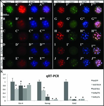
As expected, at 72 hpf we observe Oct4 and Nanog in all wild-type blastomeres as well as a few cells with markedly higher Cdx2 levels in wild-type morula (Fig. 3 A, F). Knockdown of Sf3b14 or Sf3b1 did not alter the protein localization of Oct4, Nanog or Cdx2, and we observed appropriately variable levels of Nanog and Cdx2 in individual blastomeres (Fig. 3 B, C, G, H). However, the overall levels of both Nanog and Cdx2 were reduced relative to that of wild-type controls, and many fewer ‘Cdx2 high’ blastomeres were apparent in both dsSf3b14 and dsSf3b1 embryos. Knockdown of Rpl7l1 resulted in undetectable levels of Nanog and reduced Cdx2 and Oct4 (Fig. 2 D, I). Similarly, knockdown of Rrp7a resulted in very low levels of Nanog and Cdx2, although Cdx2 high/Oct4 low cells were obvious in dsRrp7a morula (Fig. 3 J). The presence of Cdx2 high/Oct4 low blastomeres suggested that dsSf3b1 and dsRrp7a embryos have functionally initiated TE lineage specification, while dsSf3b14 and dsRrp7a embryos have not.
Lineage specification in the absence of Sf3b14, Sf3b1, Rpl7l1 and Rrp7a. Immunofluorescence (IF) analysis of key lineage specific proteins Nanog, Cdx2 and Oct4 in KD embryos (A–J). qRT-PCR analysis of the same gene mRNAs in knockdown (KD) embryos (K). Error bars represent standard error of the mean (SEM). Asterisks indicate t-test P-values <0.05.

As each of these genes is implicated in cellular functions required for molecular gene expression (splicing or translation), we also assessed Oct4, Nanog, and Cdx2 mRNA levels in KD morula (Fig. 3 K). Consistent with only minor reduction in Oct4 protein observed by IF, Oct4 mRNA is modestly reduced in all KD morula. Also consistent with the reduction in Nanog and Cdx2 observed by IF in each case, both Nanog and Cdx2 were reduced significantly in all KD embryos.
The dramatic loss of transcripts suggests that the underlying defects resulting in blastocyst failure in KD embryos is likely due to defects in essential molecular biological processes, rather than in a failure of specific developmental events. We therefore focused on the 8-cell stage to evaluate the cell biological consequences of loss of function of these genes, a developmental time at which loss of protein has already occurred (Fig. 2), but no obvious morphological phenotype is present.
RNA polymerase II activity and RNA abundance in KD embryos
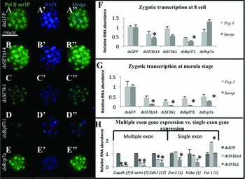
In order to assess global transcriptional activity, we performed IF with an antibody that specifically detects RNA polymerase II (Pol II) phosphorylated at serine 2 in the C-terminal domain, a post-translational modification that marks initiating polymerase (Komarnitsky et al., Reference Komarnitsky, Cho and Buratowski2000). As Sf3b14, Sf3b1, Rpl7l1 and Rrp7a proteins are implicated in post-transcriptional events (splicing or translation), we did not expect to observe differences in active Pol II levels or nuclear localization – which was the case for dsSf3b14 and dsRrp7a embryos (Fig. 4 A, B, E). Surprisingly, we observed a reduction in active Pol II in dsSf3b1 embryos (Fig. 4 C), and a near-complete absence of phospho-ser5 Pol II in dsRrpl7l1 embryos (Fig. 4 D). Combined with the unexpected nuclear localization of Rpl7l1 (Fig. 2 M), our results implicate Rrpl7l1 in either phosphorylation of Poll II, polymerase complex formation or initiation of transcription. This observation also explains the reduction in all transcripts that we observed in dsRrpl7l1 embryos; without active Pol II, a reduction in all Pol II-dependent mRNAs would be expected.
Transcription and splicing in the absence of Sf3b14, Sf3b1, Rpl7l1 and Rrp7a. Assessment of active RNA polymerase II in wild-type and KD embryos at the morula stage (A–E). Quantitative assessment of Peg3 and Snrpn zygotic transcripts in 8-cell (F) and morula (G) knockdown (KD) embryos. Quantitative analysis of multi-exon and single-exon transcripts in KD embryos (H). Error bars represent standard error of the mean (SEM). Asterisks indicate t-test P-values <0.01.

We next asked if zygotic transcription was occurring in KD embryos by assessing mRNA levels of Peg3 and Snrpn, both maternally imprinted loci expressed exclusively from the paternal genome (Szabo & Mann, Reference Szabo and Mann1995; Kuroiwa et al., Reference Kuroiwa, Kaneko-Ishino, Kagitani, Kohda, Li, Tada, Suzuki, Yokoyama, Shiroishi, Wakana, Barton, Ishino and Surani1996; Komarnitsky et al., Reference Komarnitsky, Cho and Buratowski2000). All Peg3 and Snrpn transcripts after the 2-cell stage originate from the paternal allele and therefore serve as an indication of active transcription from the zygotic genome. Reduced but appreciable levels of both Peg3 and Snrpn transcripts are present in all KD embryos at the 8-cell stage. At the morula stage both Peg3 and Snrpn levels were reduced further in dsSf3b14, dsSf3b1, dsRpl7l1 and dsRrp7a embryos. These results indicated that although transcription from the zygotic genome is occurring during cleavage stages, a steady decrease in zygotic transcript abundance occurs as development proceeds in the absence of these critical factors. The reduction of Peg3 and Snrpn is also consistent with the reduction of Nanog and Cdx2 mRNAs, further supporting the possibility that Sf3b14, Sf3b1, and Rrp7a are required after transcription during nascent RNA processing. The loss of Peg3 and Snrpn transcripts in dsRpl7l1 is consistent with the lack of active Pol II observed in KD embryos.
Pre-mRNA splicing in Sf3b14 and Sf3b1 KD embryos
As Sf3b14 and Sf3b1 are implicated in the splicing of pre-mRNAs; we compared levels of single-exon genes versus multi-exon genes in KD embryos. Previous studies have shown that defects in splicing machinery can result in reduction of multi-exon transcript abundance, while single-exon transcripts (that are not spliced) remain unchanged (Furumai et al., Reference Furumai, Uchida, Komi, Yoneyama, Ishigami, Watanabe, Kojima and Yoshida2010). To determine if Sf3b14 and Sf3b1 have a role in splicing during preimplantation embryo development, we assessed expression of additional multi-exon genes as well as several single-exon genes. Consistent with a role in production of properly spliced transcripts, each of the multi-exon genes that we examined (n = 5) showed a significant reduction in both dsSf3b14 and dsSf3b1 embryos (Oct4, Nanog, Cdx2 in Fig. 3 K, Peg3 and Snrpn in Fig. 4 F, G). However, we did not observe the same reduction in single-exon transcripts. dsSf3b14 embryos showed no significant reduction in any of the single-exon genes examined (n = 3, Zsr1, H2be and Fut1, Fig. 4 I). dsSf3b1 embryos had reduced H2be, but no reduction of Zrsr1 and Fut1. The consistent reduction in multi-exon transcripts, but absence of reduction in single-exon transcripts supports a role for both Sf3b1 and Sf3b14 in pre-mRNA splicing during preimplantation development in vivo.
Conclusions
Here we report loss of function preimplantation phenotypes when Sf3b14, Sf3b1, Rpl7l1 and Rrp7a are abrogated functionally. We report detailed embryonic expression patterns and show that each of these genes is required in the mammalian embryo for the morula to blastocyst transition. Towards functional annotation of these genes, we document for the first time: (1) essential developmental requirement of Sf3b14, Rpl7l1, and Rrp7a; (2) in vivo nuclear localization Sf3b14, Sf3b1; nucleolar localization of Rpl7l1; and cytoplasmic localization of Rrp7a; (3) in vivo data that supports a role for Sf3b14 and Sf3b1 in splicing; (4) in vivo data that implicates Rpl7l1 in initiation of Poll II and/or RNA transcription; and (5) activation of p53-dependent apoptosis in the absence of Sf3b14, Sf3b1, Rpl7l1, and Rrp7a gene functions.
Acknowledgements
This work was supported by a Massachusetts Life Sciences Grant and March of Dimes Research Grant to JM.