Introduction
Congenital heart disease (CHD) is the most common form of birth defect in the United States, affecting roughly 1% of births per year (Hoffman, Kaplan, & Liberthson, Reference Hoffman, Kaplan and Liberthson2004; Reller, Strickland, Riehle-Colarusso, Mahle, & Correa, Reference Reller, Strickland, Riehle-Colarusso, Mahle and Correa2008). Although advances in pediatric cardiology have increased survival rates, CHD remains the most likely cause of infant mortality due to birth defect (Yang et al., Reference Yang, Chen, Correa, Devine, Mathews and Honein2006). Recent studies report an increased prevalence for some forms of CHD (e.g., Tetralogy of Fallot), likely as a result of improvements in diagnostic technology (Botto, Correa, & Erickson, Reference Botto, Correa and Erickson2001; Correa et al., Reference Correa, Cragan, Kucik, Alverson, Gilboa, Balakrishnan and Chitra2007; Grech & Savona-Ventura, Reference Grech and Savona-Ventura1999). Despite improvements in immediate medical outcomes, functional concerns have been identified across the lifespan in survivors of CHD. A study of children with CHD between the ages of 5 and 10 years reported that 49% were using remedial academic services, 29% were rated in the high-risk range for hyperactivity, and 30% were rated in the high-risk range for inattention on the Attention-Deficit/Hyperactivity Disorder Rating Scale–IV (Shillingford et al., Reference Shillingford, Glanzman, Ittenbach, Clancy, Gaynor and Wernovsky2008). Responses on health-related quality of life measures have demonstrated that CHD-survivors between the ages of 8 and 18 years endorse more quality of life concerns than controls (Mellion et al., Reference Mellion, Uzark, Cassedy, Drotar, Wernovsky, Newburger and Marino2014).
Differences in Cognitive Performance
Impairments across cognitive domains have been documented in younger patients with CHD. Longitudinal studies following children during the first 3 years of life highlight the risk of significant developmental delays in cognitive abilities for samples of children with both single and biventricular CHD diagnoses (Mussatto et al., Reference Mussatto, Hoffmann, Hoffman, Tweddell, Bear, Cao and Brosig2014). Delays in language and motor skills, particularly for children with single ventricle lesions (Sananes et al., Reference Sananes, Manlhiot, Kelly, Hornberger, Williams, MacGregor and McCrindle2012) have been reported. Some preschool age (4–5 years) children with specific single ventricle lesions (e.g., hypoplastic left heart syndrome [HLHS]) have demonstrated lower functioning on estimates of IQ (Sarajuuri et al., Reference Sarajuuri, Jokinen, Mildh, Tujulin, Mattila, Valanne and Lonnqvist2012). IQ, language, memory, and executive functioning performance have been found to be lower for children with CHD (with participants with HLHS performing the lowest) than controls (Gaynor et al., Reference Gaynor, Gerdes, Nord, Bernbaum, Zackai, Wernovsky and Jarvik2010), although controlling for demographic and treatment variables reduced differences below statistical significance. Other samples of children with single and biventricular CHD have performed within the expected range on cognitive measures (Neufeld et al., Reference Neufeld, Clark, Robertson, Moddemann, Dinu, Joffe and Rebeyka2008). A recent review of the CHD literature (Tyagi et al., Reference Tyagi, Austin, Stygall, Deanfield, Cullen and Newman2014) recommended additional research with older samples to explore cognitive outcomes for adults with CHD.
Differences in White Matter Integrity
It has been suggested that reduced pre and perioperative blood flow to the brain during infancy can increase the risk of white matter abnormalities for patients with CHD (Andropoulos et al., Reference Andropoulos, Hunter, Nelson, Stayer, Stark, McKenzie and Fraser2010; Mahle et al., Reference Mahle, Tavani, Zimmerman, Nicolson, Galli, Gaynor and Kurth2002), resulting in white matter injury comparable to that found in premature infants (Miller et al., Reference Miller, McQuillen, Hamrick, Xu, Glidden, Charlton and Vigneron2007).
Over the past decade, diffusion tensor imaging (DTI) has emerged as a more sensitive technique to analyze subtle white matter differences in normal-appearing white matter (NAWM) compared to traditional structural magnetic resonance imaging (MRI) (Behrens, Johansen-Berg, et al., Reference Behrens, Johansen-Berg, Woolrich, Smith, Wheeler-Kingshott, Boulby and Matthews2003). Several DTI studies have identified wide-spread white matter differences in infants with single and biventricular CHD before and after early heart surgery (Dimitropoulos et al., Reference Dimitropoulos, McQuillen, Sethi, Moosa, Chau, Xu and Miller2013; Partridge et al., Reference Partridge, Vigneron, Charlton, Berman, Henry, Mukherjee and Miller2006; Sethi et al., Reference Sethi, Tabbutt, Dimitropoulos, Harris, Chau, Poskitt and McQuillen2013) and in adolescents with biventricular CHD following early heart surgery (Rivkin et al., Reference Rivkin, Watson, Scoppettuolo, Wypij, Vajapeyam, Bellinger and Newburger2013). These differences are often characterized by reduced fractional anisotropy (FA), a measure of microstructural white matter integrity (WMI), and lower FA in infants and adolescents with CHD has been related to increases in perioperative medical variables (e.g., number of operations and length of stay) (Dimitropoulos et al., Reference Dimitropoulos, McQuillen, Sethi, Moosa, Chau, Xu and Miller2013; Rivkin et al., Reference Rivkin, Watson, Scoppettuolo, Wypij, Vajapeyam, Bellinger and Newburger2013).
The current study presents a two-part study of adolescents and emerging adults with CHD. Study One aimed to identify white matter differences in CHD relative to demographically matched controls. Although identifying white matter differences is important, a critical second step is identifying cognitive correlates of WMI in CHD. After reviewing existing literature and the results of Study One, we designed a theoretically-driven study to examine relationships between cognitive outcomes and WMI (Study Two). The current study is among the first to apply Tract-Based Spatial Statistics (TBSS) to explore white matter differences and theory-driven cognitive correlates. These findings will help establish the utility of TBSS in revealing subtle markers of WMI disruption and associated differences in cognitive performance in CHD.
Study One: Differences in White Matter Integrity in CHD
The aim of Study One is to extend previous findings of significantly lower WMI that have been demonstrated primarily in younger samples with CHD (see recent adolescent study by Rollins et al., Reference Rollins, Watson, Asaro, Wypij, Vajapeyam, Bellinger and Rivkin2014), to a sample of adolescents and emerging adults using TBSS on DTI fractional anisotropy. TBSS allows for a whole-brain exploration of WMI differences resulting from a contrast of participants with CHD and demographically similar controls.
Method: Study One
Participants
Adolescent and emerging adult survivors of infant CHD surgery were recruited from the Children’s Healthcare of Atlanta/Emory University pediatric cardiology database. CHD conditions of interest, which were queried, were determined a priori (see the Procedures section). Exclusion criteria for all participants consisted of severe and persistent mental and/or physical disability (e.g., cerebral palsy or defined genetic syndrome), prematurity defined as less than 34 weeks gestational age, history of cerebral vascular accidents, prior history of brain tumor or traumatic brain injury, visual or hearing impairment, ferromagnetic material, a native language other than English, IQ scores<65, or residential location greater than 60 miles from central Atlanta.
Forty-five participants with CHD anatomy meeting inclusion criteria were contacted over a 6-month period. Recruitment rate was 53% (24/45). Twenty-one of those contacted did not participate for reasons including no interest and scheduling conflicts/availability. There were no demographic or medical differences between those CHD participants who participated and those who declined. Imaging data from two participants were excluded due to MRI artifact (i.e., one for motion artifact and one whose lower cerebellum was not captured during the MRI).
Healthy controls were recruited from local university research participant pools, community fliers, and word of mouth from clinical participants. Controls were selected for similar demographics with the CHD participants from a larger neuroimaging study.
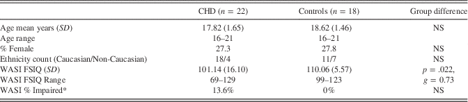
Descriptive data for both groups is presented in Table 1. The CHD sample consisted of 81.8% Caucasian, 13.6% African American, and 4.5% Asian participants. The control sample included 61.1% Caucasian, 22.2% African American, and 16.7% Asian participants. No significant differences were determined in age, sex, or racial/ethnic background between groups. Participants in the CHD group had lower IQ scores than controls, with CHD having notably larger SD.
Descriptive statistics demonstrating similar demographics between groups

Note. CHD=congenital heart disease; SD=standard deviation; NS=non-significant; WASI=Wechsler Abbreviated Scale of Intelligence-Second Edition; FSIQ-2=Full Scale Intelligence Quotient, Two-Subtest Form.
* Impairment defined as greater than −1.5 SD below average range.
Eleven subjects with CHD had single ventricle lesion diagnoses. Diagnoses for this group consisted of tricuspid atresia with normally related great vessels (n=2), combined tricuspid and pulmonary atresia (n=1), pulmonary atresia intact ventricular septum (n=1), double inlet left ventricle (n=4), double inlet ventricle with coarcation of the aorta (n=1), unbalanced atrioventricular septal defect (n=1), and hypoplastic left heart syndrome (n=1). Surgical data for this group were limited secondary to lack of available medical records. However, while the anatomy is heterogeneous, the physiology dictates one to two cardiac surgeries within the first 6 months of life, generally culminating in three cardiac operations by 3 years of age. The average number of cardiac surgeries in this group was 2.7±0.77.
The remaining 11 participants had biventricular anatomy. The anatomic diagnoses for this cohort were dextro-transposition of the great arteries (n=10), and total anomalous pulmonary venous connection (n=1). Within the biventricular group, the median age for the first cardiac operation was 4 days with a range of 1–24 days. The average number of cardiac surgeries in this group was 1±0.3.
Procedure
CHD participants were recruited via a telephone interview of the participant and/or family member in the case of those below 18 years of age. An Institutional Review Board-approved script was followed and each participant was screened for contraindications to undergo MRI procedure. Consent and assent were obtained from all participants and/or guardians. Participants were paid $50 to cover time and travel.
Cognitive assessments were completed during a single session as part of a larger neuropsychological battery immediately following the MRI scan (see Methods Study Two). MRI data were acquired on a Siemens scanner (Trio Tim, Siemens, Erlangen) at 3 Tesla with a standard RF 12-channel head coil.
MRI Acquisition Protocol
This study used DTI to determine whole-brain WMI. This method of structural imaging allows software-based visualization and indirect quantification of WMI in vivo based on a measure of water diffusion through brain tissue (Behrens, Woolrich, et al., Reference Behrens, Woolrich, Jenkinson, Johansen-Berg, Nunes, Clare and Smith2003). Diffusion weighted data was acquired during a 30-direction single-shot spin echo diffusion-weighted DTI sequence with 60 contiguous axial slices with the following specifications: repetition time/echo time (TR/TE)=7700/90 ms; b=1000 s/mm2; voxel size: 2×2×2 mm; acquisition matrix 204×204; sequence time: 8 min, 22 s. T1 weighted images (3D MPRAGE) were acquired to rule out abnormal structure or white matter: TR/TE=2250/3.98 ms, 176 contiguous sagittal slices, 1mm slice thickness, no gap, voxel size: 1.0×1.0×1.0 mm3. No intravenous contrast materials were administered.
Tract-Based Spatial Statistics on DTI Data
Post-processing of DTI data was performed using the FMRIB Software Library (FSL) version 5.1 (Jenkinson, Beckmann, Behrens, Woolrich, & Smith, Reference Jenkinson, Beckmann, Behrens, Woolrich and Smith2012; Smith et al., Reference Smith, Jenkinson, Woolrich, Beckmann, Behrens, Johansen-Berg and Matthews2004; Woolrich et al., Reference Woolrich, Jbabdi, Patenaude, Chappell, Makni, Behrens and Smith2009). The eddy current and motion correction tool from the FMRIB’s Diffusion Toolbox (FDT) was used (Behrens, Johansen-Berg, et al., Reference Behrens, Johansen-Berg, Woolrich, Smith, Wheeler-Kingshott, Boulby and Matthews2003). Diffusion tensors were calculated from each image and FA maps were generated using FDT. Individual FA maps were visually inspected for motion and other artifacts.
FA data from all subjects were aligned using the FNIRT tool (Andersson, Jenkinson, & Smith, Reference Andersson, Jenkinson and Smith2007). TBSS scripts allowed for the creation of a mean FA image (Smith et al., Reference Smith, Jenkinson, Johansen-Berg, Rueckert, Nichols, Mackay and Behrens2006). The TBSS registration and tract skeletonization process was performed using a threshold FA of 0.2 to control for cross-subject variability and prevent inclusion of non-white matter skeleton voxels (Smith et al., Reference Smith, Jenkinson, Johansen-Berg, Rueckert, Nichols, Mackay and Behrens2006). Non-brain voxels were extracted using the Brain Extraction Tool (Smith, Reference Smith2002) with a factor of 0.32.
FA data obtained using TBSS allowed for voxel-wise cross-group statistical analysis. To account for the non-Gaussian distribution of FA data demonstrated in previous studies (Jones, Reference Jones2008), a between-group, nonparametric two-sample permutation test was performed using the Randomize tool (Nichols & Holmes, Reference Nichols and Holmes2002) with 5000 permutations. Threshold-free Cluster Enhancement (TFCE) allowed for enhancement of cluster structures without additional data smoothing (Smith & Nichols, Reference Smith and Nichols2009). The significance threshold was set to p<.01 (uncorrected) and a cluster size of >30 voxels. A fslmaths script was used to create a mask over the resulting significantly different clusters. The JHU ICBM-DTI-81 White Matter and JHU White Matter Tractography atlases were used to label significant clusters. The fslmeants script allowed for extraction of each participant’s FA within each cluster. FA values range from 0 to 1. Greater values are considered to be higher WMI.
Results: Study One
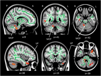
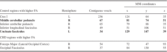
TBSS identified clusters of statistically significant differences in FA (p<.01) between CHD and control groups. To reduce family-wise error, we only considered significant clusters larger than 30 contiguous voxels for the current analyses (see Table 2). Overall, the CHD group showed larger regions with lower FA compared to the control group. Decreased FA values were found within the left hemisphere uncinate fasciculus (UF) and cerebral crus I. Within the right hemisphere, lower FA was identified in the middle cerebellar peduncle (MCP), inferior cerebellar peduncle (ICP), and inferior longitudinal fasciculus (ILF). Decreased FA values for the UF and MCP for the CHD group are highlighted in Figure 1. The control group showed lower FA compared to the CHD group within the right hemisphere along the forceps major (FM) and the occipital fasciculus (OF).
Significant group differences in white matter regions of middle cerebellar peduncles (MCP) and uncinate fasciculus (UF). Group white matter skeleton in green overlaid on a T1 image. Selected clusters of significantly reduced FA for congenital heart disease group in red/yellow. p<.01 (uncorrected).

White matter clusters with significantly different FA between groups

Note. CHD, congenital heart disease; FA, fractional anisotropy; MNI: Montreal Neurological Institute, p<.01 (uncorrected) for all regions, bolded text highlights selected clusters.
CHD treatment factors including years since last cardiac operation (r=−.04), number of operations (r=−.08), and number of current medications (r=.05) were not related to UF FA. Similarly, no relationships was determined between years since last cardiac operation (r=.23), number of operations (r=−.33), current medications (r=−.24) and MCP FA.
Summary: Study One
Consistent with previous literature showing disrupted white matter development in CHD participants, we identified lower FA along the left UF and cerebral crus I, and along the right MCP, ILF, and ICP in participants with CHD compared to controls. Two previous studies found decreased FA in CHD samples using whole-brain analyses (Rivkin et al., Reference Rivkin, Watson, Scoppettuolo, Wypij, Vajapeyam, Bellinger and Newburger2013; Rollins et al., Reference Rollins, Watson, Asaro, Wypij, Vajapeyam, Bellinger and Rivkin2014). Both studies used voxel-based analyses (VBA) that are equally robust, but lack the TBSS-specific white matter skeletonization step that has been suggested to improve FA analyses without additional alignment and spatial smoothing (Jones, Symms, Cercignani, & Howard, Reference Jones, Symms, Cercignani and Howard2005). However, these studies provide general regions of lower WMI (e.g., voxels within the right cerebellum vs. voxels along the right MCP tract) that may correspond with our current findings.
Our findings expand upon the structural MRI work of von Rhein et al. (Reference von Rhein, Scheer, Loenneker, Huber, Knirsch and Latal2011), which revealed white matter differences in 21% of their adolescent sample. Von Rhein et al. contrasted this lower proportion of white matter abnormalities with the higher prevalence of abnormality reported in previous MRI studies and suggested that this finding was due to gliotic repair and reorganization. TBSS analyses used in the current study appear to be more sensitive to microstructural differences.
Study Two: Relationships Between White Matter Differences and Cognitive Performance
Despite considerable evidence of cognitive differences in infants and adolescents with CHD, there have been only four studies to date that have investigated long-term cognitive functioning outcomes concurrently with neuroanatomic abnormalities in participants with CHD. Using traditional MRI analyses, Bellinger et al. (Reference Bellinger, Wypij, Rivkin, DeMaso, Robertson, Dunbar-Masterson and Newburger2011) described structural differences (e.g., brain mineralization and T2 hyperintensities) and performance on a neuropsychological battery in a sample of 16 year old adolescents with mixed CHD diagnoses. Although participants with CHD scored below the average range on measures of achievement, report of executive functioning, ADHD symptomatology, and social functioning, no significant relationships were identified between structural differences and these measures.
Von Rhein et al. (Reference von Rhein, Scheer, Loenneker, Huber, Knirsch and Latal2011) demonstrated cerebral abnormalities (e.g., focal T2 hyperintensities, white matter atrophy, stroke, embolism, and cortical volume loss) in 21% of a sample of adolescents (11.4 to 16.9 years of age) with mixed CHD diagnoses using standard structural MRI. IQ, motor abilities, visuomotor integration and perception, and nonverbal memory for CHD survivors were found to be lower than same-age controls. Furthermore, participants in the CHD group with anatomic lesions scored lower than those without pathology on all domains. However, no specific relationships between MRI findings and cognitive domains were reported.
In a follow-up study, von Rhein et al. (Reference von Rhein, Buchmann, Hagmann, Huber, Klaver, Knirsch and Latal2014) determined cortical and subcortical thickness, calculated total brain volume, and performed a voxel-based morphometry analyses in a sample of adolescents with mixed CHD diagnoses (11.4–16.9 years of age) and controls (9–16.9 years of age). These measures were correlated with cognitive performance. For participants with CHD, IQ estimates correlated with total brain and hippocampal volumes, total white matter volume correlated with verbal comprehension and motor performance, and cerebellar volume correlated with working memory. These findings established relationships between cognitive outcomes and structural abnormalities in brain macrostructure and gross morphometry in CHD.
In the only combined DTI and neurocognitive outcome study in CHD to date, Rollins et al. (Reference Rollins, Watson, Asaro, Wypij, Vajapeyam, Bellinger and Rivkin2014) explored relationships between white matter microstructure using a whole-brain VBA and a neuropsychological battery in a sample of adolescents (16.0–16.4 years of age) with dextro-transposition of the great arteries, a biventricular diagnosis, and controls (14.2–16.1 years of age). Fourteen regions were identified in which participants with CHD demonstrated lower FA than control participants. A secondary exploratory analysis with eight cognitive measures revealed six positive correlations and four correlations in which lower FA related to better cognitive performance. The authors indicate that multiple comparison corrections were not made given the exploratory nature and interrelatedness of cognitive functions and white matter networks.
Along with findings within the CHD literature, research across a wide range of other clinical populations has suggested relationships between lower WMI in specific brain regions and poorer functioning on cognitive measures (Ellmore, Li, Xue, Wong, & Frye, Reference Ellmore, Li, Xue, Wong and Frye2013; Genova, DeLuca, Chiaravalloti, & Wylie, Reference Genova, DeLuca, Chiaravalloti and Wylie2013; Nestor et al., Reference Nestor, Kubicki, Gurrera, Niznikiewicz, Frumin, McCarley and Shenton2004; Smith, King, Jayakar, & Morris, Reference Smith, King, Jayakar and Morris2014). Given these findings, it is possible that reduced microstructural connectivity resulting from early disruption could be related to cognitive performance in emerging adults with CHD. Therefore, the aim of Study Two was to examine two theory-driven cognitive performance correlates with areas of lower WMI identified in participants with CHD via whole-brain TBSS.
We chose two of the five tracts identified in Study One as having significantly lower FA for CHD participants based on previous research. The two tracts selected were the uncinate fasculus (UF) and the middle cerebellar peduncle (MCP). The UF has been related to memory and the MCP has been linked to attention. We also hypothesized that the converse relationships would not be significant to highlight the specificity of the relationships. We limited our analyses to these two white matter regions and specific cognitive constructs to conduct a neuropsychological theory-driven study of these relationships.
Recent studies have identified poor performance on both immediate and delayed verbal memory measures in participants with diverse clinical concerns and highlighted relationships between those deficits and differences in white matter structure along the UF (Fujie et al., Reference Fujie, Namiki, Nishi, Yamada, Miyata, Sakata and Murai2008; Niogi et al., Reference Niogi, Mukherjee, Ghajar, Johnson, Kolster, Lee and McCandliss2008; Papagno et al., Reference Papagno, Miracapillo, Casarotti, Romero Lauro, Castellano, Falini and Bello2011; Voets et al., Reference Voets, Adcock, Stacey, Hart, Carpenter, Matthews and Beckmann2009; von Der Heide, Skipper, Klobusicky, & Olson, Reference von Der Heide, Skipper, Klobusicky and Olson2013). Based on these findings and regions empirically identified via TBSS in the current study, we hypothesized that WMI of the UF would be positively correlated with verbal memory function.
Relationships between white matter of the UF and verbal memory may be specifically relevant for survivors of CHD as fetal hypoxia has been found to have negative effects on the development of the hippocampus, a structure theorized to be important in short to long-term memory consolidation (Li, Gonzalez, & Zhang, Reference Li, Gonzalez and Zhang2012). The uncinate fasciculus is a major component of the white matter network that connects the hippocampus with other subcortical structures. However, the connectivity provided by the UF may not be specific to verbal memory consolidation. A recent summary of studies examining UF function has suggested that the UF may be responsible for storing a broad range of social-emotional and mnemonic information (von Der Heide et al., Reference von Der Heide, Skipper, Klobusicky and Olson2013).
A relationship between measures of lower WMI along the middle cerebellar peduncle (MCP) and difficulties with attention has been suggested in children and adolescents with ADHD relative to controls (Ashtari et al., Reference Ashtari, Kumra, Bhaskar, Clarke, Thaden, Cervellione and Ardekani2005; Bechtel et al., Reference Bechtel, Kobel, Penner, Klarhofer, Scheffler, Opwis and Weber2009; Kobel et al., Reference Kobel, Bechtel, Specht, Klarhofer, Weber, Scheffler and Penner2010). Betchel et al. suggested that lower MCP FA could indicate lower axonal myelination and related deficient communication between cerebellar and prefrontal regions involved in attention. Similar findings described in the current study may be an initial step toward identifying a biological mechanism underlying difficulties with attention previously reported by both parent report and performance measures in younger survivors of CHD (Bellinger et al., Reference Bellinger, Wypij, duPlessis, Rappaport, Jonas, Wernovsky and Newburger2003; Hövels-Gürich et al., Reference Hövels-Gürich, Konrad, Skorzenski, Herpertz-Dahlmann, Messmer and Seghaye2007; Mahle et al., Reference Mahle, Clancy, Moss, Gerdes, Jobes and Wernovsky2000). Therefore, we predicted that WMI of the MCP would be positively correlated with auditory attention performance.
Methods: Study Two
All participants and MRI imaging data from Study One were used in Study Two.
Cognitive Performance Measures
Verbal memory was assessed using the Long Delay Free Recall trial (LDFR) from the California Verbal Learning Test-Second Edition (Delis, Kaplan, Kramer, & Ober, Reference Delis, Kaplan, Kramer and Ober2000). Participants were scored on their ability to recall items from a word list presented verbally five times after a 30-min delay. The LDFR trial has been widely used to measure verbal memory function in participants with health concerns (White et al., Reference White, Moinuddin, McKinstry, Noetzel, Armstrong and DeBaun2006).
Auditory attention span was assessed with the Digits Forward subtest (DF) of the Wechsler Memory Scale-Third Edition (Wechsler, Reference Wechsler1997). The DF subtest required participants to repeat verbally presented series of numbers in correct order. DF has been used recently with CHD participants (Matos et al., Reference Matos, Sarmento, Moreira, Pereira, Quintas, Peixoto and Areias2013) and other populations as a measure of auditory attention span (Santamarina-Perez et al., Reference Santamarina-Perez, Freniche, Eiroa-Orosa, Llobet, Saez, Alegre and Jacas2011).
The two-subtest version of the Wechsler Abbreviated Scales of Intelligence (WASI) (Wechsler, Reference Wechsler2009) was used as a measure of overall cognitive performance comprised of a measure of vocabulary and nonverbal reasoning. This measure has been used to assess cognitive function in multiple populations, including children with congenital heart disease (Lande et al., Reference Lande, Gerson, Hooper, Cox, Matheson, Mendley and Flynn2011; Quartermain et al., Reference Quartermain, Ittenbach, Flynn, Gaynor, Zhang, Licht and Wernovsky2010). Of note, IQ performance correlated with attention, r(38)=.43, p<.01, and memory r(38)=.49, p<.01. However, we did not consider IQ as a covariate given the theoretical and statistical cautions about overcorrection in studies of neurodevelopmental disorders, raised by Dennis et al. (Reference Dennis, Francis, Cirino, Schachar, Barnes and Fletcher2009).
Statistical Analysis
Group mean differences on cognitive measures were computed using independent-samples t tests. To examine hypothesized relationships between WMI and cognitive measures, four bivariate correlations were conducted between TBSS-identified FA values along the MCP and UF and LDFR and DF measures across groups. We also examined these relationships within groups. A false discovery rate (FDR) procedure was applied to adjust for multiple comparisons (Benjamini & Hochberg, Reference Benjamini and Hochberg1995). In addition, secondary analyses to examine the potential role of CHD severity were conducted by dividing CHD into single and biventricular anatomy subgroups. Independent samples t tests explored FA differences between these subgroups. Additional secondary analyses to examine the influence of medical variables on specific regions included bivariate correlations between the five non-hypothesized WMI clusters and three medical variables.
Results: Study Two
There was a significant group difference on verbal memory performance. Thirty-two percent of CHD participants were impaired on LDFR compared to 6% of the controls (see Table 3).
Mean white matter integrity and cognitive performance values between groups

Note. CHD=congenital heart disease; FA=fractional anisotropy; SD=standard deviation; NS=non-significant; Verbal Memory=measured by the California Verbal Learning Test, Second Edition (CVLT-II) Long Delay Free Recall Subscale (LDFR); Attention measured by raw scores on the Wechsler Memory Scale (WMS) Digits Forward (DF) subtest.
* Impairment defined as greater than −1.5 SD below average range.
# Impairment defined as recalling below 5 digits (Miller, Reference Miller1956; Shiffrin & Nosofsky, Reference Shiffrin and Nosofsky1994).
Primary Analyses: Relationships between white matter differences and cognitive performance
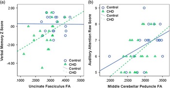
As predicted, a significant positive correlation between UF FA and verbal memory was found in the overall sample, r(38)=.40, p=.01 (uncorrected), and FDR-corrected P critical threshold of .025 (q=0.05), but no significant relationship was identified between UF FA and auditory attention span. A positive correlation between the MCP FA and attention span performance was found, r(38)=.50, p<.01 (uncorrected), and FDR-corrected P critical threshold of .013 (q=0.05), and no relationship was found between MCP FA and memory (see Figure 2). These findings together demonstrate a correlational double dissociation of brain-behavior relationships in the overall sample.
Correlations between white matter region fractional anisotropy (FA) and cognitive performances for each group separately. (a): Specific correlations identified for congenital heart disease (CHD) group uncinate fasciculus and verbal memory (uncorrected), but not for control group. (b): A significant correlation identified for CHD group middle cerebellar peduncle and auditory attention span (uncorrected), but not for control group.

In addition, these relationships were explored for each group separately. The relationship between FA along the UF and verbal memory performance r(20)=.42, p=.049 (uncorrected) was significant, but did not meet a FDR-corrected P critical threshold of .025 (q=0.05). Likewise, the relationship between MCP FA and attention span r(20)=.47, p=.027 (uncorrected) was significant, but did not meet a FDR-corrected P critical threshold of .013 (q=0.05). Within the control group, no significant relationships are identified.
Secondary Analyses: CHD Anatomy Subgroup Differences
Group differences were revealed between single and biventricular CHD anatomy subgroups. Participants with single ventricle lesions demonstrated lower UF FA (M=0.27; SD=0.05) compared to controls (M=0.33; SD=0.06), t(27)=2.90, p=.007, g=1.03 and lower MCP FA (M=0.27; SD=0.03) than controls (M=0.30; SD=0.04), t(27)=2.5, p=.017, g=0.80. Participants with single ventricle lesions had lower IQ scores (M=94.09; SD=16.14) compared to controls (M=110.06; SD=5.57), t(27)=3.17, p=.009, g=1.41, and lower memory scores (M=−1.05; SD=1.52) than controls (M=0.25; SD=0.94), t(27)=2.84, p=.008, g=1.05. In contrast, participants with biventricular anatomy did not differ significantly from controls on FA along the UF (g=0.71), MCP (g=0.27), memory (g=0.65), attention span (g=0.39), or IQ (g=0.20). These findings likely reflect the increased severity of single ventricle lesions on both white matter and cognition.
Secondary Analyses: CHD Treatment Factors and Cognition
CHD treatment factors including years since most recent cardiac operation (r(20)=−.22), number of operations (r(20)=.15), and number of current medications (r(20)=.06) were not related to verbal memory. Similarly, no relationship was determined for years since cardiac operation (r(20)=.34), number of operations (r(20)=.15), current medications, (r(20)=−.07) and auditory attention.
Discussion
This study of emerging adults with CHD is the first, of which the authors are aware, to use TBSS and test theory-driven hypotheses of specific white matter and cognitive relationships. TBSS identified several areas along white matter tracts where FA differed between the CHD and control group. Five white matter regions where identified where the CHD group demonstrated lower FA than the control group, and two regions were identified where controls evidenced lower FA compared with the CHD group. Several regions of white matter identified in the current study are consistent with previous studies describing broad areas of decreased white matter for participants with CHD (Partridge et al., Reference Partridge, Vigneron, Charlton, Berman, Henry, Mukherjee and Miller2006; Rivkin et al., Reference Rivkin, Watson, Scoppettuolo, Wypij, Vajapeyam, Bellinger and Newburger2013; Rollins et al., Reference Rollins, Watson, Asaro, Wypij, Vajapeyam, Bellinger and Rivkin2014), suggesting that the current method is a valid approach for examining white matter differences in CHD, and particularly sensitive to microstructural differences when participants present with NAWM on standard MRI. This is supported by previous research describing TBSS as an improvement over standard voxel-based approaches when analyzing group differences in DTI data (Jones et al., Reference Jones, Symms, Cercignani and Howard2005). Of note, recent work has suggested methods to improve the accuracy of VBA-based analyses (Sage et al., Reference Sage, Van Hecke, Peeters, Sijbers, Robberecht, Parizel and Sunaert2009; Schwarz et al., Reference Schwarz, Reid, Gunter, Senjem, Przybelski, Zuk and Jack2014).
As higher FA values are usually attributed to higher WMI, current findings of higher FA for the CHD group along two tracts within the occipital cortex were unexpected. Higher FA in clinical samples theorized to have lower WMI has been described in other populations, including ADHD (Davenport, Karatekin, White, & Lim, Reference Davenport, Karatekin, White and Lim2010) and Williams syndrome (Hoeft et al., Reference Hoeft, Barnea-Goraly, Haas, Golarai, Ng, Mills and Reiss2007). As suggested in these studies, current higher FA values may be due to differences in white matter structure (e.g., the presence of larger but fewer axons due to reduced branching), to which FA may not be as sensitive. Future studies with larger sample sizes could explore these findings through calculation of radial diffusivity and axial diffusivity; subcomponents of FA that may provide additional data to better interpret these findings.
The current study extends previous findings of associations between brain macrostructure (von Rhein et al., Reference von Rhein, Buchmann, Hagmann, Huber, Klaver, Knirsch and Latal2014) and microstructure (Rollins et al., Reference Rollins, Watson, Asaro, Wypij, Vajapeyam, Bellinger and Rivkin2014) to specific white matter tracts that are independently associated with performance on memory and attention tasks. The significant difference on verbal memory performance noted in the current study is consistent with previous literature describing parent report of difficulties with verbal memory and lower performance on verbal memory measures compared to controls (Miatton, De Wolf, François, Thiery, & Vingerhoets, Reference Miatton, De Wolf, François, Thiery and Vingerhoets2007, Reference Miatton, De Wolf, François, Thiery and Vingerhoets2008). In addition, the relationship between verbal memory and left UF WMI is consistent with a growing body of literature (Christidi et al., Reference Christidi, Zalonis, Kyriazi, Rentzos, Karavasilis, Wilde and Evdokimidis2014; Diehl et al., Reference Diehl, Busch, Duncan, Piao, Tkach and Lüders2008; Mabbott, Rovet, Noseworthy, Smith, & Rockel, Reference Mabbott, Rovet, Noseworthy, Smith and Rockel2009).
We did not demonstrate a group difference on auditory attention span. However, a medium effect size was determined (see Table 3). A recent study by Matos et al. described a sample of CHD survivors that performed significantly worse than controls using the same measure (Matos et al., Reference Matos, Sarmento, Moreira, Pereira, Quintas, Peixoto and Areias2013). It is possible that the larger CHD sample size (n=77) in Matos et al. provided enough statistical power to detect this difference. Although CHD participants did not perform significantly lower than controls on a measure of auditory attention span, a relationship between performance on this task and FA in the right MCP was identified.
Our findings are in line with previous literature reporting significantly lower WASI IQ on average in CHD survivors (Schaefer et al., Reference Schaefer, von Rhein, Knirsch, Huber, Natalucci, Caflisch and Latal2013; von Rhein et al., Reference von Rhein, Scheer, Loenneker, Huber, Knirsch and Latal2011). There was considerably more variability and a wider range of IQ scores within the CHD group versus control group. Despite this, only 13.6% of the CHD group (n=3) fell below the average range. In addition, 13.6% of CHD group were in the superior range (IQ above 120) and only one control participant (0.06%) scored in this range. Therefore, we likely have a higher functioning CHD sample due to our inclusion criteria. Although the current sample may reflect CHD in emerging adults with optimal outcomes, FA from DTI TBSS was able to identify white matter differences relative to controls. Future studies may consider recruiting a homogenous CHD sample including those with more severe cognitive impairments. Although this approach may identify additional white matter disruptions, it will be critical to rule out all the potential confounds that may co-exist such as genetic conditions, prematurity and intraventricular hemorrhage. Although we recruited a heterogeneous CHD group, we explicitly excluded many of these potential co-morbid confounds. This approach may also be why our medical variables did not correlate with cognitive or white matter variables.
We identified two highly specific theory-driven brain-behavior relationships: (1) WMI of the left UF and verbal memory, and (2) WMI of the right MCP and attention span. Further strengthening these findings was the lack of correlation between each tract and the alternate cognitive performance (UF and attention span; MCP and memory). This correlational double dissociation demonstrates that lower FA was not related to overall lower cognitive performance. Likewise, poor cognitive performance was not related to all areas of lower WMI. The approach to explore independent brain-behavior relationships guided by theory-driven hypotheses is a strength of the current study. Similar to dissociations established in earlier studies with adults with mild traumatic brain injury (Niogi et al., Reference Niogi, Mukherjee, Ghajar, Johnson, Kolster, Lee and McCandliss2008), our correlational double dissociation is an innovative contribution to the CHD literature.
Our results should be discussed in the context of current limitations. In comparison with other DTI approaches, TBSS is generally considered to be an exploratory measure. Although contrasting whole-brain white matter pathways provides a wide lens to identify group differences, TBSS revealed specific ROIs along portions of white matter pathways. These ROIs overlap with specific areas of the white matter tract and do not yield information about differences in WMI that may be occurring along the entire tract. White matter tractography can be used to address these questions. Future studies may consider the ROIs highlighted by the current study as seed points for white matter tractography to further explore structural connectivity in these specific regions.
Conclusion
In summary, this is the first study to our knowledge to report TBSS-derived white matter differences in emerging adults with CHD relative to demographically matched controls. We examined two selected regions from those identified using TBSS. Furthermore, we established distinct brain-behavior relationships in CHD with the uncinate fasciculus and verbal memory and between the middle cerebellar peduncle and attention span. Together these findings suggest that TBSS is a robust technique useful for quantifying white matter disruption in emerging adults with CHD. Future multi-site studies with larger samples may find that fractional anisotropy has the potential to be used as a sensitive and quantitative biomarker for cognitive outcomes in adolescents and young adults with CHD.
Acknowledgments
This research was supported by grants from the Children’s Healthcare of Atlanta Cardiovascular Biology Research Center Seed Grant (W.M., PI) and a Research Scholar Grant from the American Cancer Society (#114044-RSGPB-07-170-01-CPPB; King, PI). R.B. is supported by the Georgia State University Second Century Initiative (2CI) Fellowship in Human Neuroimaging. We are indebted to the participants who gave willingly of their time to make this research possible. The authors acknowledge Stacey Berry for helping with data acquisition and the King Developmental Neuropsychology Research Team for data management. Funding sources had no influence on study design, recruitment, data collection, analyses, or data interpretation. The authors declare no conflicts of interest. Dr. Brewster and Mr. King contributed equally to this work.