Statement of Research Significance
Research Question(s) or Topic(s): Reading is a complex cognitive process involving distributed cortical networks, but the white matter pathways that support it are less understood. This neurosurgical case report examines the role of the vertical occipital fasciculus in reading and provides the first real-time spatial localization of injury to this tract resulting in alexia. Main Findings: During awake resection of an occipitotemporal metastasis, the patient developed alexia with letter-by-letter reading as a tubular retractor was advanced toward the lesion. Tractography confirmed the retractor tip intersected the vertical occipital fasciculus at symptom onset. Study Contributions: This is the first lesion-based case to anatomically localize the vertical occipital fasciculus’ role in reading in real time, strengthening evidence for its contribution to reading networks. We discuss the potential contributions of the vertical occipital fasciculus in reading and clinical considerations during surgical planning and mapping in this area.
Introduction
Reading is a complex process that involves decoding written symbols or orthographic representations to derive meaning. Accumulating evidence from functional magnetic resonance imaging (fMRI) and lesion studies implicates an area in the left fusiform gyrus in orthographic decoding. Specifically, Cohen and colleagues (Reference Cohen, Dehaene, Naccache, Lehéricy, Dehaene-Lambertz, Hénaff and Michel2000) identified an area in the lateral occipitotemporal sulcus, which they labeled the visual word form area (VWFA), that showed left lateralized activation (corresponding to Talairach coordinates: x = −42, y = −57, z = −6) during word reading independent of the side of visual field presentation. Subsequent fMRI studies demonstrated that activation in the VWFA is not affected by variations in the visual features of the presented letters, such as font size or letter case (Dehaene et al., Reference Dehaene, Naccache, Cohen, Bihan, Mangin, Poline and Rivière2001). The activation is largely independent of the semantic content of the stimuli (Dehaene et al., Reference Dehaene, Jobert, Naccache, Ciuciu, Poline, Le Bihan and Cohen2004; Dehaene et al., Reference Dehaene, Le Clec’H, Poline, Le Bihan and Cohen2002), and it is tuned to letter sequencing probability or sublexical orthographic structure (Binder et al., Reference Binder, Medler, Westbury, Liebenthal and Buchanan2006). Such findings suggest that this area is responsible for forming more abstract orthographic representations of word form stimuli. Lesions to or near the VWFA can result in alexia (Binder & Mohr, Reference Binder and Mohr1992; Leff et al., Reference Leff, Spitsyna, Plant and Wise2006), an inability to read. Additionally, focal electrical stimulation to the VWFA with stereoencephalography (sEEG) results in pure alexia with no disruption in phonological or lexical-semantic processes (Sabsevitz et al., Reference Sabsevitz, Middlebrooks, Tatum, Grewal, Wharen and Ritaccio2020).
The VWFA has functional connectivity with primary and higher-order visual areas as well as perisylvian language areas (Bouhali et al., Reference Bouhali, de Schotten, Pinel, Poupon, Mangin, Dehaene and Cohen2014), making it an ideal interface between ventral visual recognition and orthographic-to-phoneme and orthographic-to-semantic mapping. The specific white matter pathways supporting the VWFA are still being elucidated. Yeatman and colleagues (2013) used whole-brain tractography with a deterministic streamline tracking algorithm in 27 healthy participants to identify the white matter connections to the VWFA. The authors found that the inferior longitudinal (ILF), inferior frontal occipital (IFOF), and vertical occipital fasciculus (VOF) consistently passed within close proximity of fMRI functionally defined VWFA. There is evidence from clinical lesion studies implicating the ILF, a tract that connects the occipital lobe to anterior and medial temporal lobe areas, in reading. Epelbaum et al. (Reference Epelbaum, Piel, Gaillard, Delmaire, Perrin, Dupont, Dehaene and Cohen2008) reported on a patient who developed pure alexia following a small surgical resection near the VWFA. Analysis of the diffusion tensor imaging (DTI) and fMRI data showed that the VWFA was anatomically preserved; however, the resection resulted in progressive and selective degeneration of the ILF posterior to the VWFA, essentially disconnecting the VWFA from occipital inputs and accounting for the alexia. Similarly, Zemmoura et al. (Reference Zemmoura, Herbet, Moritz-Gasser and Duffau2015) showed that surgical resections involving the posterior portion of the left ILF resulted in enduring and severe alexia in a small series of brain tumor patients, while stimulation or resection of more anterior portions of the ILF did not result in alexia. Still, a different study showed that more anterior ILF damage can impact performance on a variety of orthographic tasks (Wang et al., Reference Wang, Li, Huang, Ding, Song and Han2020). Intraoperative electrical stimulation of the ILF in low-grade glioma patients can also result in reading deficits (Sarubbo et al., Reference Sarubbo, De Benedictis, Merler, Mandonnet, Balbi, Granieri and Duffau2015).
Less is known about the VOF and its role in reading. The VOF is a major fiber bundle that connects the VWFA to primary and higher-order visual areas in the lateral occipital-parietal cortex. Unlike the ILF and IFOF, it has specific endpoints in the VWFA (Yeatman et al., Reference Yeatman, Rauschecker and Wandell2013). The VOF ascends from the occipital temporal sulcus, where the VWFA is located, to the transverse occipital sulcus and posterior intraparietal sulcus (Yeatman et al., Reference Yeatman, Weiner, Pestilli, Rokem, Mezer and Wandell2014). The connectomics of the VOF make it an ideal, critical white matter tract for supporting reading; however, to our knowledge there have been only two case reports describing the functional consequences of damaging the VOF. Greenblatt et al. (Reference Greenblatt1973) described a patient with a glioblastoma that involved the posterior white matter, including the VOF, who presented with pure alexia. In another case report, Greenblatt et al. (Reference Greenblatt1976) described a patient who developed transient alexia following surgical resection of a vascular malformation in the VOF. The main limitation of these case reports is the inability to precisely localize and differentiate the involvement of the VOF from nearby white matter tracts (e.g., ILF, IFOF). Further support for the VOF’s role in alexia comes from electrical stimulation studies where ESM of the VOF was more associated with pure alexia compared to phonological or semantic type alexia errors (Ng et al., Reference Ng, Moritz-Gasser, Lemaitre, Duffau and Herbet2021).
In this case report, we describe a patient who developed an alexia with letter-by-letter reading in real time during an awake brain surgery for resection of a left occipitotemporal metastasis. The patient underwent continuous neuropsychological testing during insertion of a tubular retractor. Stealth imaging enabled localization of the tubular retractor at the exact onset of the alexia and correlating this with tractography showed that the tubular retractor was in the VOF. This case report is the first to provide precise spatial localization of injury to the VOF with resulting alexia, supporting its important role in reading.
Methods
All study procedures were approved by the Mayo Clinic IRB and were conducted in accordance with the Helsinki Declaration. The patient provided written consent to publish this case report.
Clinical history
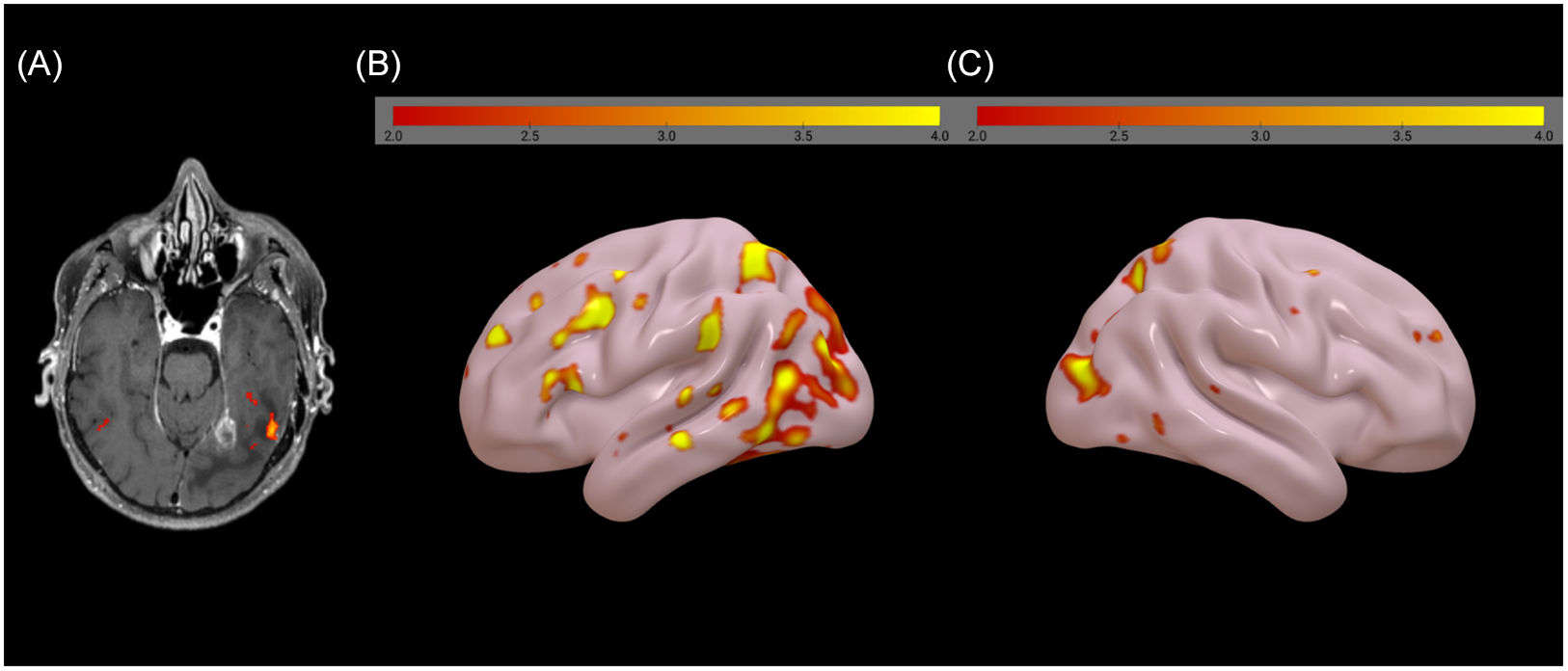
The patient is a 65-year-old, right-handed, monolingual English-speaking, master’s level educated male who was in his usual state of health until he began experiencing headaches several weeks prior to presenting to our hospital. He went on vacation and noticed that he was having difficulty reading the menu at a restaurant and text messages on his phone. He reported that he could see the individual letters but they “did not fit together” and did not make sense. Upon returning home from vacation, he went to a local emergency room where MRI revealed a contrast-enhancing lesion in the left ventral medial occipitotemporal region with significant surrounding vasogenic edema and intralesional hemorrhage, and a smaller enhancing lesion in the left cerebellum, concerning for metastases. He was started on dexamethasone, which alleviated his headaches and produced noticeable improvement in his reading abilities. He presented to our institution for further workup and management. Computed tomography (CT) scans of the chest, abdomen, and pelvis were negative. Skin exam was unrevealing. An awake craniotomy and surgical resection of the larger left occipitotemporal lesion was recommended to obtain tissue for diagnosis and reduce dependency for steroids given that he was symptomatic to the lesion. Preoperative fMRI language mapping was performed using a visual sentence reading completion task contrasted with viewing random consonant strings. Language mapping demonstrated some bilateral activation but overall strong left hemisphere dominance. An area of functional activation in response to the reading paradigm was found lateral to the occipitotemporal lesion in the area of vasogenic edema (see Figure 1). Preoperative diffusion-weighted images were acquired on a 3T Prisma (Siemens Healthineers, Forchheim, Germany) using a 64-channel receive head and neck coil. Images were acquired with a 2 mm isotropic resolution consisting of 64 diffusion directions at b = 1500 s/mm2 and 6 at b = 0 s/mm2. The diffusion data underwent standard preprocessing of realignment and eddy current correction with ODF modeling using a DTI model in DSI Studio (https://dsi-studio.labsolver.org/). A semi-automated tracking algorithm was used in DSI studio. Tractography was generated using the following parameters: the anisotropy threshold was randomly selected between 0.5 and 0.7 otsu threshold, the angular threshold was randomly selected from 45 degrees to 90 degrees, step size was set to voxel spacing, tracks with length shorter than 30 and longer than 300 mm were discarded. Visualization of the tracts showed superior displacement and likely involvement of the ILF and IFOF.
(A) Sentence comprehension fMRI activation overlaid on preoperative MRI T1 with contrast, shown in radiologic convention (right is left), (B) & (C) Left and right hemisphere sentence comprehension fMRI, respectively.

Cognitive evaluation
The patient underwent a comprehensive neuropsychological evaluation the day before surgery that included the following tests: Wechsler Test of Adult Reading (WTAR) (Wechsler, Reference Wechsler2001), Wechsler Adult Intelligence Scale - Fourth Edition (WAIS-IV) digit span, symbol search, and coding subtests (Wechsler et al., Reference Wechsler, Coalson and Raiford2008), Wechsler Memory Scale - Third Edition (WMS-III) spatial span subtest (Wechsler, Reference Wechsler1997), Hopkins Verbal Learning Test - Revised (HVLT-R) (Brandt & Benedict, Reference Brandt and Benedict2001), Brief Visual Memory Test (BVMT-R) (Benedict, Reference Benedict1997), Boston Naming Test (Kaplan et al., Reference Kaplan, Goodglass and Weintraub1983), Trail Making Test (Reitan, Reference Reitan1955), Delis–Kaplan Executive Function System (D-KEFS) (Delis et al., Reference Delis, Kaplan and Kramer2001), grooved pegboard (Kløve, Reference Kløve1963), and the Quick Aphasia Battery (QAB) (Wilson et al., Reference Wilson, Eriksson, Schneck and Lucanie2018).
The patient was also baselined preoperatively on a series of intraoperative cognitive paradigms using NeuroMapper, a tablet-based testing platform for extra- and intraoperative cognitive mapping developed by the first author of this paper (DSS). The primary task of interest was a visual adaptation of an auditory naming task developed by Hammeke et al. (Reference Hammeke, Kortenkamp and Binder2005), where the patient was instructed to read aloud descriptions (e.g., “A zoo animal with a long neck”) and then name what is being described (e.g., giraffe). This allowed for simultaneous testing of orthographic or reading processes, mapping that information onto conceptual semantics, and lexical or name retrieval. The sentences contained on average 4.97 words (SD = 1.01) and 22.90 (SD = 4.13) letters. Item-level accuracy was collected and importantly only items that were correctly answered at baseline were selected for intraoperative testing. Errors were categorized as a reading error if the patient misread any of the words in the sentence and as a naming error if he read the sentence correctly but could not name or produced an incorrect name for the item. The QAB was administered 24 hours postoperatively using an alternate form. The entire preoperative neuropsychological battery and QAB were administered again 2 weeks after surgery. Alternate forms were used where available.
Surgical procedure
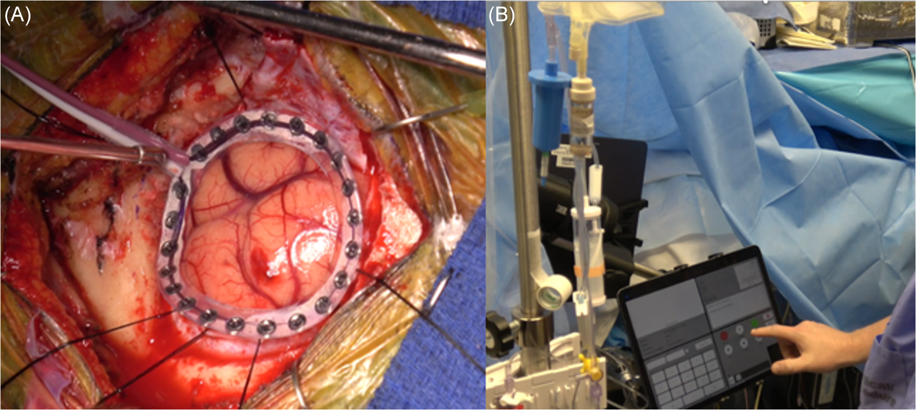
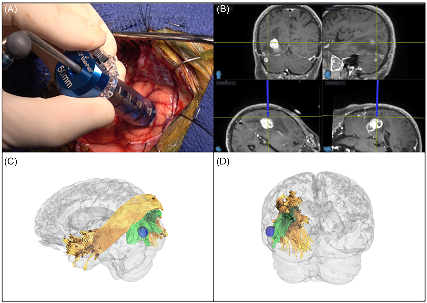
The patient underwent an ultrasound-guided scalp block prior to entering the operating room. He was then taken to the operating room and placed in the supine position and a Mayfield 3-pin headframe was attached. The patient was lightly sedated with 0.3 mcg/kg/hr dexmedetomidine during the craniotomy. A craniotomy was performed, and the dura was opened in a cruciate fashion. He was awake and conversational during the craniotomy and tolerated it well. Anesthesia was discontinued approximately 15 minutes before mapping was performed. A circular EEG grid (Freund et al., Reference Freund, Sherman, Sabsevitz, Middlebrooks, Feyissa, Garcia, Grewal, Chaichana, Quinones-Hinojosa and Tatum2023; ReFaey et al., Reference ReFaey, Chaichana, Feyissa, Vivas-Buitrago, Brinkmann, Middlebrooks, McKay, Lankford, Tripathi, Bojaxhi, Roth, Tatum and Quiñones-Hinojosa2019) was then placed on the surface over the area of the planned trajectory (see Figure 2). Several trials of the reading task were administered prior to stimulation mapping to ensure the patient was functioning at his presurgical baseline. The patient underwent surface bipolar electrical stimulation mapping using the Natus stimulator, with the following stimulation parameters: 50 Hz frequency, pulse width of 500 microseconds, and starting at 2 mA, increasing in 0.5 mA to 1 mA increments to a maximum of 10 mA. The exposed convexity of the temporo-occipital region was mapped using the reading-adapted auditory naming task described above. A corticectomy was made and a 13.5 mm diameter, 50 mm length NICO BrainPath tubular retractor (Indianapolis, Indiana, USA) was slowly inserted while continuous neuropsychological testing was performed (see Figure 3A). The tubular retractor was used to minimize damage to surrounding white matter tracts. Surgical entry was through the lateral middle occipital gyrus and the tubular retractor was inserted in an anterior medial inferior trajectory to reach the lesion. The retractor path was posterior to fMRI-defined VWFA.
(A) Placement of a circular grid that provides 360 degree coverage to capture any seizure activity around the surgical field. (B) Intraoperative neuropsychological mapping and monitoring, performed using the NeuroMapper platform.

(A) Insertion of the tubular retractor, (B) Stealth Navigation System coordinates indicating where reading errors started, (C) & (D) Representations of tip of the tubular retractor (blue) in relation to the vertical occipital fasciculus (green) and inferior longitudinal fasciculus (yellow).

Results
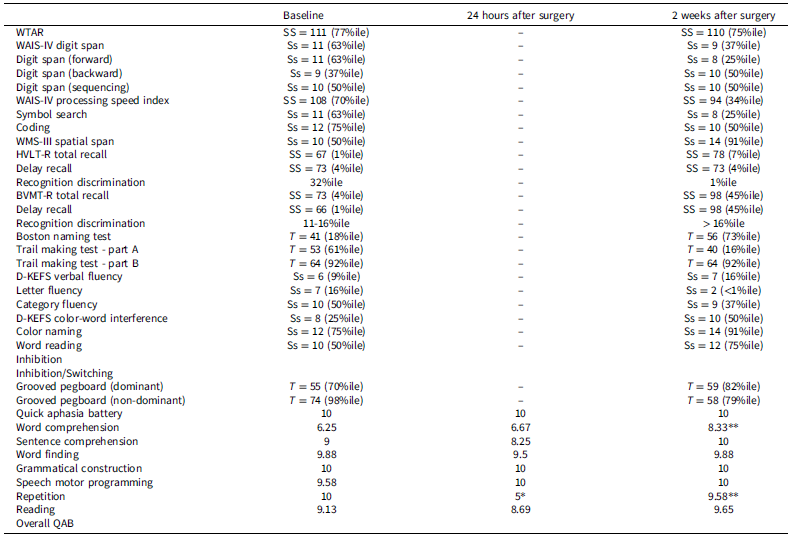
Presurgical neuropsychological testing revealed weaknesses in learning and memory retrieval for verbal (HVLT-R total recall SS = 67; delay recall SS = 73) and visual information (BVMT-R total recall SS = 73; delay recall SS = 78) but importantly irregular/exception word reading and sentence reading were normal on the WTAR (SS = 111; high average) and QAB (reading score = 10/10), respectively. There were no signs of aphasia on the QAB (overall score = 9.13). Speech was fluent and articulate (speech motor programing score = 10) and grammatically well formed (grammatical construction score = 9.88). Phonological processes were intact with normal repetition of words and short sentences (repetition score = 9.58). Single-word comprehension was normal (word comprehension score = 10). He had mild difficulty comprehending spoken sentences that had syntactically more complex structure (sentence comprehension score = 6.25). Lexical retrieval was normal on picture naming (word-finding score = 9). On the reading adaptation of the auditory naming test, the patient made 3 reading-only errors (85/88, 97% accuracy), 15 naming only errors (73/88, 83% accuracy), and 1 error on both reading and naming. His reading errors included reading long as large, referees as references and dropping a plural ending on a word. His naming errors were predominately anomic (unable to name) with several incorrect semantic word substitutions (e.g., polar bear for panda bear, kangaroo for rabbit). In total, there were 69 items where he both correctly read the sentence and accurately named the item within 5 s of presentation. Only these 69 items were retained for intraoperative testing to ensure that any errors would represent a true decline from baseline performance attributable to intraoperative stimulation or resection. See Table 1 for baseline cognitive results.
Cognitive performance at the presurgical baseline, 24 hours after surgery, and 2 weeks after surgery

Note: For the QAB, a reliable change index (RCI) of 0.8, which approximately represents the amount of change that would exceed the 95% confidence interval for test-retest variability in individuals with stable aphasia (Wilson et al., Reference Wilson, Eriksson, Schneck and Lucanie2018) was used
* Denotes a significant change from baseline to 24 hours after surgery using this RCI cutoff
** Denotes a statistically significant change from 24 hours after surgery to 2 weeks after surgery using the same RCI cutoff.
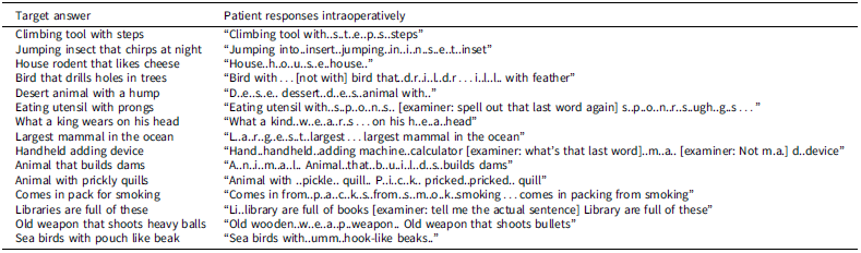
In surgery, prior to surface electrical stimulation mapping, the patient was administered 4 trials of the adapted auditory naming task using the NeuroMapper platform to determine whether he was functioning at his preoperative baseline before proceeding with mapping. He read and named these trials correctly. Surface mapping was then performed starting at 2 mA and increasing to 10 mA. The patient made a few variable reading errors when the occipito-temporal cortical surface was mapped but these errors were not consistent or reproducible. Surgical entry was made through the cortex with negative mapping. After a small corticectomy was made, the tubular retractor was inserted (see Figure 3A). The patient was tested continuously as the retractor was being inserted. The patient started making reading errors (see Table 2 for examples). The surgeon stopped inserting the retractor and the location of the tip of the retractor at the onset of reading errors was recorded on the Stealth Navigation System (see Figure 3B). Using DSI Studio, we examined the tracts that passed through a region of interest corresponding to the tip of the BrainPath device at the exact onset of the alexia (see Figure 3C and D). Seeding the whole brain and filtering by ROA, 98% of the fibers in the ROI were from the VOF, while 1% was from the IFOF and 1% from parietal aslant tract. The patient continued to make consistent reading errors and resorted to a letter-by-letter reading strategy on some trials. He also made several semantic paralexias. Visual fields were tested and deemed intact based on his ability to perceive flashing dots in all visual fields on the tablet screen while fixating on a center point and based on his ability to visually search and identify a target letter among an array of distractor letters and symbols in all quadrants of the screen. Despite struggling to read the individual words, when prompted to do so, he was able to rapidly identify how many words were in a sentence. The reading deficit persisted throughout the resection, but naming abilities remained intact as indicated by his ability to name all the items that he eventually read via a letter-by-letter strategy or were corrected and read aloud to him.
Intraoperative reading errors

Note: represent corrections provided to the patient by the examiner.
Twenty-four hours postoperatively, reading abilities were again tested with the QAB Reading Aloud subtest. The patient read only 1 of 4 single words and 1 of 2 sentences correctly, which is in stark contrast to his performance preoperatively where he read all words and sentences correctly. He again resorted to letter-by-letter reading on several of the reading items. He had no difficulty on other visual language-based tasks, such as picture naming and word-to-picture matching.
Two weeks postoperatively, the neuropsychological battery and QAB were re-administered. Compared to his reading abilities 24 hours postoperatively, there was a significant improvement in reading performance such that word and sentence reading returned to baseline performance levels on the QAB and his WTAR word reading was similar to his presurgical level. There were no signs of aphasia on the QAB (overall score = 9.65), which is consistent with baseline performance. His comprehension of spoken sentences of syntactically more complex structure improved significantly compared to his performance at baseline and 24 hours after surgery. Other cognitive functions were largely unchanged to improved. He continued to show some mild inefficiencies in his learning and recall of verbal and visual material with overall good storage of this information into memory.
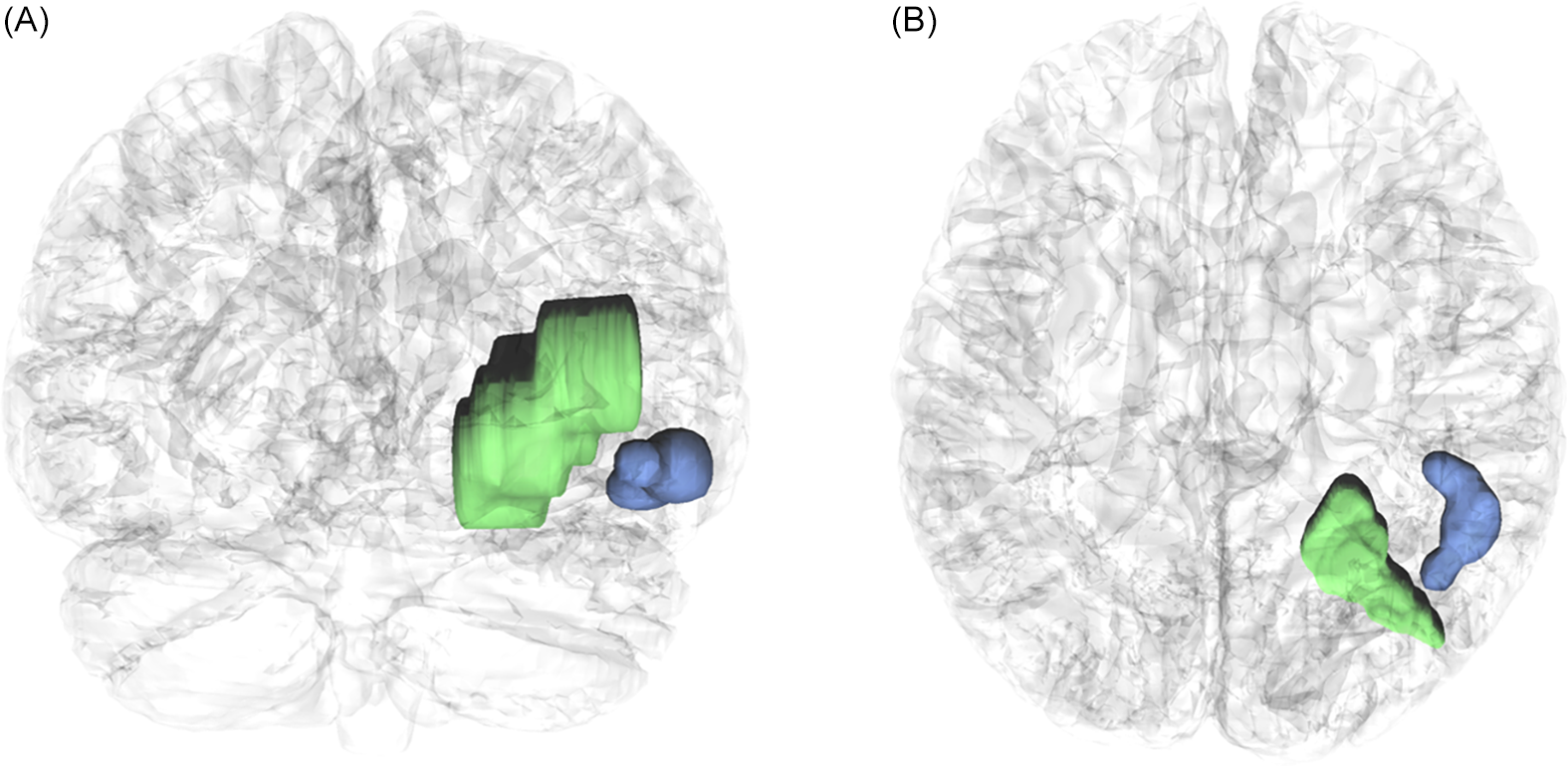
MRI of the brain the day after surgery revealed a gross total resection of the occipital lesion. The area in the ventral temporo-occipital junction, which showed fMRI BOLD activation in response to reading-based language tasks (presumed VWFA), was not involved in the surgical resection. Figure 4 shows the postoperative MRI and reconstruction showing the surgical resection in relation to fMRI-identified VWFA.
(A) Coronal and (B) Axial 3D reconstruction of the tubular retractor path and surgical resection (green) in relation to fMRI-identified VWFA (blue).

Discussion
The neuroanatomy of reading involves a complex network of cortical structures and subcortical white matter connections that work together to transform written text into meaningful language. The role of the VWFA in the dominant ventral temporo-occipital junction is well established in reading, although its exact computational functions are not fully understood. Damage or disruption to this region results in alexia or the inability to read. The VWFA has strong connectivity with a distributed network of language brain regions in frontal, temporal, and parietal cortices (Chen et al., Reference Chen, Wassermann, Abrams, Kochalka, Gallardo-Diez and Menon2019) and likely communicates orthographic information to these areas for mapping onto phonological and semantic representations. Much less is known about the white matter tracts that support reading. Several tracts have close proximity to the VWFA, including the ILF and IFOF (Yeatman et al., Reference Yeatman, Rauschecker and Wandell2013) with the ILF being the most studied and having the most support from lesion and electrical stimulation studies for its role in reading. The ILF connects occipital to temporal regions of the brain, and based on its connectomic properties one can infer that it may be involved in transferring visual input from the occipital lobe to the VWFA for orthographic processing. Further downstream, the ILF connects the VWFA to the ventral temporal cortex and anterior temporal lobe, structures known to be involved in semantic processing, as well as the superior temporal sulcus, which is involved in phonological processing, likely reflecting critical pathways for orthographic-to-semantic and orthographic-to-phonological mapping, respectively. The posterior arcuate fasciculus (pAF) also likely supports reading, likely through a phonological stream connecting temporal and supramarginal gyrus areas and a semantic stream connecting temporal and angular gyrus areas, and electrical stimulation studies have implicated the pAF in phonological and semantic type alexic errors (Ng et al., Reference Ng, Moritz-Gasser, Lemaitre, Duffau and Herbet2021). The VOF runs in a vertical orientation lateral to the ILF and posterior to the AF and connects the VWFA to the lateral occipital parietal junction and the posterior angular gyrus. Yablonski et al. (Reference Yablonski, Karipidis, Kubota and Yeatman2024) showed that the VWFA can be subdivided into two distinct regions. The posterior VWFA or VWFA-1, modulated by spatial attention (White et al., Reference White, Palmer, Boynton and Yeatman2019), appears sensitive to visual features and likely helps encode visual properties of written text (Kay & Yeatman, Reference Kay and Yeatman2017; Lerma-Usabiaga et al., Reference Lerma-Usabiaga, Carreiras and Paz-Alonso2018). It is connected with visual and attention regions in the occipital and parietal cortex. The anterior VWFA or VWFA-2 is thought to process higher-level language information and is connected to language regions in the frontal lobe. Tractography studies have shown that VWFA-1 and VWFA-2 are connected to their cortical endpoints by the VOF and the AF, respectively (Kubota et al., Reference Kubota, Grotheer, Finzi, Natu, Gomez and Grill-Spector2023; Lerma-Usabiaga et al., Reference Lerma-Usabiaga, Carreiras and Paz-Alonso2018) and suggest that VWFA-1 processes visual and orthographic information, which then undergoes further processing and integration with the language system in VWFA-2 (Yablonski et al., Reference Yablonski, Karipidis, Kubota and Yeatman2024).
There are limited case reports on the functional consequences of damaging the VOF. To our knowledge, only two case reports exist (Greenblatt, Reference Greenblatt1973, Reference Greenblatt1976) and both lack precise localization of damage and are unable to differentiate damage to the VOF from adjacent white matter tracts, such as the ILF and IFOF. Ours is the first lesion study with precise anatomical localization that supports the role of the VOF in reading. Here we describe a patient who underwent awake craniotomy for surgical resection of a left occipitotemporal metastasis. A tubular retractor was used to access the deep lesion and while the tubular retractor was being inserted into the brain the patient underwent careful neuropsychological examination with specific focus on reading. The patient developed alexia with evidence of letter-by-letter reading as the tubular retractor was being advanced. The location of the tubular retractor tip was localized using stealth imaging and using tractography this point in space was localized to the VOF. Seeding the whole brain and filtering by ROI, 98% of the fibers were from the VOF, while 1% was from the IFOF and 1% from parietal aslant tract, providing compelling evidence that this tract conveys important information for reading.
Our patient resorted to a letter-by-letter reading strategy. Letter-by-letter reading is a compensatory strategy seen in some patients with pure alexia and involves identifying and processing each letter sequentially to construct a word, rather than recognizing whole words. Research on split-brain (Baynes & Eliassen, Reference Baynes and Eliassen1998) and left hemispherectomy (Patterson et al., Reference Patterson, Vargha-Khadem and Polkey1989) patients have shown that the right VWFA has the capacity to support the identification of letters. Functional imaging studies have shown that the right VWFA responds to alphabetic stimuli, although not more than checkerboard stimuli, in normal subjects and the right VWFA can take on a pattern of activation normally specific to the dominant VWFA in patients with pure alexia from left VWFA damage (Cohen et al., Reference Cohen, Martinaud, Lemer, Lehéricy, Samson, Obadia and Dehaene2003). Cohen and colleagues (Cohen et al., Reference Cohen, Henry, Dehaene, Martinaud, Lehéricy, Lemer and Ferrieux2004) proposed that during letter-by-letter reading letters are identified in the intact right-hemispheric visual system, letters are serially transferred to the left hemisphere through the intact segment of the corpus callosum, and word identity is recovered by the left hemisphere through an effortful verbal working memory process. We suspect that in our case the insertion of the tubular retractor resulted in the disconnection of the VWFA on the left by transiently damaging the VOF, and our patient was able to read via a letter-by-letter strategy through the intact right VWFA. It is also notable that our patient was able to read some words without using letter-by-letter reading. One explanation for this is that the retractor did not completely disconnect the VWFA so he might have been getting some signal through this pathway. Reading also involves the ILF, which was not disrupted by the tubular retractor at the onset of the alexia. The patient’s intact ILF may have supported his ability to read some words.
Tubular retractors, such as the BrainPath device, are designed to minimize neurological injury during brain surgery by more gently displacing, rather than cutting white matter tracts to create a working corridor for surgeons. By utilizing a tapered and cylindrical design, the BrainPath device reduces pressure on surrounding tissue, decreasing the risk of damage to critical neural structures. Studies have shown that tubular retractors can lead to improved postoperative outcomes, including reduced swelling, faster recovery times, and lower rates of neurological deficits (Mansour et al., Reference Mansour, Echeverry, Shapiro and Snelling2020). Our patient developed a pure alexia in real time during awake brain surgery that persisted in severe form 24 hours postoperatively, but it remarkably resolved when evaluated 2 weeks postoperatively. We suspect that his recovery was due to the result of only minimal permanent damage to the VOF through the use of the tubular retractor.
Another realization from this case study is the importance of mapping and sparing the VOF during surgical resections when taking a surgical approach through this area. Electrical stimulation mapping with reading tasks (e.g., single-word and sentence-level reading) should be used when resecting through this subcortical space. In this case, we did not perform electrical stimulation mapping as we used a tubular retractor and there are logistical and technical challenges of stimulating through the closed cylinder and obturator during insertion. We used an adaptation of an auditory naming task, which allowed us to monitor reading but also lexical and semantic processes. We would recommend consideration of reading of regular, irregular words, and pseudowords to help better differentiate between phonological and semantic aspects of reading.
While this study advances our understanding of the anatomical and functional relevance of the VOF, future work is needed to clarify the specific contribution of the VOF to reading and how it functional differs from other major occipito-temporal tracts, particularly the ILF and IFOF, during reading and visual recognition tasks. The connectivity pattern of the VOF, linking more posterior ‘visual’ VWFA with visual attention systems seem to favor an attentional role of some kind, perhaps providing the substrate for directing and maintaining visual foveation on the stimulus, or moving fixation from word to word during sentence reading. We are currently leveraging sEEG in combination with tractography to explore whether these tracts serve distinct, complementary, or redundant roles in the transformation of visual input into linguistic representations. Critically, this approach can help determine the domain specificity of these pathways, i.e., whether their involvement in reading is unique or reflects more general-purpose visual integration and top-down attentional mechanisms. Such insights will be essential for refining neurobiological models of reading and may have translational value in disorders affecting literacy and visual cognition. This line of inquiry also has critical implications for pre-surgical planning, and intraoperative mapping.
Funding statement
This research did not receive any specific grant from funding agencies in the public, commercial, or not-for-profit sectors.
Competing interests
No conflicts of interest.
No part of the study procedures or analyses was pre-registered prior to the research being conducted.