Introduction
The sphenoid bone is the main skeletal support of the central skull base and comprises one of the most complex and variable anatomies.Reference Laine, Nadel and Braun1 Its remarkable anatomical variations can be attributed to the extent of pneumatisation and the presence of septa. The sphenoid sinus is often approached during minimally invasive procedures, such as functional endoscopic sinus surgery and endoscopic transsphenoidal techniques.Reference Tomovic, Esmaeili, Chan, Shukla, Choudhry and Liu2 It is surrounded by the internal carotid artery, the optic nerve, the maxillary nerve and the vidian nerve.Reference Anusha, Baharudin, Philip, Harvinder, Shaffie and Ramiza3 In an extensively pneumatised sphenoid sinus, protrusion or dehiscence of these neurovascular structures make them vulnerable to injury during surgical procedures.Reference Sirikci, Bayazit, Bayram, Mumbuç, Güngör and Kanlikama4 In this regard, injury to the optic nerve is one of the most feared complications of skull base endoscopic surgery, which could lead to complete vision loss.Reference Rene, Rose, Lenthall and Moseley5
The optic canal typically courses on the superolateral wall of the sphenoid sinus. As it passes adjacent to the sinus, it might produce a protrusion or indentation in the sphenoid wall and can even become dehiscent at different points.Reference Itagi, Adiga, Kalenahalli, Goolahally and Gyanchandani6 Previous studies addressing the anatomical variants of the optic canal have shown heterogeneous data, which can be the result of poorly defined criteria to distinguish protrusion, indentation or dehiscence of the optic canal and the inherent variability between different ethnic populations. Prevalence of protrusion of the optic canal into the sphenoid sinus has been observed to range from 2 to 35 per cent,Reference Anusha, Baharudin, Philip, Harvinder, Shaffie and Ramiza3,Reference Gibelli, Cellina, Gibelli, Cappella, Oliva and Termine7,Reference Asal, Bayar Muluk, Inal, Şahan, Doğan and Arıkan8 and its dehiscence has been reported to range from 4 to 30 per cent.Reference Asal, Bayar Muluk, Inal, Şahan, Doğan and Arıkan8–Reference Fujii, Chambers and Rhoton10 These variations and the presence of Onodi cells have been considered predisposing factors for intra-operative injury.Reference Stankiewicz11–Reference DeLano, Fun and Zinreich13 A detailed computed tomography (CT) examination to detect these anatomical variations is crucial in the pre-operative surgical planning for endoscopic procedures.Reference Bhatti, Schmalfuss and Mancuso12,Reference Melhem, Oliverio, Benson, Leopold and Zinreich14,Reference Chong, Fan, Lau and Sethi15
During the last few years, there has been a continuously growing scope of endoscopic skull base surgery, which highlights the importance of developing anatomical studies to increase the array of approaches to the skull base while decreasing the surgical risks. Reported data in other ethnicities have shown a high degree of variance in the anatomical relations of the optic canal within the sphenoid sinus.Reference Cellina, Gibelli, Floridi, Toluian, Valenti and Martinenghi16 There is increasing but limited data on Hispanic populations; this study aimed to describe and analyse the anatomical variants of the optic canal in the sphenoid sinus in a Hispanic population, evaluate the presence of protrusion and dehiscence of the optic canal, and analyse their relationship to the pneumatisation and septation patterns of the sphenoid sinus.
Materials and methods
This retrospective study was conducted in the University Hospital ‘Dr. Jose E. González' in Monterrey, México. The research protocol was approved by the local research and institutional ethics committee (key: 320942034). The authors assert that all procedures contributing to this work comply with the ethical standards of the relevant national and institutional guidelines on human experimentation and with the Helsinki Declaration of 1975, as revised in 2008.
Patients
We analysed a sample of the 160 paranasal sinus CT images from March 2021 to July 2021 of patients complying with inclusion and exclusion criteria. Only Mexican patients over 18 years were included. Patients with a previous history of acute or chronic sinusitis, skull base fracture or trauma, sinonasal polyposis or tumours, facial deformity, and skull base or sinonasal surgery were excluded from the study. All scans were obtained from the database of the University Hospital ‘Dr. Jose E. González’ Diagnostic Radiology Department.
Imaging and analysis
Scans were obtained with the conventional protocol for paranasal sinus CT in the supine position, without contrast or sedation during the procedure. The images were acquired using a 64-slice CT (GE Medical Systems light speed VCT, Waukesha, USA) with a tube voltage of 120 kV, effective milliampere-seconds of 18 and a field of view of 142 × 278 mm. Acquired images contained 1.25 mm axial sliced and sagittal and coronal reconstructions from the vertex of the cranium to the level of the hyoid bone in a high-definition bone window.
Images were transferred to the Diagnostic Radiology Department workstation and were evaluated individually by three investigators, a neuroradiologist and two otolaryngologists. Reviewers were blinded to each other's responses. Data were classified at the end according to the investigator's evaluation. In the case of three different responses, a second neuroradiologist reviewed the CT to reach a final consensus.
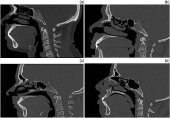
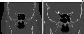
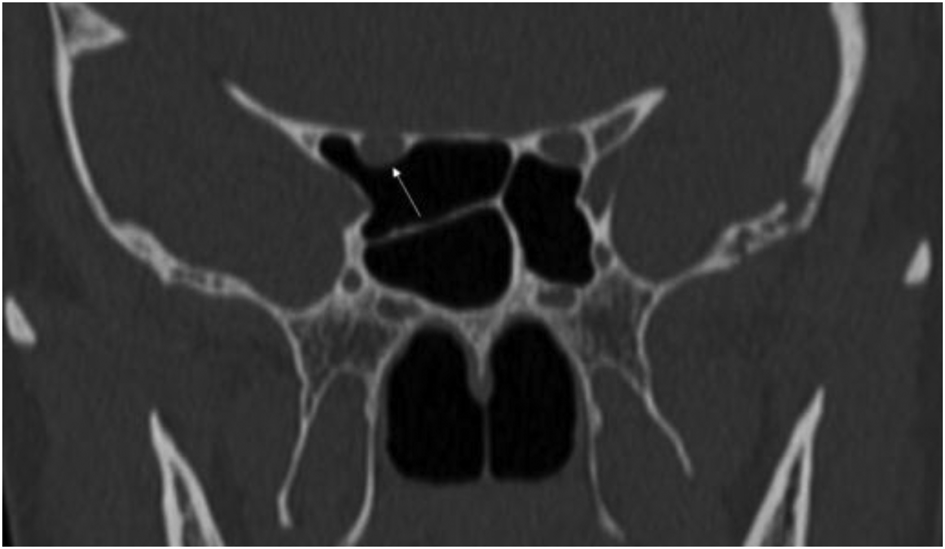
The following variables were evaluated. (1) Sphenoid sinus pneumatisation: it was classified as conchal, presellar, sellar or postsellar.Reference Sirikci, Bayazit, Bayram, Mumbuç, Güngör and Kanlikama4 The conchal type is characterised by the presence of solid bone anterior to the sellae without any pneumatisation. The presellar type presents limited pneumatisation of the region anterior to a plane parallel to the anterior sellar wall. In the sellar type, the pneumatisation extends beyond the anterior wall but not past the posterior sellar wall. The pneumatisation in the postsellar type extends beyond the posterior wall of the sellaeReference Sirikci, Bayazit, Bayram, Mumbuç, Güngör and Kanlikama4,Reference Batra, Citardi, Gallivan, Roh and Lanza17 (Figure 1). (2) Insertion of main or accessory septa on optic canal was classified as present or absent. (3) Pneumatisation of the anterior clinoid process and Onodi cells was categorised as present or absent. (4) Optic canal type was categorised according to DeLano´s classification. A type 1 optic canal presents no indentation into the sphenoid sinus wall. A type 2 optic canal is defined by an indentation of the optic canal into the sphenoid sinus wall (less than 50 per cent of its circumference projecting into the sphenoid sinus). A type 3 optic canal is defined by a protrusion of the optic canal into the sphenoid sinus (more than 50 per cent of its circumference projecting into the sphenoid sinus). A type 4 optic canal extends laterally to the sphenoid sinus and a posterior ethmoid cell (Onodi cell)Reference DeLano, Fun and Zinreich13 (Figure 2). (5) Dehiscence of the optic canal was defined as the absence of bone density layer at some point of the optic canal within its course around the sphenoid sinus (Figure 3).
Sagittal plane computed tomography images showing different types of pneumatisation of the sphenoid sinus: (a) conchal, (b) presellar, (c) sellar and (d) postsellar.

Coronal plane computed tomography images showing different types of optic canal according to DeLano´s classification. (a) Right side type 4 optic canal, separated from the sphenoid sinus by an Onodi cell (*), and left side type 1 optic canal, with no indentation into the sphenoid sinus wall. (b) Right side type 3 optic canal, with protrusion of the optic canal into the sphenoid sinus and surrounded by pneumatisation of the anterior clinoid process (short arrow) and left side type 2 optic canal, with insertion of the main septum over the optic canal (long arrow).

Coronal plane computed tomography image showing dehiscence of the optic canal with exposure of the optic nerve within the right sphenoid sinus.

Results
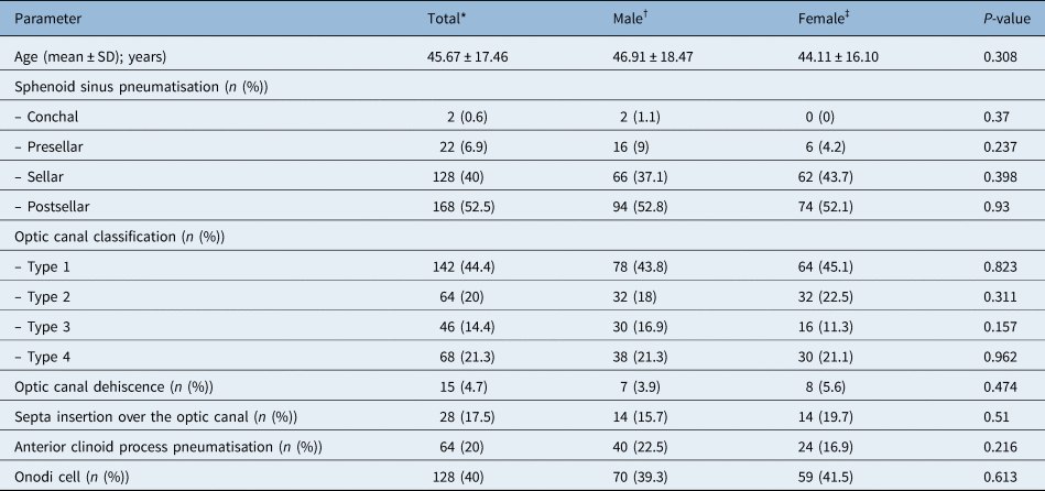
A total of 320 sphenoid sinuses in paranasal sinus CT images of adult patients were evaluated. Patient age ranged from 18 to 88 years, with a mean of 45.67 years (standard deviation, 17.46 years). A total of 178 (55.6 per cent) of the analysed sphenoid sinus scans were from males, and 142 (44.4 per cent) were from females.
The postsellar pneumatisation was the most frequent type of sphenoid sinus, observed in 52.5 per cent (n = 168) of the analysed sinuses. A sellar type sphenoid sinus was found in 40 per cent (n = 128) of the sinuses. Conchal and presellar sinuses represented a less frequent type of pneumatisation, with a prevalence of 0.6 per cent (n = 2) and 6.9 per cent (n = 22), respectively. A total of 17.5 per cent (n = 56) of the analysed sphenoid sinuses had a main or accessory septum inserted on the optic canal. A type 1 optic canal was the most prevalent type of optic canal (44 per cent, n = 142). Dehiscence of the optic canal within the sphenoid sinus was observed in 4.7 per cent (n = 15). Onodi cells were observed in 40.3 per cent (n = 129) of the sinuses and anterior clinoid process pneumatisation in 20 per cent (n = 64). There were no significant differences among the assessed variables between males and females (Table 1).
Sphenoid sinus characteristics and differences between genders

*n = 320; †n = 178; ‡n = 142. SD = standard deviation
We analysed the frequency of the different types of optic canal according to the type of sphenoid sinus pneumatisation. A type 1 optic canal was the most common among all types of pneumatisation of the sphenoid sinus. Type 3 and 4 optic canals were significantly more frequent among postsellar sphenoid sinuses compared with other types of sphenoid sinus pneumatisation (p = 0.018, p = 0.002). Dehiscence of the optic canal was more frequently observed in the postsellar sphenoid sinus (7.1 per cent, n = 12), without reaching statistical difference when compared with other types of sphenoid sinus (p = 0.17; Table 2).
Frequency of different types of optic canal and its dehiscence, according to type of sphenoid sinus pneumatisation

*n = 2; †n = 22; ‡n = 128; **n = 168; §Statistically significant according to chi-square test
The frequency of anterior clinoid process pneumatisation, septa insertion over the optic canal, optic canal dehiscence and Onodi cells was analysed according to the types of optic canal. Type 4 optic canals had a significantly higher rate of septa insertion (17.6 per cent, n = 12; p = 0.014). Type 3 optic canals had a significantly higher frequency of related anterior clinoid process pneumatisation (54.3 per cent, n = 25; p < 0.001). Optic canal dehiscence was more frequent among type 3 optic canals (8.7 per cent, n = 4), without reaching statistical difference when compared with other optic canal types (p = 0.201; Table 3).
Association between different types of optic canal and sphenoid sinus characteristics

*n = 142; †n = 64; ‡n = 46; **n = 68; §Statistically significant according to chi-square test
The statistical analysis was performed using SPSS® (version 24.0) statistical analysis software.
Categorical and continuous variables were reported as percentages and frequencies or means and standard deviations, respectively. We used the chi-squared test for categorical variables, and the Student's t-test and Mann–Whitney tests were performed for continuous variables. Statistically significant differences in the prevalence of optic canal type between gender were evaluated through chi-squared testing. Dependence between variables was investigated by the chi-squared test. Differences in volume between type of optic canal were assessed through the one-way analysis of variance test. A p-value less than 0.05 was statistically significant.
Discussion
In the present study, we described and analysed the configuration of the optic canal and its relationship with different features of the sphenoid sinus in a Hispanic group of patients. The analysed variables are important to consider before undergoing endoscopic sinus surgery, especially in the context of skull base approaches, to minimise the operative risks of iatrogenic injury to the optic nerve.
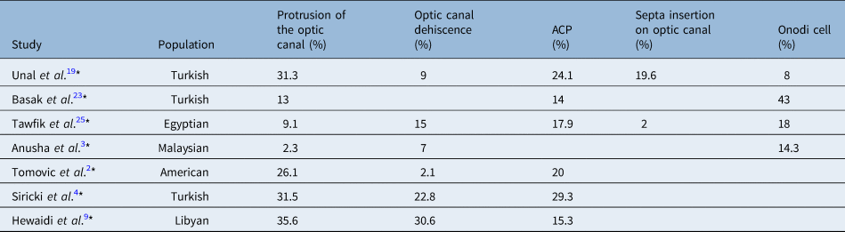
The optic canal has been established as one of the most critical structures neighbouring the sphenoid sinus; in some cases it indents or protrudes into the sphenoid sinus.Reference Peele18 DeLano et al. were among the first to study the optic canal's relation to the sphenoid sinus and propose a classification system based on the degree of protrusion of the optic canal into the sphenoid sinus wall.Reference DeLano, Fun and Zinreich13 In our study, the most common type of optic canal, according to DeLano's classification, was type 1 (no indentation), followed by type 2 (indentation into sphenoid sinus wall), similar to previously reported data.Reference Itagi, Adiga, Kalenahalli, Goolahally and Gyanchandani6,Reference DeLano, Fun and Zinreich13 Optic canal protrusion has been observed to vary widely among different ethnicities, being observed in 35 per cent of Indians,Reference Itagi, Adiga, Kalenahalli, Goolahally and Gyanchandani6 up to 31 per cent in Turkish patientsReference Sirikci, Bayazit, Bayram, Mumbuç, Güngör and Kanlikama4,Reference Asal, Bayar Muluk, Inal, Şahan, Doğan and Arıkan8,Reference Unal, Bademci, Bilgili, Batay and Avci19 and 69 per cent in AsiansReference Tan and Ong20 (Table 4 and 5). The heterogeneity of these results has been attributed to ethnic backgrounds and the different criteria used to define the variables.Reference Hewaidi and Omami9 Type 3 optic canal (protrusion into the sphenoid sinus) was observed in 14 per cent of the assessed sphenoid sinuses of our Hispanic population. We observed that postsellar sphenoid sinus configurations, which give better access for transsphenoidal endoscopic approaches, are associated with a higher frequency of the type 3 and type 4 optic canal, both of which are related to an increased operative risk and injury to the optic nerve.
Reported prevalence of optic canal types and related anatomical variants of the sphenoid sinus among populations in several computed tomography based studies

ACP = anterior clinoid process
Reported prevalence of optic canal protrusion and dehiscence and sphenoid sinus variations among populations in computed tomography based studies

* Studies that did not use DeLano´s classification and only reported the optic canal as protruding or not protruding into the sphenoid sinus. In some studies, no particular data were reported and spaces were left blank. CT = computed tomography; ACP = anterior clinoid process pneumatisation
Another anatomical variation that predisposes the optic nerve to an iatrogenic injury and warrants pre-operative consideration includes the dehiscence of the bony optic canal.Reference Meyers and Valvassori21 We identified optic canal dehiscence in 5 per cent of the evaluated sphenoid sinuses, lower than what was previously reported in Turkish and Libyan populations.Reference Itagi, Adiga, Kalenahalli, Goolahally and Gyanchandani6,Reference Asal, Bayar Muluk, Inal, Şahan, Doğan and Arıkan8,Reference Hewaidi and Omami9 Itagi et al.Reference Itagi, Adiga, Kalenahalli, Goolahally and Gyanchandani6 observed an association between the protrusion and a higher frequency of optic canal dehiscence. In our study, the rate of optic canal dehiscence was higher in type 2 and type 3 optic canals; however, this relation was not statistically significant.
During transseptal sphenoidectomy, excessive manipulation of a septum inserted on the wall of the optic canal might result in excessive bleeding, retrobulbar haematoma, proptosis or vision loss.Reference Unal, Bademci, Bilgili, Batay and Avci19 We observed a rate of 17.5 per cent of septa insertion over the optic canal, which falls among the reported prevalence in the published literature of 5.9–19.6 per cent.Reference Unal, Bademci, Bilgili, Batay and Avci19,Reference Elwany, Elsaeid and Thabet22,Reference Başak, Karaman, Akdilli, Mutlu, Odabasi and Erpek23
The air cells in the sphenoid sinus may extend and pneumatise the anterior clinoid process, which places the internal carotid artery and the optic canal in close proximity to the sinus. Anterior clinoid process pneumatisation is related to the presence of an optic-carotid recess, a small space in the superolateral portion of the sphenoid sinus limited superiorly by the optic nerve and inferiorly by the internal carotid artery. In our study, the rate of pneumatisation was 20 per cent, which falls among the reported range of 4–43.7 per cent for different populations.Reference Tomovic, Esmaeili, Chan, Shukla, Choudhry and Liu2,Reference Anusha, Baharudin, Philip, Harvinder, Shaffie and Ramiza3,Reference Itagi, Adiga, Kalenahalli, Goolahally and Gyanchandani6,Reference Asal, Bayar Muluk, Inal, Şahan, Doğan and Arıkan8,Reference DeLano, Fun and Zinreich13,Reference Treviño-Gonzalez, Maldonado-Chapa, Becerra-Jimenez, Soto-Galindo and Morales-del Angel24 We also observed that anterior clinoid process pneumatisation was significantly higher among evaluated sinuses with optic canal type 3 than other types. This finding is supported by previous research, which has shown an association between pneumatisation of the anterior clinoid process with protrusion and dehiscence of the optic nerve in the sphenoid sinus.Reference Sirikci, Bayazit, Bayram, Mumbuç, Güngör and Kanlikama4,Reference Itagi, Adiga, Kalenahalli, Goolahally and Gyanchandani6,Reference DeLano, Fun and Zinreich13,Reference Tawfik, El-Fattah, Nour and Tawfik25
• The sphenoid sinus is approached during minimally invasive procedures, such as sinus surgery and transsphenoidal techniques
• Dehiscence and protrusion of the optic canal into the sphenoid sinus have been considered risk factors for intra-operative injury
• Prevalence of protrusion and dehiscence of the optic canal into the sphenoid sinus varies widely among populations, with no previous reports in Hispanic patients
• This study observed dehiscence and protrusion of the optic canal in 4.7 per cent and 14.4 per cent of cases, respectively
• A postsellar pneumatisation of the sphenoid sinus is associated with an increased rate of dehiscence of the optic canal
• A detailed computed tomography examination is crucial in planning endoscopic procedures, as high-risk anatomic variations are highly prevalent among the Hispanic population
Onodi cells, also known as sphenoethmoidal cells, are posterior ethmoid cells extending through the superior portion of the sphenoid sinus. When present, they are closely related to the optic canal. According to DeLano's classification, an optic canal that travels lateral to an Onodi cell is classified as type 4. They represent a risk for endoscopic sinus surgery when intending to enter the sphenoid sinus by entering an Onodi cell during ethmoidectomy. Bony walls separating these cells from the optic canal can be as thin as 0.3 mm.Reference Cellina, Gibelli, Floridi, Toluian, Valenti and Martinenghi16 The 40 per cent rate of Onodi cells in our population was higher than those reported in other populations.Reference Anusha, Baharudin, Philip, Harvinder, Shaffie and Ramiza3,Reference Tawfik, El-Fattah, Nour and Tawfik25,Reference Dal Secchi, Dolci, Teixeira and Lazarini26 Similar to Tawfik et al.,Reference Tawfik, El-Fattah, Nour and Tawfik25 we found a significant association between the presence of Onodi cells and optic nerve protrusion.
Our study has several methodological strengths that should be considered for future studies in the same topic to provide comparable results. First, it was CT-based with fine axial slice thickness, allowing an accurate evaluation of the studied variables. Second, up to four specialists blinded to each other's responses were included. Third, standardised criteria were used to categorise variables, such as DeLano's classification and dehiscence of the optic canal. Finally, we studied a population that, to our knowledge, was not addressed in previous reports. Some limitations ought to be mentioned. Our sample is relatively small and was performed in a single centre. Our data is insufficient to establish anatomical variants among children and patients with established skull base pathologies.
Conclusion
This study described and analysed optic canal variants in a Mexican population, which supplements published literature addressing optic canal configurations in other ethnicities. A pre-operative analysis of the CT scan prior to sphenoid sinus surgery is of utmost importance to avoid iatrogenic injuries to the optic nerve because variants related to a higher risk of injury are highly prevalent among our population. Further studies with uniform criteria and larger sample sizes are needed to provide an objective comparison among studied populations.
Competing interests
None declared