The mucopolysaccharidoses are a group of rare genetic diseases with mendelian inheritance, each caused by deficiency of different lysosomal enzymes involved in the degradation of glycosaminoglycans. They are divided on the basis of various alphanumeric subgroupings, and most of them are also known eponymously (Text Box 1). Progressive lysosomal accumulation of the glycosaminoglycans affects the connective tissues in many organs, including the heart.Reference Neufeld and Muenzer1 The natural history of all types of the disease is characterized by chronic and progressive multisystemic clinical features, with a variable degree of severity and life expectancy. Severe mental retardation is characteristic for the more severe forms of disease falling within the general heading, particularly for type I, known as Hurler’s syndrome, for the severe variant of type II, known also as Hunter’s syndrome type A, and for the third type, known as Sanfilippo syndrome. Clinical cardiac symptoms, including anatomical and functional abnormalities of the cardiac valves, myocardial hypertrophy, thickened tendinous cords, and narrowing of the coronary arteries, develop in most patients, and may have a relevant impact on their state of health.Reference Schieken, Kerber, Ionasescu and Zellweger2–Reference Hayflick, Rowe, Kavanaugh-McHugh, Olson and Valle13 Early diagnosis is extremely important, permitting earlier detection of any cardiac lesions by means of echocardiographyReference Schieken, Kerber, Ionasescu and Zellweger2–Reference Rigante and Segni11 prior to the onset of cardiac symptoms,Reference Donaldson, Pennock, Berry, Duncan, Cawdery and Leonard12, Reference Hayflick, Rowe, Kavanaugh-McHugh, Olson and Valle13 and to start promptly adequate treatment. Several therapeutic options are now available for children affected by the mucopolysaccharidoses. Transplantation of haematopoietic stem cells was reported to improve cardiac function and overall clinical outcomes in individuals with Hurler syndrome.Reference Ortega, Dominguez and Olive14–Reference Peters and Steward20 Enzyme replacement therapy is now available for the first, third, and fourth types, and data on possible beneficial effects on the heart are emerging.Reference Wraith, Clarke and Beck21–Reference Harmatz, Ketteridge and Giugliani23 The aim of our study was to analyze the frequency and severity of cardiac anomalies and their evolution with age in a large cohort of patients with different types of mucopolysaccharidosis who had been followed at our centre for metabolic diseases.
Box 1 Classification of the mucopolysaccharidoses ( Modified from Muenzer J, J.Pediatr 2004, 144, S27-S34)

Patients and methods
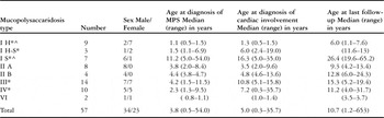
From 1984 to 2005, we studied a total of 57 patients with various types of mucopolysaccharidosis, 34 males and 23 females. Our cohort included 19 patients with type I, 12 with type II, 14 with type III, 10 with type IV, and 2 with type VI. The age at diagnosis of the disease, confirmed by enzymatic and/or molecular analysis, was from 6 months to 54 years, with a median of 3.8 years. We have summarised their characteristics in Table 1.
We followed 55 of the 57 patients for a median duration of 4.6 years, and a range 0.9 to 21.2 years. In 2 instances, patients with the Hurler and Scheie variants of the first type of the disease had a single echocardiogram performed at the first evaluation, with normal findings, and were subsequently lost to follow-up. During follow-up, 5 patients have died. The median age of the 50 patients who were alive at their last visit was 10.7 years, with a range from 1.2 to 65.3 years.
Transplantation of haematopoietic stem cells was performed in 7 patients with Hurler disease at a median age of 1.8 years, with a range from 1.2 to 2.9 years. They have been followed for a median period of 5.5 years, with a range from 1.5 to 7.5 years. We have replaced the enzymes in 9 patients, 7 with the first type, 2 of these having the Hurler-Scheie variant of the disease and 5 the Scheie variant, and 2 with the attenuated variant of Hunter’s syndrome. The treatment was started at a median age of 23.4 years, with a range from 6.2 to 64 years, and the median follow-up was 2.7 years, with a range from 2 to 5 years.
Design of the study
The protocol for cardiac monitoring included a thorough baseline assessment at diagnosis or at the first presentation at our centre, including echocardiography. During follow-up, the patients were examined at yearly intervals, or more frequently according to the findings and the clinical need.
Echocardiography was performed according to standard criterions, and included cross-sectional, M-mode, and Doppler imaging using the Ultramark 9 machine built by the Advanced Technology Laboratory, Bothell, Washington. Since 1990, colour Doppler imaging has been performed using Acuson 128 XP/10 and Acuson Sequoia 512 machines. We used probes of either 3.5 or 5 megaherz depending on the weight of the patients. Routine echocardiographic measurements were obtained and functional parameters calculated. Echocardiographic measurements were analyzed and compared with normal standard values.Reference Henry, Gardin and Ware24 We used previously published criterions for classification of mitral and aortic regurgitation.Reference Miatake, Izumi and Okamoto25, Reference Berger, Hecht, Van Tosh and Lingam26 Examinations were performed by three of the authors (VF, SM, PLR) who edited the reports, using the criterions cited above.Reference Henry, Gardin and Ware24–Reference Berger, Hecht, Van Tosh and Lingam26 There was no relevant interobserver variability noted when retrospective evaluation of the videotapes recordings was performed by the other two operators.
Pathological studies were performed using specimens collected from one patient with Hurler syndrome obtained at postmortem, and in another patient with Scheie syndrome obtained at cardiac surgery.
A scoring system was devised to summarize the echocardiographic data at baseline and at the last examination. We scored the severity of cardiac involvement according to the mild, moderate or severe nature of valvar insufficiency or stenosis, hypertrophy of the septum, and/or left ventricular wall, hypokinesis of the left ventricle, pericardial effusion, and pulmonary hypertension, giving 1 point for mild involvement, 2 for moderate findings, and 3 for severe changes. We gave 4 points should a valve have been replaced surgically. We added a half point for intermediate conditions defined as mild to moderate, or moderate to severe. Mitral and aortic insufficiencies were defined as mild when the regurgitant flow was evident within a third of the left atrium or left ventricle, moderate when the flow reached to half of the chamber, and severe when beyond this level. The evaluation of the left ventricular hypertrophy was based upon the measurement of the diastolic thicknesses of the septum and posterior wall assessed using M-mode interrogation according to the normal standard values, grading changes as mild when less than 2, moderate for 2 to 3, and severe when more than 3 standard deviations from the norm. Pericardial effusion was considered mild when the pericardial free space was less than 5 millimetres, moderate for 5 to 8 millimetres, and severe when over 8 millimetres. Criterions for aortic valvar stenosis were based upon the gradient as assessed using continuous wave Doppler, grading the findings as mild when the gradient was less than 50 mmHg, moderate for 50 to 80 mmHg, and severe when over 80 mmHg. Scoring of pulmonary hypertension was based upon the gradient of tricuspid regurgitation, and considered mild when between a third and a half of the systemic pressure, moderate when between the half and two-thirds, and severe when over two-thirds. The grading of the left ventricular hypokinesis was based upon the left ventricular shortening fraction. Values from 24 to 27% were considered mild, those from 18 to 24% as moderate, and those less than 18% as severe. The delta score was calculated by subtracting the score at baseline from the final score to show the progression of the cardiac anomalies.
Statistical analysis
The STATISTIX package version 8 was used for the statistical computations. For each patient, echocardiographic findings at the first and last visit were scored and compared. The types, extent and frequency of cardiac involvement were analyzed for each of the subgroups of the disease. Differences between the initial and final scores were compared using Student’s t-test for paired data in each individual, in the whole series, and within each of the subgroups of the disease. Pearson’s correlation was used for correlation of scores with age. Linear regression analysis of the relation between the initial and final scores, and the age at presentation, was performed for the subgroups. The differences in the means of the initial and final scores, and of the delta scores between subgroups, were analyzed by one-way ANOVA or Kruskall-Wallis non-parametric testing as required. Post-hoc comparisons were performed using the Bonferroni test or non-parametric tests. A probability value of p less than 0.05 was considered statistically significant.
Results
Onset of cardiac anomalies
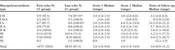
Cardiac anomalies were observed in 34 of the 37 patients (59.6%) at the first echocardiographic study, performed at a median age of 5.0 years, with a range from 0.3 to 35.7 years, and in 48 of 55 patients (87.3%) at the last echocardiographic study, carried out at the median age of 10.7 years, with a range from 1.2 to 65.3 years. The median age at the diagnosis of cardiac involvement in the overall cohort was 5.0 years, with a range from 0.3 to 35.7 years. Cardiac involvement at baseline was more frequent in those falling in the first and second types of the disease. It was noted at an earlier age in the Hurler subgroup of type I, at a median age of 1.3 years, with a range from 0.5 to 1.5 years, in the severe variant of Hunter’s syndrome, with a median age of 3.5 years and a range from 2.0 to 9.6 years, and in those with the attenuated variant of Hunter’s syndrome, for whom the median age of cardiac involvement was 4.8 years, with a range from 4.6 to 13.6 years when compared to those having disease of the other types (Tables 1 and 2). Cardiac anomalies were the first presenting signs of disease in 4 children at the ages of 1.2 years for a patient with Hurler syndrome, 3 years for one with Hunter’s syndrome, 9 years for a subject with Sanfilippo syndrome, and 0.3 years for a patient with Morquio syndrome. Cardiac changes also brought to our notice 2 adults with the Scheie variant of type I disease, at the ages of 19 and 35 years. These two latter patients were correctly diagnosed as having Scheie’s disease many years after the initial cardiac clinical signs were noted, with diagnostic delays of 13 and 18 years, respectively.
Characteristics and diagnoses of the patients.

Legend: MPS – mucopolysaccharidosis, H – Hurler, H-S – Hurler-Scheie, S – Scheie.
*5 patients died during follow-up (2 patients with type I Hurler, 1 with type I Hurler-Scheie, 1 with type III, 1 with type IV) and ^ other 2 were lost to follow-up (1 type I Hurler, 1 type I Scheie).
Patients with cardiac anomalies at the first and last echocardiographic studies.
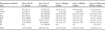
Legend: MPS – mucopolysaccharidosis, H – Hurler, H-S – Hurler-Scheie, S – Scheie, N – number, N – number, Score 1: echocardiography score at baseline, Score 2: echocardiography score at last examination.
*1 patient lost at follow-up.
Types and progression of cardiac anomalies
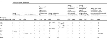
The types of cardiac anomalies observed at baseline and at the last echocardiographic examination are presented in Table 3. The most common finding at both occasions was mitral valvar insufficiency, variable in degree among patients from mild to severe, with dysplastic and thickened leaflets. Mitral insufficiency was detected in almost half of all patients (49.1%) at baseline, and in over four-fifths (81.8%) at the last examination. Such lesion was therefore found in 45 of 48 (93.7%) patients with cardiac involvement. They were often associated with aortic valvar insufficiency, but also with asymmetric septal hypertrophy or hypertrophic cardiomyopathy with concentric left ventricular hypertrophy. Aortic insufficiency was found in 20 of the 57 patients (35.1%) at baseline, and in 31 of 55 patients (56.4%) at the last echocardiographic examination. It was detected in almost two-thirds (64.5%) of cases with cardiac involvement at the last examination.
Types of cardiac anomalies in the patients at their first and last echocardiographic studies.

Legend: PE – pericardial effusion, PHT – pulmonary hypertension, AS – aortic stenosis, Tr – tricuspid regurgitation, H – Hurler, H-S – Hurler-Scheie, S – Scheie.
Insufficiency of both mitral and aortic valves was already present at baseline in 17 patients (29.8%), and had involved 29 patients (52.7%) at the last examination. Both valves were abnormal in 29 of the 48 patients with abnormal echocardiographic findings at follow-up. A reduced velocity of the diastolic slope, indicative of the presence of mild mitral stenosis in addition to mitral insufficiency, was detected in one infant with Hurler syndrome at 2 years of age.
Asymmetric septal hypertrophy or hypertrophic cardiomyopathy were seen in 6 patients at baseline, and in 18 at the last echocardiographic study. These anomalies were noted in association with valvar lesions in 17 cases during the final study, and as an isolated anomaly in 1 patient.
Echocardiographic scores
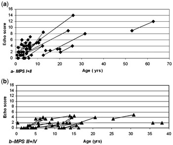
In Table 2, we show the median echocardiographic scores at baseline and at last examination for the whole cohort, as well as for the various subgroups of the disease. Cardiac lesions progressively worsened in patients with all types of the disease during follow-up. Overall, we found a statistically significant correlation of the scores with age (p less than 0.0001, Pearson r equal to 0.55 at baseline; p less than 0.0003, Pearson r equal to 0.48 at last echocardiography). The median ratings for the whole cohort increased from 2, with a range from 1 to 9 at baseline, to 4 at the last examination, the range increasing from 1 to 14. There was a significant difference (p less than 0.0001) between these scores. Scores at the first and last studies were both higher for those with disease of the first and second types than for those falling into the third and fourth subgroups of the disease. The difference between subgroups was tested considering together the first and second groups with respect to the third and fourth groups. No significant difference was noted for the initial score 1 (p less than 0.06 ), but the difference became significant (p less than 0.017) for the final score. The delta scores for the first and second groups were also significantly different when compared to those for the third and fourth groups (p less than 0.00001 – Fig. 1).
The scores (a) at follow-up in the first and second subgroups of mucopolysaccharidosis, and the comparable scores (b) for the third and fourth subgroups.

Therapeutic interventions and outcomes
Standard cardiovascular therapy: Out of 48 cardiologic patients, 16 (33.3%) received standard therapy at some stage for haemodynamic complications caused by valvar lesions or cardiac failure. Of these patients, 4 showed left ventricular dysfunction during the acute phase of failure, associated with mild pericardial effusion and with respiratory infections in 3. Medical therapy was able to be discontinued in 1 patient with Hurler syndrome 2 years after transplantation of haematopoietic stem cells.
Surgical interventions: In 3 patients with the Scheie variant of the first type of the disease, and in another with the attenuated variant of Hunter’s syndrome, the mitral and aortic valves were replaced because of severe worsening of their lesions at ages ranging from 13 to 53 years. Of this group, 3 patients remain free of signs of congestive cardiac failure about 10 years after surgery. The fourth patient, a young woman with the Scheie variant, had moderate residual right ventricular dilation and pulmonary hypertension at the last visit, 2 1/2 years after cardiac surgery.
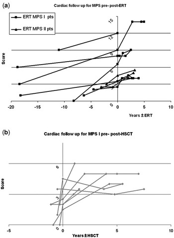
Enzyme replacement therapy: Of the patients who started enzyme replacement therapy, 1 has yet to have an examination as part of the follow-up. For the 8 patients examined subsequent to enzyme replacement therapy, their scores prior to treatment varied from 2.5 to 8, with a median of 4. At the last evaluation, after intervals varying from 2 to 5 years, with a median of 2.7 years, the scores ranged from 4 to 14, with a median of 5.25. Of these patients, 4 are deemed stable from a cardiologic point of view, while the valvar lesions have progressed in the other 4 (Fig. 2a). One of these latter patients was the young lady with Scheie syndrome who required replacement of both valves 2 years after the start of enzyme replacement therapy.
The scores at follow-up (a) in those with type I disease before and after enzyme replacement therapy, and in those with type I disease before and after haematopoietic stem cell transplantation (b).

Transplantation of haematopoietic stem cells: Of the 7 patients who received haematopoietic stem cells, 6 are alive at the median age of 7.0 years, with a range from 2.1 to 8.0 years, with median follow-up of 5.5 years and a range from 1.5 to 7.5 years. They all have stable complete or partial chimerism. Levels showing activity of the enzymes are normal. Their median score prior to treatment was 2.5, with a range from 1.5 to 4.5. At the last evaluation, from 1.5 to 7.5 years after the transplantation, with a median of 5.5 years, their scores varied from 3.5 to 5, with a median score of 4 (Fig. 2b). Cardiac function appears to worsen immediately after the transplantation of the stem cells, but then stabilizes with longer follow-up. It has remained unchanged in the sixth patient during a short period of follow-up.
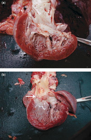
Deaths: During follow-up, 5 patients have died. A boy with Hurler syndrome, who did not have abnormal findings at echocardiography, died at the age of 6 years due to respiratory problems. A girl, also with Hurler syndrome, died one year after transplantation of haematopoietic stem cells at the age of 2.8 years due to pulmonary haemorrhage following immune thrombocytopenia and autoimmune anaemia during a period of rejection. Major cardiac abnormalities were seen at autopsy (Fig. 3). A young woman with the Hurler-Scheie variant, who had mild-to-moderate cardiac involvement, died of obstructive sleep apnoea at the age of 19 years. A 40 year woman with Morquio syndrome, and with mild cardiac involvement, died in a car accident. The final death occurred in a 9 year old girl with Sanfilippo syndrome, who previously did not show echocardiographic abnormalities, but who died suddenly at home. No recent cardiac evaluation was available at the time of death.
The autopsy specimen (a) shows a dysplastic and thickened mitral valve with a thickened subvalvar apparatus and parietal left ventricular hypertrophy. The specimen was obtained from a girl with Hurler disease who died at the age of 2.8 years 1 year after rejection of transplanted haemotopoietic stem cells. Panel b shows the dysplastic aortic valve of the same patient.

Pathological studies
Postmortem examination, performed in the girl with Hurler syndrome who died 12 months after transplantation of haematopoietic stem cells at the age of 2.8 years, revealed grossly thickened mitral and aortic valvar leaflets and hypertrophy of the septum, left ventricular wall, and of the subvalvar apparatus (Fig. 3a, b). Similar macroscopic observations were made from valvar tissues obtained during replacement of both valves in a woman aged 24 with Scheie syndrome, who was receiving enzyme replacement therapy over 2 years, as discussed above.
Discussion
Our findings underline the well accepted fact that progressive cardiac involvement is common in patients with the various forms of mucopolysaccharidosis.Reference Gross, Williams, Caprioli, Dominguez and Howell4, Reference Wippermann, Beck, Schranz, Huth, Michel-Behnke and Jungst8–Reference Rigante and Segni11 Cardiac involvement was present at diagnosis in more than a half of our patients, and was progressive in the majority. Our data confirms that mitral regurgitation is the most common lesion, and is usually the first sign of cardiac involvement to appear in all types of the disease,Reference Wippermann, Beck, Schranz, Huth, Michel-Behnke and Jungst8–Reference Rigante and Segni11 but that all four valves can be involved. The frequency of mitral valvar involvement in nine-tenths of those with cardiac abnormalities at their last echocardiogram, is even higher than previously reported.Reference Wippermann, Beck, Schranz, Huth, Michel-Behnke and Jungst8–Reference Rigante and Segni11 Aortic valvar changes were also detected in two-thirds of these patients, and seemed to occur preferentially in association with other cardiac structural lesions, and not as an isolated anomaly. Mitral regurgitation was previously been said to be more frequent in those with Hurler syndrome, and aortic regurgitation in those with Hunter’s and Morquio syndromes.Reference Mohan, Hay, Cleary, Wrath and Patel10 In contrast to these observations, but in agreement with another report,Reference Dangel9 we did not find any lesion to be characteristic for the different types of the overall disease. Only 2 out of our 8 patients with the fourth type, Morquio syndrome, had isolated aortic insufficiency.
As we observed in pathologic studies in 2 cases, the leaflets of both valves undergo diffuse thickening, with shortening of the tendinous cords of the mitral valve. The leaflets may assume a nodular appearance and become dysplastic.Reference Renteria, Ferrans and Roberts27 Valvar damage can progress to stenosis, even of a severe degree.
In contrast to the majority of the published reports concerning this disease,Reference Wippermann, Beck, Schranz, Huth, Michel-Behnke and Jungst8–Reference Mohan, Hay, Cleary, Wrath and Patel10 we had the opportunity to follow a relatively large number of patients for a long period of time, and to document that valvar morphology and function continued to deteriorate in all types of the disease with age, and that the number of cardiac anomalies increased. A positive correlation between age and the development of abnormalities of the mitral and aortic valves has previously been noted in patients with different types of the disease,Reference Mohan, Hay, Cleary, Wrath and Patel10 but not by all observers,Reference Wippermann, Beck, Schranz, Huth, Michel-Behnke and Jungst8 presumably due to the less severe and less progressive cardiac involvement in their older patients, and to the variability between individual patients.
The scoring system that we devised to assess the complexity of cardiac anomalies at baseline and at the last evaluation enabled us to confirm that worsening of cardiac involvement occurs significantly faster in those with the first type, comprising the Hurler, Hurler/Scheie, and Scheie syndromes, and the second type, including the severe and attenuated variants of Hunter’s syndrome, compared to those falling into the third and fourth types (Sanfilippo and Morquio groups). The patients with the Hurler variant of the first type had in our series the earliest onset of cardiac lesions, around the age of one year. In those with Sanfilippo and Morquio syndromes, the heart is usually affected at a later age, and to a lesser degree, although the extent and severity of cardiac progression may vary between patients.Reference Schieken, Kerber, Ionasescu and Zellweger2, Reference Johnson, Vine, Cottrill and Noonan3, Reference Tan, Schaff, Miller, Edwards and Karnes7–Reference Rigante and Segni11 The differences in progression between patients falling in the different types were obvious, in spite of the fact that some of the patients in our series with the first two types received enzyme replacement therapy or transplantation of haematopoietic stem cells. These treatments may probably have slowed down or stabilized the natural course of cardiac involvement in the disease in individual patients as shown in Figure 2.
Other structural cardiac abnormalities, such as left ventricular hypertrophy, cardiomyopathy, and coronary arterial anomalies with associated ischaemia or infarction have been reported.Reference Donaldson, Pennock, Berry, Duncan, Cawdery and Leonard12, Reference Hayflick, Rowe, Kavanaugh-McHugh, Olson and Valle13, Reference Renteria, Ferrans and Roberts27 All these findings may contribute to the development of cardiac failure. We have observed asymmetric septal hypertrophy or mild to moderate concentric left ventricular hypertrophy in one-third of our patients, usually associated with valvar lesions. A few of our patients developed cardiac failure, usually secondary to the haemodynamic impact of valvar lesions, but had only mild left ventricular dysfunction. In contrast, left ventricular dysfunction and cardiac failure were reported frequently in one series, with subsequent death in a considerable number of cases.Reference Mohan, Hay, Cleary, Wrath and Patel10 Deaths from other cardiac causes, such as hypertrophic cardiomyopathy and cardiac failure have also been reported.Reference Dangel9 The deaths of 5 patients in our cohort were, in most instances, not directly caused by cardiac complications, although the patient who died following immune complications during rejection after transplantation of haematopoietic stems had cardiac impairment. As for the patient who died suddenly, arrhythmia cannot be excluded as a causal factor, since sudden death has been reported in other patients with mucopolysaccharidoses, albeit without specific findings at autopsy.Reference Renteria, Ferrans and Roberts27
Due to an increased awareness of cardiac involvement in mucopolysaccharidoses, patients are nowadays examined at an earlier stage, and monitored more thoroughly. Patients with the mucopolysaccharidoses may benefit from earlier cardiac therapies, including prophylaxis of endocarditis in those with valvar lesions. We may expect that this may influence positively the progression of the cardiac involvement. Cardiac surgery also seems to give much better results than in the past,Reference Dangel9 as we could see in our 4 patients undergoing surgery.
New therapeutic strategies, specifically enzyme replacement therapy and transplantation of haematopoietic stem cells, are expected to improve the course of the disease.Reference Ortega, Dominguez and Olive14–Reference Harmatz, Ketteridge and Giugliani23 Enzyme replacement therapies have been developed for treatment of the first,Reference Wraith, Clarke and Beck21 second,Reference Muenzer, Wraith and Beck22 and sixthReference Harmatz, Ketteridge and Giugliani23 types of the disease. Both enzyme replacement therapy and transplantation of haematopoietic stem cells have been reported to have beneficial effects on those with the first and sixth types of the disease, preserving the cardiac function, with regression of hypertrophy in some patients.Reference Herskhovitz, Young and Rainer17, Reference Braunlin, Berry and Whitley28 Valvar lesions, nonetheless, may progress despite a good engraftment.Reference Viñallonga, Sanz, Balaguer, Miro, Ortega and Casaldaliga15–Reference Braunlin, Stauffer and Peters19 As described, we have found a progression of valvar lesions in half of our patients receiving enzyme replacement therapy, albeit only for a median duration of 2.4 years. These patients, however, were old when starting their treatment, and had severe disease. This may explain the lack of demonstrated efficacy. Out of the 6 patients receiving haematopoietic stem cells, after an initial period of deterioration, 4 have stabilized their cardiac anomalies and normalized their asymmetric septal hypertrophy, allowing discontinuation of cardiac medications in one case. In conclusion, in agreement with the observations of others,Reference Braunlin, Stauffer and Peters19, Reference Braunlin, Berry and Whitley28, Reference Wraith29 our results are encouraging, but it is still too early to draw definite conclusions on the efficacy of the new treatments in blocking the progression of the cardiac involvement in those with the various forms of mucopolysaccharidosis.
Acknowledgements
We thank the non-profit organizations Fondazione Pierfranco e Luisa Mariani, Milano, Comitato Maria Letizia Verga, Monza, and the Italian Mucopolysaccharidoses Association for their continuous financial support. We also thank Hans Ebels, from Genzyme Europe BV, for his editorial support. Financial disclosure: The authors did not receive financial support for preparation of the manuscript and do not have financial interests related to it. Hans Ebels, Genzyme Europe BV, provided some editorial support (Genzyme Corporation, Cambridge, MA is manufacturer of Aldurazyme for treatment of MPS I).