Introduction
Orbital apex pathologies include orbital tumours, such as haemangioma and schwannoma.Reference Vohra, Escott, Stevens and Branstetter1 The surgical approach for orbital tumours must provide optimal visualisation and maximum safety, especially for the highly vulnerable optic nerve. The approach is chosen mainly based on the location, the extension and the type of lesion.Reference Schick, Dott and Hassler2–Reference Lee, Ramakrishnan, Chiu, Palmer and Gausas4 Access to the orbital apex has traditionally been accomplished through conventional open orbitotomy or neurosurgical approaches,Reference Khan and Varvares5 the latter of which is seldomly used nowadays. The advent of endoscopic surgery and intra-operative image guidance has offered a less invasive and more precise approach to the orbital apex without crossing the optic nerve.Reference Cavallo, Messina, Cappabianca, Esposito, de Divitiis, Gardner and Tschabitscher6–Reference Stokken, Gumber, Antisdel and Sindwani7 From reviewing the literature, it appears that only a few studies report using the endoscopic approach for managing apical lesions. This study aimed to present a series of orbital apex lesions managed purely by the endoscopic transnasal approach in order to improve the current knowledge of this approach and to report its long-term clinical, visual and radiological outcomes.
Materials and methods
Two tertiary hospitals (Sheba Medical Center and Tel-Aviv Medical Center, Israel) participated in this study. Patients with isolated orbital apex lesions who underwent transnasal endoscopic surgery were included. This approach was deemed feasible for the tumours situated inferiorly to the anterior skull base and medially to the infraorbital nerve. Apical lesions managed by transorbital approach or irradiation were excluded. In addition, patients who underwent biopsy as a sole procedure were excluded too. Demographic data, clinical data, visual fields, surgical reports, pre- and post-operative images, data on adjuvant therapy, complications, and follow-up information were retrieved from a database.
The pre-operative diagnostic workup included computed tomography, magnetic resonance imaging (MRI) scans, visual field and a neuro-ophthalmological evaluation in all but one case. All patients scheduled for the endoscopic approach were informed about the possibility of switching to an open approach.
A complete ophthalmic examination was performed at each follow-up visit, including best-corrected visual acuity, intraocular pressure, ocular motility, Hertel exophthalmometer measurements, anterior segment, fundoscopy, computerised visual field (Humphrey visual field) and Ishihara colour plates.
Surgical technique
Although the surgery varied slightly in each patient according to the site of origin and extent of the lesion, the surgery included six main surgical steps. (1) Registration and calibration of an intra-operative navigation system. (2) Resection of the middle turbinate sparing its skull base attachment and tail, followed by a middle antrostomy, complete ethmoidectomy and optional sphenoidotomy. (3) Bony orbital wall removal inferior to the level of the anterior ethmoidal artery and medial to the infraorbital nerve, when required. (4) Opening of the periorbit and exposure of the inferior and medial rectus, enabling access to the lesion. (5) To get good access to the intraconal lesion, these two muscles were retracted apart before starting the resection of the lesion itself. An additional tool can be introduced through the contralateral nostril by creating a temporary cut in the nasal septum, allowing binasal intraorbital manipulation. (6) At the end of the surgery, exposed orbital fat was covered with a vascularised pedicled mucoperichondrial flap harvested from the ipsilateral side of the nasal septum and bolstered with dissolvable nasal tamponade in order to reduce post-operative enophthalmos and to facilitate surgical cavity re-epithelialisation in three of the cases. In the other four cases, according to the institutional policy, silicone sheath was placed laterally over the orbital content to avoid adhesions without creating a mucoperichondrial flap.
Peri-operative management and adjuvant therapy
Nasal packing was gradually removed within 48 hours. An antibiotic agent (a second-generation cephalosporin or amoxicillin-clavulanate) was given for at least seven days until the silicon sheath removal. Oral steroids were started on the day of surgery and tapered down rapidly. Nasal irrigation with saline solution twice daily was recommended for at least one month. All patients were followed clinically and with MRI imaging.
Ethics committee
The research was overseen by the formal ethics committee of both medical centres and adhered to the tenets of the Helsinki declaration.
Statistical analysis
Categorical variables were summarised according to their frequency and percentage. Continuous variables were evaluated for normal distribution. Statistical tests were two-sided, and p < 0.05 was considered significant. Non-parametric tests were used for the comparison of numerical variables, such as the logarithm of the minimum angle of resolution for visual acuity and enophthalmos measurements. SPSS® statistical software statistical analysis was used (version 24).
Results
Baseline characteristics
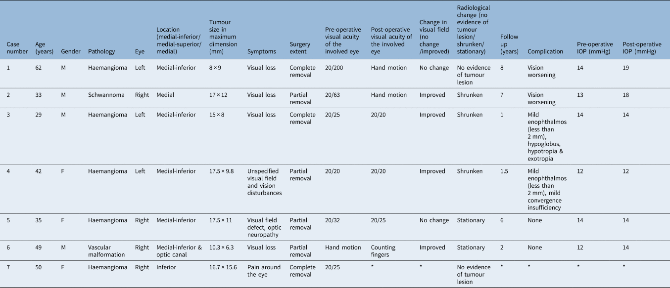
From 2011 to 2019, seven patients with orbital apex lesions were treated purely by the endoscopic transnasal approach at two tertiary hospitals (Sheba and Tel-Aviv medical centres). The study included 4 males (57.1 per cent) and 3 females (42.9 per cent). Five patients underwent surgery for an orbital apex haemangioma, one for an orbital apex schwannoma and one for a suspected vascular malformation. The distribution of tumours was similar in the right and left eyes. Histology confirmed the diagnosis post-operatively. The mean (± standard deviation) age was 42.9 years (± 12 years), with a range of 29–62 years. The distribution of the anatomical location of lesions is presented in supplementary Figure 1, available on The Journal of Laryngology & Otology website. All patients presented with visual acuity, visual field deterioration or both except for one patient, patient number 7, who presented with infraorbital pain. The clinical characteristics are presented in Table 1.
Baseline characteristics of patients

*Minimum, maximum. SD = standard deviation
Post-operative results
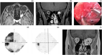
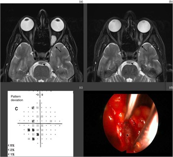
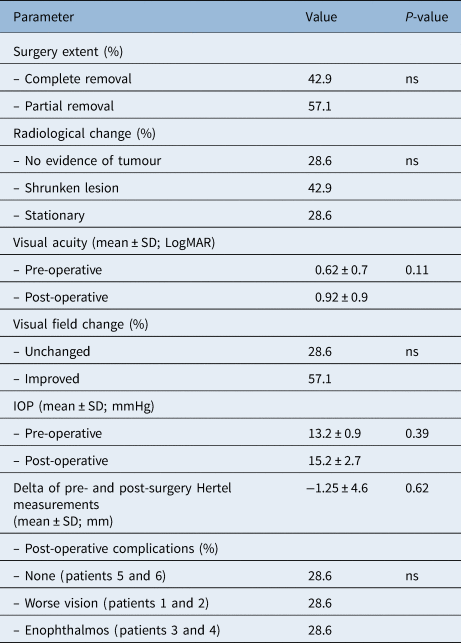
The tumour was completely resected in 3 patients (42.9 per cent) with haemangiomas, and partial removal was performed in the remaining 4 patients (57.1 per cent). Two patients (28.6 per cent) had no radiological evidence of tumour lesions post-surgery. The lesions of three patients (42.9 per cent) significantly shrunk post-surgery, and two patients (28.6 per cent) had no significant radiological change post-surgery. The radiological outcomes of patients 3, 4 and 5 are presented in Figures 1 and 2 and supplementary Figure 2 (available on The Journal of Laryngology & Otology website), respectively. One patient (patient number 7) was lost to follow up after the complete removal of the tumour. Therefore, no visual acuity and visual field results were documented.
A patient with left orbital apex cavernous haemangioma (patient 3). (a) Pre-operative magnetic resonance image (axial T1-weighted sequence with gadolinium). A black arrow points to the tumour. (b) Pre-operative magnetic resonance image (coronal T1-weighted sequence with gadolinium). A white arrow points to the tumour. (c) Intra-operative endoscopic view. Asterisk indicates the tumour. (d) Pre-operative computerised visual field (greyscale) of the patient's left eye showing the visual field defect. (e) Post-operative computerised visual field of the patient's left eye showing significant improvement. (f) Post-operative magnetic resonance image (coronal T1-weighted sequence with gadolinium), showing orbital fat prolapse into the adjacent paranasal sinuses. MRM = medial rectus muscle; IRM = inferior rectus muscle.

A patient with left orbital apex cavernous haemangioma (patient 4). (a) Pre-operative magnetic resonance imaging (axial T2-weighted sequence with gadolinium). The black arrow points to the tumour. (b) Post-operative magnetic resonance imaging (axial T2-weighted sequence with gadolinium) showing the shrunken tumour post-partial resection. The black arrow points to the tumour. (c) Pre-operative computerised visual field (greyscale) of the patient's left eye showing the visual field defect. The post-operative visual field was normal. (d) Intra-operative endoscopic view. The asterisk indicates the tumour in the orbital apical intraconal fat.

A one to eight-year (4.25 ± 3.1 years) follow up was performed. The overall best-corrected visual acuity for distance improved in three patients, visual acuity remained stable in one patient and two patients had decreased visual acuity post-operatively. Visual acuity improved from 20/32 pre-operatively to 20/25 post-operatively in one patient, 20/25 to 20/20 in the second patient and improved from hand motion to counting fingers in the third patient. Visual acuity remained stable in one patient (20/20) and decreased in the other two (from 6/60 to hand motion and 6/20 to hand motion).
There was no statistically significant difference in visual acuity before (0.62 ± 0.7 logarithm of the minimum angle of resolution, Snellen chart, 20/80) and after (0.92 ± 0.9 logarithm of the minimum angle of resolution, Snellen chart, 20/160) surgery (p = 0.11). The visual field improved in 4 patients (57.1 per cent) and remained stable in 2 patients (28.6 per cent). The visual field outcome of patient number 3 is presented in Figure 1, demonstrating a normal visual field post-surgery. There was no statistically significant difference in intraocular pressure before (13.2 ± 0.9 mmHg) and after surgery (15.2 ± 2.7 mmHg; p = 0.39). Post-operative outcomes are presented in Table 2.
Post-operative results

ns = not significant; SD = standard deviation; LogMAR = logarithm of the minimum angle of resolution; IOP = intraocular pressure
Major complications in our study group included vision worsening down to hand motion detection in two patients (patient 1 with schwannoma and patient 2 with haemangioma) as a result of compressive optic neuropathy because of a rapid tumour progression rate. Mild complications included minimal enophthalmos (less than 2 mm) and diplopia in 2 cases of haemangioma lesions. One of these cases (patient 3, Figure 1) experienced hypoglobus, hypotropia and exotropia of his left eye. He was scheduled to undergo orbital wall repair with a custom-made implant to correct both hypoglobus and enophthalmos. The second patient, who underwent partial haemangioma removal, suffered from mild enophthalmos and convergence insufficiency treated successfully with eye exercises only. There was no statistically significant difference in Hertel measurements pre- and post-operatively, with a mean delta of −1.25 ± 4.6 mm (p = 0.62). Complete removal of the tumour in patient 5 failed because of severe bleeding and a tight adherence of the lesion to the optic nerve. Thus, apex decompression was performed in order to preserve sight. During a six-year follow-up period, this patient experienced no complications and remained stable. Table 3 summarises the patient data.
Patients' clinical characteristics and outcome

*Data were not obtained. IOP = intraocular pressure; M = male; F = female
Discussion
Cavernous haemangioma, the most common vascular tumour of the orbit, is a benign tumour that tends to occur in adulthood as a relatively stable or slowly progressive lesion producing painless proptosis.Reference Ferdousi, Sheibani and Baghini8 Schwannoma, a benign tumour of the peripheral nerve sheath,Reference Ducatman, Scheithauer, Piepgras, Reiman and Ilstrup9 can cause proptosis and diplopia.Reference Gupta, Sharma and Singh10 When located at the orbital apex, these pathologies are challenging for diagnosis and treatment. Biopsy or tumour resection must be executed in a manner that does not harm critical structures, including the optic nerve, the ophthalmic artery, the ciliary ganglion, and the extraocular muscles and their motor innervation. Violating these structures carries the risk of morbidities, such as loss of vision, mydriasis or diplopia. Minimising manipulation of the orbital tissues decreases complications. Thus, the surgeon should balance the need for adequate exposure to allow for dissection of the tumour and conducting surgery that is as minimally invasive as possible.
Our study demonstrated that complete removal of orbital apex tumours could be performed using the transnasal endoscopic technique only. The lack of neurovascular retraction and the absence of skin incision make the transnasal endoscopic procedure preferable for infero-medial apical lesions. Nevertheless, it should be considered that vision impairment after the procedure can occur, as seen in two of our cases. These patients had a rapid tumour progression rate; thus, it is reasonable to assume that without the procedure, they would have lost their vision entirely because of the disease. Yet, haemangiomas usually progress slowly,Reference Ferdousi, Sheibani and Baghini8 and patients without significant vision deficits can be observed. Those with signs of progression or severely symptomatic patients, such as the patients included in our study, should be offered surgery. Complete tumour removal should be weighed against the risk of damage to the optic nerve because preservation of vision is our primary goal. The possibility of partial resection should be considered to reduce this potential visual damage (as was demonstrated in two patients in this study; Table 2). In addition, a deep medial decompression itself enables the preservation of vision. This option should be offered to patients with tight tumour adherence to the optic nerve (patient 5).
The review by Curragh et al. suggested that the endoscopic transnasal endoscopic route should be considered a preferable approach in selected orbital lesion excisions, such as for haemangiomas in the medial orbit.Reference Curragh, Halliday and Selva11 Yoshimura et al. reported a case of a 48-year-old patient with a 1.5 × 1.1 cm mass lesion extending from the medial-inferior part of the left orbital apex to the pterygopalatine fossa. He presented with right hemianopsia of the left eye. At the six-month follow-up examination, the entire lesion was successfully removed via the endoscopic transnasal approach, with resolution of all symptoms and signs and no radiological evidence of recurrence on MRI.Reference Yoshimura, Kubo, Yoneda, Hasegawa, Tominaga and Yoshimine12 A similar location of a haemangioma lesion was found in patient number 7 in our study, who also underwent complete removal of the tumour using the endoscopic transnasal approach. The patient was lost to follow up, and post-surgery data were not documented. However, an improvement in symptoms was noted post-surgery.
In the study of Ruiz et al., an endoscopic transnasal approach was undertaken to completely remove a venous malformation from the orbital apex. The tumour was successfully removed in a single specimen without injury to the nearby orbital anatomy. The patient experienced resolution of her presenting diplopia three weeks after the procedure with no adverse effects.Reference Ruiz and Rebeiz13 In addition, in the retrospective chart review of three endoscopic orbital apex surgeries using posterior nasal septectomy, Murchison et al. demonstrated that this procedure improved visualisation and surgical access to the orbital apex and periorbital skull base. There were no complications noted in these cases.Reference Murchison, Rosen, Evans and Bilyk14
In the study by Karaki et al., an orbital apex cavernous haemangioma was removed using an endoscopic transethmoidal approach. In this patient, both visual-field examination and visual acuity were normal before the procedure. No complications were noted after the procedure.Reference Karaki, Kobayashi and Mori15 Locatelli et al. described the case of a 59-year-old woman who was operated on with an endoscopic endonasal transphenoidal transmaxillary transethmoidal approach for a large cavernous haemangioma of the orbital apex. Two months after the procedure, the patient had no neurological symptoms, had intact vision and an MRI of the orbit showed that the tumour had been completely removed.Reference Locatelli, Carrabba, Guastella, Gaini and Spagnoli16 Karaki et al. reported similar results of removing an intraconal orbital apex haemangioma using the endoscopic transethmoidal approach.Reference Karaki, Kobayashi and Mori15 Stokken et al.Reference Stokken, Gumber, Antisdel and Sindwani7 reviewed 18 patients with primary orbital apex lesions and 9 patients with sinonasal lesions predominantly involving the medial orbital apex. All the cases were performed endoscopically using surgical navigation. Sphenoethmoidectomy was performed in cases with isolated orbital apex lesions. They found complications in three patients during a follow-up of four years, including myocardial infarction, deep venous thrombosis and vision loss. The only case of visual decline in this series was in an elderly patient with aggressive B-cell lymphoma who had rapid deterioration to light perception only in that eye pre-operatively and had a decline to no light perception post-operatively. This may be because of the inflammatory nature of lymphoma.
Based on our results and the literature review, we can conclude that the endoscopic transnasal approach is an excellent technique for resecting orbital tumours located inferiorly, medially and infero-medially to the optic nerve. Even when a complete resection cannot be accomplished, this approach alleviates most if not all presenting symptoms. In some patients, complete resection is not the best option. Arai et al.Reference Arai, Kawahara, Yokoyama and Oridate17 and Haruna et al.Reference Haruna, Tukidate, Konno, Fukami and Nakajima18 describe cases where a complete resection of the tumours using this approach would damage critical surrounding tissues, such as the extraocular muscle, optic nerve and ophthalmic artery. In these cases, partial removal of the tumour should be considered. In our study, four patients had partial removal of their tumours. In three of them, it alleviated presenting symptoms, and visual acuity improved at the follow-up appointment up to seven years after the operation. Thus, when malignancy is ruled out, a complete resection not compromising sight or a partial resection by the endoscopic transnasal approach are appropriate options.
In our study, we conducted a follow up of one to eight years. Other than the vision impairment in two patients discussed above, no other major complications were noted in this long period. A few minor complications were noted, including two cases of enophthalmos and strabismus.
The strength of our study is that it is a case series of seven cases that adds to the limited available knowledge regarding this approach and the vision loss complication that could occur in such cases. In addition, a follow up of 1 to 8 years (4.25 ± 3.1 years) is a relatively long period for documentation of any complications resulting from this approach.
• Access to the orbital apex has traditionally been accomplished through open orbitotomy or neurosurgical approaches
• Endoscopic surgery offers a less invasive and more precise approach to the orbital apex
• Endoscopic transnasal approach to the orbital apex is a good technique for resecting tumours located inferiorly and medially to the optic nerve
• When malignancy is ruled out, a complete resection not compromising sight or a partial resection by this approach should be considered
The main disadvantage of our study is that the data were retrospectively obtained. A larger prospective series is required to confirm the outcomes of this approach. Nevertheless, based on our results and the studies presented here, we believe that the transnasal approach is a valuable tool for surgeons who are experienced in the endoscopic technique.
Conclusion
Based on this case series and the literature review, we can conclude that the transnasal approach to infero-medial orbital apex lesions in selected cases may provide a rational alternative to transorbital surgery. Complete tumour removal should be weighed against the risk of damage to the optic nerve.
Supplementary material
The supplementary material for this article can be found at https://doi.org/10.1017/S0022215122001323.
Competing interests
None declared