Introduction
During the last decade, the prevalence of porcine circovirus type 2 (PCV2) has been recorded in several parts of the world including North America, Europe, Australia, Asia and South America. PCV2 has emerged as an important cause of production losses to the pork industry in all these geographical locations (Allan et al., Reference Allan, McNeilly, Kennedy, McNeilly, Ellis, Clark, Harding, Espuna, Botner and Charreyre1998b, Reference Allan, Mc Neilly, Meehan, Kennedy, Mackie, Ellis, Clark, Espuna, Saubi, Riera, Botner and Charreyre1999; Choi et al., Reference Choi, Chae and Clark2000; Fenaux et al., Reference Fenaux, Halbur, Gill, Toth and Meng2000; Rodriguez-Arrioja et al., Reference Rodriguez-Arrioja, Segales, Rosell, Rovira, Pujols, Plana-Duran and Domingo2003b; Castro et al., Reference Castro, Cortez, Ruiz, Leomil, Moreno, Doto and Richtzenhain2004; Banks et al., Reference Banks, Grierson, Tucker, Bailey, Donadeau, Sargent, King and Mellencamp2006; Csagola et al., Reference Csagola, Kecskemeti, Kardos, Kiss and Tuboly2006).
Based on the characteristic clinical signs of wasting, reduced weight gain, jaundice, respiratory complications and lymphoid depletion in 8–15-week-old piglets, the disease syndrome caused by PCV2 was first described as post-weaning multisystemic wasting syndrome (PMWS) (Clark, Reference Clark1996; Harding and Clark, Reference Harding and Clark1997; Ellis et al., Reference Ellis, Hassard, Clark, Harding, Allan, Willson, Strokappe, Martin, McNeilly, Meehan, Todd and Haines1998). With the manifestations of other syndromes associated with PCV2 infection such as reproductive disorders, enteric signs and porcine dermatitis and nephropathy syndrome (PDNS), PMWS was recently renamed as porcine circovirus-associated disease (PCVAD) in the USA and porcine circovirus disease (PCVD) in Europe (Opriessnig et al., Reference Opriessnig, Meng and Halbur2007b).
PCV2 is one of the smallest viruses that are known to affect animals. Its single-stranded DNA genome of 1768 bp (or 1767 bp for PCV2b isolates) in length has a simple, circular, genomic organization with two known functional genes (Hamel et al., Reference Hamel, Lin and Nayar1998). Several milestones have been achieved in understanding the pathogenesis of PCV2. Yet other aspects of PCV2 biology are complex and even paradoxical in nature. For example, although the first clinical cases of PCVAD emerged in the late 1990s, retrospective studies showed that the virus had been in circulation since the 1960s (Grierson et al., Reference Grierson, King, Sandvik, Hicks, Spencer, Drew and Banks2004). The ensuing debate about whether PCV2 is a new virus or an evolved form of an old virus has not been resolved, probably because the minor changes in genetic sequences that occur between strains do not appear to correlate with changes in pathogenicity (Cheung et al., Reference Cheung, Lager, Kohutyuk, Vincent, Henry, Baker, Rowland and Dunham2007; Olvera et al., Reference Olvera, Cortey and Segales2007). While it is now known that PCV2 is a ubiquitous virus and a primary pathogen of pigs, not all pigs that are positive for PCV2 develop PMWS (Balasch et al., Reference Balasch, Segales, Rosell, Domingo, Mankertz, Urniza and Plana-Duran1999). Moreover, pigs that are co-infected with other pathogens such as porcine reproductive and respiratory syndrome virus (PRRSV), swine influenza virus (SIV) and Mycoplasma hyopneumoniae experience increased severity of PCVAD (Dorr et al., Reference Dorr, Baker, Almond, Wayne and Gebreyes2007). However, it is not known whether the trigger for PCVAD manifestations in cases of co-infections with other pathogens arises from PCV2 or the co-infecting pathogen. In the same vein, it is known that PCV2 is immunosuppressive. However, immunostimulation by other pathogens or even certain vaccines and adjuvants has an exacerbatory effect on PCV2 (Darwich et al., Reference Darwich, Segales and Mateu2004). Although high levels of PCV2-specific antibodies are found in both PMWS- and non-PMWS-affected pigs, reduced T and B lymphocyte populations are characteristic of PCVAD (Larochelle et al., Reference Larochelle, Magar and D'Allaire2003). This review is a summary of recent findings that have shed light on these and other questions. Areas that require better understanding are also identified.
PCVAD
Etiology
Porcine circovirus (PCV), a small, non-enveloped DNA virus that belongs to the family Circoviridae, was discovered as a contaminant of the porcine kidney PK-15 cell line in the mid-1970s (Tischer et al., Reference Tischer, Rasch and Tochtermann1974). Approximately 77–95% of pigs in Berlin were found to have high levels of antibody titers to PCV but did not show any signs of disease (Tischer et al., Reference Tischer, Mields, Wolff, Vagt and Griem1986). The virus was later identified in Canada and Britain (Dulac and Afshar, Reference Dulac and Afshar1989; Edwards and Sands, Reference Edwards and Sands1994) leading to the conclusion that it was ubiquitous in nature. PCV was considered to be a non-pathogenic virus because inoculation of pigs with the PK-15 cell contaminant virus did not cause any disease (Tischer et al., Reference Tischer, Mields, Wolff, Vagt and Griem1986). The virus could be easily cultured in PK-15 cells and Vero cells, and was found to infect macrophages and monocytes of porcine and bovine origins (Allan et al., Reference Allan, McNeilly, Foster and Adair1994b). PCV had a sedimentation coefficient of 57 s, and was found to be resistant to disinfection at pH 3.0, chloroform, and 70°C for 15 min (Allan et al., Reference Allan, Phenix, Todd and McNulty1994c).
It was not until 1997 that a variant strain of PCV was found to be associated with a wasting disease in piglets (LeCann et al., Reference LeCann, Albina, Madec, Cariolet and Jestin1997; Nayar et al., Reference Nayar, Hamel and Lin1997; Saxena et al., Reference Saxena, Nayar and Kapil1997; Allan et al., Reference Allan, Meehan, Todd, Kennedy, McNeilly, Ellis, Clark, Harding, Espuna, Botner and Charreyre1998a). Affected piglets experienced clinical signs including weight loss, enlarged lymph nodes, jaundice and un-thriftiness. On necropsy, characteristic histopathological lesions were seen in lymphatic tissue, where severe lymphoid depletion and infiltration with histiocytes with basophilic inclusion bodies were found. Subsequent isolation and characterization of the virus from symptomatic pigs revealed that it was distinct from the previously known type of PCV derived from the PK-15 cells. Sequence analysis revealed that the newly isolated virus had only about 68% nucleotide sequence identity to the existing PCV strain (Hamel et al., Reference Hamel, Lin and Nayar1998). Reactivity of the new isolate to a panel of monoclonal antibodies against PCV was negligible (Allan et al., Reference Allan, Mackie, McNair, Adair and McNulty1994a). The newly discovered pathogenic virus from diseased pigs was designated as PCV2 and the non-pathogenic virus from PK-15 cells was called PCV type 1 (PCV1) (Allan et al., Reference Allan, Meehan, Todd, Kennedy, McNeilly, Ellis, Clark, Harding, Espuna, Botner and Charreyre1998a, Reference Allan, McNeilly, Kennedy, McNeilly, Ellis, Clark, Harding, Espuna, Botner and Charreyre1998b; Hamel et al., Reference Hamel, Lin and Nayar1998; Meehan et al., Reference Meehan, McNeilly, Todd, Kennedy, Jewhurst, Ellis, Hassard, Clark, Haines and Allan1998).
Viruses of the family Circoviridae are small, non-enveloped, single-stranded DNA viruses that affect birds, swine and other mammals. The family Circoviridae is divided into two genera: (i) the genus Gyrovirus, consisting solely of the chicken anemia virus, and (ii) the genus Circovirus, consisting of transfusion transmitted virus (TTV) in humans, the psittacine beak and feather disease virus, circoviruses of other avian species like pigeon, duck and goose and PCV1 and PCV2 (Studdert, Reference Studdert1993).
A closely related family of plant viruses called Nanoviridae has been implicated as the genetic ancestor of PCV1 due to the similarity in the N-terminal regions of their replicase proteins. It is believed that a recombination event with a RNA virus, probably a picorna-like virus, in a mammalian host gave rise to the functional replicase gene (rep) of PCV1 and affected the host switch to a vertebrate species (Gibbs and Weiller, Reference Gibbs and Weiller1999). However, it is not clear how the pathogenic form of the virus, PCV2, evolved. A genetic or antigenic correlation for the acquisition of virulence has not yet been established (Larochelle et al., Reference Larochelle, Magar and D'Allaire2002). It has also been shown that PCV is most closely related to a bovine circovirus isolate and that it can replicate well in bovine leukocytes. However, no further analysis of the connection between bovine and PCVs has been made (Allan et al., Reference Allan, McNeilly, Foster and Adair1994b; Nayar et al., Reference Nayar, Hamel, Lin, Sachvie, Grudeski and Spearman1999). Retrospective analysis of serum samples in Canada (Magar et al., Reference Magar, Muller and Larochelle2000b) and tissue samples with characteristic PMWS lesions from Japan (Mori et al., Reference Mori, Sato, Akachi, Asahi, Taniguchi and Narita2000) indicated that PMWS had been prevalent in pig populations for several years before the first clinical case was diagnosed. The reasons for the sudden emergence of PCV2 as an important swine pathogen are still under speculation.
Molecular genetics of PCVs
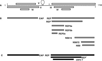
The single-stranded DNA genomes of PCV1 and PCV2 share about 68–76% nucleotide sequence identity and are 1758 (Tischer et al., Reference Tischer, Gelderblom, Vettermann and Koch1982; Fenaux et al., Reference Fenaux, Opriessnig, Halbur, Xu, Potts and Meng2004c) and 1767 or 1768 nucleotides in length, respectively. PCV2 isolates from Europe and the USA had greater than 96% nucleotide sequence identity. The two major genes encoded by the viral genome included the 942 bp rep gene on the positive strand and the 702 bp capsid gene (cap) on the negative strand (Fig. 1). The rep gene is highly conserved between PCV1 and PCV2 with about 83% nucleotide sequence identity, whereas the cap gene has about 67–70% identity (Mankertz et al., Reference Mankertz, Caliskan, Hattermann, Hillenbrand, Kurzendoerfer, Mueller, Schmitt, Steinfeldt and Finsterbusch2004).
Pictorial representation of the genomic, transcriptomic and proteomic organization of PCV2. (A) Putative ORFs of PCV2 as identified by computer algorithms. ORF I, Rep; ORF II, Cap; ORFs III–VI, unknown. The 1768 bp genome with the conserved stem–loop structure is shown. (B) Experimentally identified transcripts of PCV2. The rep and rep′ along with other smaller transcripts are indicated (with permission from Cheung, 2003c). (C). Identified proteins of PCV2. The known Cap, Rep and Rep′ proteins are indicated. The existence of the ORF3 protein remains to be verified.

Transcriptional analysis
Based on initial computerized predictions, six open reading frames (ORFs) were predicted for PCV2 and four of them had sequence homology to the corresponding ORFs in PCV1. A conserved stem–loop structure containing a nonanucleotide motif which was present in the origin of replication of all the members of the Circoviridae family was also identified in PCV2 (Mankertz et al., Reference Mankertz, Persson, Mankertz, Blaess and Buhk1997; Meehan et al., Reference Meehan, Creelan, McNulty and Todd1997, Reference Meehan, McNeilly, Todd, Kennedy, Jewhurst, Ellis, Hassard, Clark, Haines and Allan1998). Transcriptional analysis of PCV1 revealed the presence of three functional mRNAs (Mankertz et al., Reference Mankertz, Buhk, Blaess and Mankertz1998b): two were splice variants of the rep gene and one pertained to the cap gene. Further characterization of ORF1 showed that it encoded a replicase protein which was essential for viral replication. The Rep protein was found to contain sequence motifs that were characteristically associated with rolling circle replication and a putative dNTP-binding box (Mankertz et al., Reference Mankertz, Mankertz, Wolf and Buhk1998a). However, subsequent transcriptional analysis of PCV2 and PCV1 by Cheung revealed nine and 12 mRNA transcripts in each, respectively (Fig. 1). The PCV2 transcripts consisted of one cap gene transcript, five rep splice variants and three non-structural gene transcripts (Cheung, Reference Cheung2003c). In contrast to the findings of Mankertz et al. (Reference Mankertz, Mankertz, Wolf and Buhk1998a), PCV1 had eight rep-associated transcripts besides the capsid and non-structural transcripts (Cheung, Reference Cheung2003a). Mutational analysis helped us to determine that rep and rep′ are the only transcripts that were essential for DNA replication (Cheung, Reference Cheung2003b). SDS-PAGE analysis of purified PCV2 virus indicated that the virus was composed of three major proteins of 28 kDa (Cap), 28.5 kDa (Rep′) and 35 kDa (Rep). The 38 kDa band cross-reacted to PCV1-specific serum, while the reactivity of the 28 kDa band remained specific to each genotype (Pogranichniy et al., Reference Pogranichnyy, Yoon, Harms, Swenson, Zimmerman and Sorden2000) (Fig. 1).
It appears that rep, rep′ and cap genes are the only major genes involved in completing the replicative cycle of the virus. However, it has recently been reported that a new protein encoded by the ORF3 is involved in host cell apoptosis (Liu et al., Reference Liu, Chen and Kwang2005), although others could not independently confirm this report. Carrying out a more detailed, comparative antigenic and proteomic analysis of PCV1 and PCV2 using advanced techniques that are now available will go a long way in improving our understanding of the pathogenesis of PCVAD.
DNA replication
It has been proposed that PCV replicates via a rolling circle melting-pot mechanism (Cheung, Reference Cheung2004b). A 111 bp intergenic region between the cap and rep genes contains the PCV origin of replication (Mankertz et al., Reference Mankertz, Persson, Mankertz, Blaess and Buhk1997). Sequence and secondary structure, which are critical for replication initiation by binding of rep and rep′, include a conserved nonamer at the proximal part of a stem–loop structure, followed by four repeats of a hexameric sequence, both located in the origin of replication (Steinfeldt et al., Reference Steinfeldt, Finsterbusch and Mankertz2001). Mutational analysis showed that one hexamer on the left of the stem–loop is sufficient for supporting replication, but the presence of tandem repeats confers stability on the interaction (Cheung, Reference Cheung2006). Similarly, it was determined that the conformation of the stem–loop, but not the exact nucleotide sequence, was important for binding for the Rep proteins (Steinfeldt et al., Reference Steinfeldt, Finsterbusch and Mankertz2001). The conserved octanucleotide sequence was determined to be indispensable for replication although some nucleotide substitutions were tolerated (Cheung, Reference Cheung2004a).
The promoter of the rep gene was negatively regulated by the full-length Rep protein but not by Rep′, whereas the cap gene did not appear to be regulated by any viral protein. Rep and Rep′ co-localized to the nucleus and formed homo- and heteromeric complexes (Mankertz and Hillenbrand, Reference Mankertz and Hillenbrand2002). Rep, rep′, cap and the origin of replication were found to be interchangeable between PCV1 and PCV2 (Mankertz et al., Reference Mankertz, Mueller, Steinfeldt, Schmitt and Finsterbusch2003; Fenaux et al., Reference Fenaux, Opriessnig, Halbur, Elvinger and Meng2004a). It was determined that Rep and Rep′ proteins have nicking and resealing properties, and three conserved amino acid motifs were found to be essential for the execution of this function (Steinfeldt et al., Reference Steinfeldt, Finsterbusch and Mankertz2006, Reference Steinfeldt, Finsterbusch and Mankertz2007). Further analysis of transcriptional regulation of PCV1 and PCV2 in the context of viral infectivity and host cell responses to infection may shed more light on the complex biology of PCV2.
Phylogenetic analysis and genetic determination of virulence
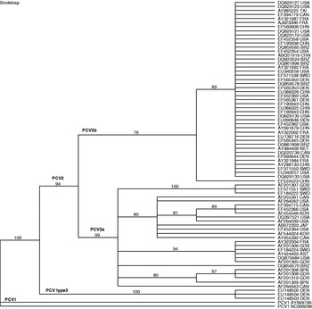
Phylogenetic analyses of porcine, avian and plant circoviruses demonstrated that PCV was most closely related to the psittacine beak and feather disease virus and other avian circoviruses, followed by the plant nanoviruses and geminiviruses. The chicken anemia virus was most distantly related to PCV (Bassami et al., Reference Bassami, Berryman, Wilcox and Raidal1998; Niagro et al., Reference Niagro, Forsthoefel, Lawther, Kamalanathan, Ritchie, Latimer and Lukert1998; Phenix et al., Reference Phenix, Weston, Ypelaar, Lavazza, Smyth, Todd, Wilcox and Raidal2001). Porcine circovirus isolates are grouped into two major genotypes: PCV1 and PCV2. PCV2 is further divided into two major groups, which are now designated as PCV2 group 1 or PCV2b and PCV2 group 2 or PCV2a (Fig. 2). Group 1 viruses cluster into three clades and group 2 into five clades. The PCV2b isolates had a predominantly European origin, whereas the PCV2a isolates had a North American origin (Fig. 2). However, both types were closely related to each other with about 93–100% nucleotide sequence identity (Fenaux et al., Reference Fenaux, Halbur, Gill, Toth and Meng2000). Since the rep gene was highly conserved and more sequence diversity was detected in the cap gene, it is accepted that the cap gene is a suitable candidate for phylogenetic analysis of PCV (Olvera et al., Reference Olvera, Cortey and Segales2007).
A phylogenetic tree of PCV isolates based on the full-length genomic sequences of selected PCV isolates. The phylogenetic grouping of recent PCV2 isolates drawn from GenBank into PCV2a, PCV2b, and PCV2 type 3 isolates is depicted. The tree was generated by the neighbor joining method and bootstrap analysis of 1000 replicates. Numbers above each branch are the bootstrap values. The major branches, namely PCV1, PCV2 and PCV type3 and the subtypes PCV2a and PCV2b, are indicated. Abbreviations to indicate the geographical location of the isolates include: KOR, Korea; CAN, Canada; AST, Austria; GDR, Germany; JAP, Japan; SPN, Spain; CHN, China; BRZ, Brazil; DEN, Denmark; FRA, France; SWD, Sweden; and NET, the Netherlands.

Examination of archived tissues in the UK suggested that PCV2, rather than PCV1, was the predominant circulating genotype at that time and that PCV2 had been present in swine for nearly 30 years before the first clinical case was recognized (Grierson et al., Reference Grierson, King, Sandvik, Hicks, Spencer, Drew and Banks2004). Subsequently, several studies attempted to correlate genetic sequence information with disease status by examination of PCV2 isolates recovered from pigs with or without PMWS (Kim and Lyoo, Reference Kim and Lyoo2002; Larochelle et al., Reference Larochelle, Magar and D'Allaire2002). The general consensus was that no specific geographical or amino acid sequence changes could be correlated with pathogenicity. However, it was found that changes in the immunogenic epitope regions of the Cap protein were more frequent in pathogenic isolates. More recent analysis of several hundred PCV2 isolates confirmed these findings (Olvera et al., Reference Olvera, Cortey and Segales2007). A recent study found that PMWS-associated genotypes belonged to group 1a and non-PMWS genotypes clustered in groups 2c and 2d as classified by Olvera et al. (Reference Olvera, Cortey and Segales2007). PCV2 genotype 1a was never detected in non-PMWS cases in this study. The authors attribute the discrepancy between their study and previous findings to the fact that they detected more than one genotype in the same pig and that studies derived from GenBank data may not be sufficiently annotated with regard to disease status of the pigs from which the isolates were derived (Grau-Roma and Segales, Reference Grau-Roma and Segales2007). The presence of more than one strain of PCV2 existing at the same time in pigs has recently been confirmed. It was also found that recombination in the rep gene can occur between the coexisting strains (Hesse et al., Reference Hesse, Kerrigan and Rowland2008). However, the biological implications of such a recombination have not been investigated.
Recent emergence of more severe PCVAD outbreaks in certain areas in the USA and Canada has been attributed to the emergence of the European-like PCV2b (or PCV2 group 1) isolates in these areas (Gagnon et al., Reference Gagnon, Tremblay, Tijssen, Venne, Houde and Elahi2007). It has recently been shown that PCV2a and PCV2b isolates are cross-protective. However, experimental reproduction of the virulence exhibited by these isolates in the field was not possible (Lager et al., Reference Lager, Gauger, Vincent, Opriessnig, Kehrli and Cheung2007; Opriessnig et al., Reference Opriessnig, Ramamoorthy, Madson, Patterson, Pal, Meng and Halbur2007a). Cheung et al. (Reference Cheung, Lager, Kohutyuk, Vincent, Henry, Baker, Rowland and Dunham2007) identified a unique amino acid sequence motif which could be used to distinguish between PCV2a and PCV2b isolates. Minor antigenic differences have been demonstrated between PCV2 strains using a panel of monoclonal antibodies (Lefebvre et al., Reference Lefebvre, Costers, Van Doorsselaere, Misinzo, Delputte and Nauwynck2008). However, an antibody that can consistently distinguish between PCV2a and PCV2b has not been identified so far. A recent analysis of archived tissue samples in Denmark has revealed that a third PCV2 genotype existed before the appearance of clinical PMWS (1980–1990). It was found that PCV2a isolates were in circulation between 1993 and 1996 in Denmark, followed by PCV2b isolates thereafter, suggesting that genotype switching of PCV2 isolates occurs in a given geographical area with time (Dupont et al., Reference Dupont, Nielsen, Baekbo and Larsen2008).
It has previously been shown that small differences in the amino acid sequence of PCV2 can result in major changes in pathogenicity, as two amino acid changes in the cap gene were sufficient for attenuating a virus (Fenaux et al., Reference Fenaux, Opriessnig, Halbur, Elvinger and Meng2004b; Opriessnig et al., Reference Opriessnig, McKeown, Zhou, Meng and Halbur2006d). While phylogenetic analysis is useful in classification of new strains, it is evident that a more functional genetic approach is required to establish the relationship between sequence changes and pathogenicity.
Epidemiology
Geographical distribution
Since the discovery of PCV1 as a contaminant of the PK-15 cells in 1974 (Tischer et al., Reference Tischer, Rasch and Tochtermann1974) and the subsequent identification of PCV2 as the cause of PMWS in Canada in 1997 (Meehan et al., Reference Meehan, McNeilly, Todd, Kennedy, Jewhurst, Ellis, Hassard, Clark, Haines and Allan1998), both viruses have been found to have a worldwide distribution. The prevalence of PCV2 and PCVAD has been recorded in the USA, European countries such as Ireland, Germany, the Netherlands, Hungary, Spain, Switzerland, Greece and the UK, in Japan, Korea, Taiwan, and in countries of South America, and later in Australia (Allan et al., Reference Allan, McNeilly, Kennedy, McNeilly, Ellis, Clark, Harding, Espuna, Botner and Charreyre1998b; Morozov et al., Reference Morozov, Sirinarumitr, Sorden, Halbur, Morgan, Yoon and Paul1998; Gresham et al., Reference Gresham, Jackson, Giles, Allan, McNeilly and Kennedy2000; Kiss et al., Reference Kiss, Kecskemeti, Tuboly, Bajmocy and Tanyi2000; Rosell et al., Reference Rosell, Segales, Rovira and Domingo2000b; Kawashima et al., Reference Kawashima, Katsuda and Tsunemitsu2007; Olvera et al., Reference Olvera, Cortey and Segales2007). Therefore, PCV2 can be considered to be enzootic in most parts of the world and to acquire epizootic status whenever there is a significant increase in mortality when compared with previous history.
Prevalence
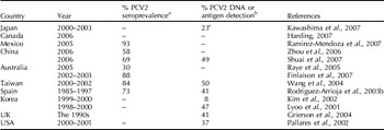
Tischer et al. (Reference Tischer, Mields, Wolff, Vagt and Griem1986) determined that 77–95% of pigs in Berlin were seropositive to PCV in 1986. However, subsequent studies that were conducted after the emergence of PMWS indicated that PCV2, but not PCV1, was the predominant circulating strain even a decade earlier (Quintana et al., Reference Quintana, Segales, Rosell, Calsamiglia, Rodriguez-Arrioja, Chianini, Folch, Maldonado, Canal, Plana-Duran and Domingo2001). The rates of seroprevalence of PCV2 in affected countries like Canada (Magar et al., Reference Magar, Muller and Larochelle2000b), Spain (Rodriguez-Arrioja et al., Reference Rodriguez-Arrioja, Segales, Rosell, Rovira, Pujols, Plana-Duran and Domingo2003b), Taiwan (Wang et al., Reference Wang, Huang, Huang, Tu, Jong, Lin and Lai2004) and Australia (Finlaison et al., Reference Finlaison, Kirkland, Luong and Ross2007) have varied between 40 and 80%. However, the prevalence of PCV2 as shown by the presence of PCV2 antigen or viral DNA in tissue ranged from about 23% in Japan (Kawashima et al., Reference Kawashima, Katsuda and Tsunemitsu2007), 50% in Taiwan (Wang et al., Reference Wang, Huang, Huang, Tu, Jong, Lin and Lai2004), 8% in Korea (Kim and Lyoo, Reference Kim and Lyoo2002), 30–40% in archived tissue samples from 1970–1997 in Britain (Grierson et al., Reference Grierson, King, Sandvik, Hicks, Spencer, Drew and Banks2004) and 10% in the USA (Pallares et al., Reference Pallares, Halbur, Opriessnig, Sorden, Villar, Janke, Yaeger, Larson, Schwartz, Yoon and Hoffman2002) (Table 1). In several of these studies, co-infection with other swine bacterial and viral agents was detected. The difficulty in obtaining exact data on the incidence of PCV2 may be due to the sensitivity of the detection methods and cross-identification of PCV1 and PCV2 in early studies. Although the morbidity rates of PCV2 infection are low, mortality rates are as high as 80% in some affected herds (D'Allaire et al., Reference D'Allaire, Moore and Cote2007).
Prevalence of PCV2 in different geographic regions as determined by detection of PCV2 antibody, antigen or nucleic acid.

a PCV2 antibody detected by serology methods.
b PCV2 DNA detected by restriction fragment length polymorphism (RFLP) or PCR.
c PCV2 antigen detected by immunohistochemistry (IHC).
Transmission
PCV is a non-enveloped virus that is resistant to pH 3, chloroform and temperatures of 70°C for 15 min (Allan et al., Reference Allan, Phenix, Todd and McNulty1994c). Therefore, PCV is environmentally very stable, which plays an important role in promoting transmission and hindering viral elimination. PCV2 is transmitted predominantly by the oro-nasal route. In a controlled study, it has been shown that point source exposure of naïve pigs to PCV2-infected pigs resulted in asymptomatic seroconversion of naïve pigs (Bolin et al., Reference Bolin, Stoffregen, Nayar and Hamel2001). The widespread distribution of the virus in the lymphatic system, the respiratory system, the urogenital system and the gastrointestinal system indicated that the virus would be shed in most of the excretions and secretions of the infected pigs (Rosell et al., Reference Rosell, Segales, Plana-Duran, Balasch, Rodriguez-Arrioja, Kennedy, Allan, McNeilly, Latimer and Domingo1999; Okuda et al., Reference Okuda, Ono, Yazawa and Shibata2003; Krakowka et al., Reference Krakowka, Ellis, McNeilly, Waldner and Allan2005). Experimental confirmation that the virus was present in oro-nasal swabs, feces, urine and blood has been reported (Shibata et al., Reference Okuda, Ono, Yazawa and Shibata2003). Thereafter it has also been shown that the virus is present in colostrum (Shibata et al., Reference Shibata, Okuda, Kitajima and Asai2006) and semen (Larochelle et al., Reference Larochelle, Bielanski, Muller and Magar2000; Kim et al., Reference Kim, Han, Choi and Chae2001; McIntosh et al., Reference McIntosh, Harding, Parker, Ellis and Appleyard2006b). However, experimental proof that insemination or infection of piglets via colostrum is a means of transmission is still lacking to date.
The occurrence of reproductive failure in PCV2 cases led to the investigation of vertical transmission of PCV2. Experimental infection of intra-uterine fetuses through maternal PCV2 inoculation has been demonstrated (Pensaert et al., Reference Pensaert, Sanchez, Ladekjaer-Mikkelsen, Allan and Nauwynck2004; Park et al., Reference Park, Kim, Ha, Jung, Choi, Lim, Kim and Chae2005). However, it appears that natural transmission via this route is rare (Stevenson et al., Reference Stevenson, Kiupel, Mittal, Choi, Latimer and Kanitz2001). It is known that, under both field and experimental conditions, PCV2 can cause a persistent infection characterized by viremia in blood, tissues and semen that lasts for several weeks in carrier piglets, regardless of whether they suffered from PCVAD or not (Rodriguez-Arrioja et al., Reference Rodriguez-Arrioja, Segales, Calsamiglia, Resendes, Balasch, Plana-Duran, Casal and Domingo2002; Larochelle et al., Reference Larochelle, Magar and D'Allaire2003). The exact mechanism and cell types involved in viral persistence are unknown.
PCV2 virus has also been detected in the wild boar. Sequence analysis indicated that the boar isolates were similar to the existing PCV2 strains (Schulze et al., Reference Schulze, Neumann, Grutze, Engelhardt, Mirle, Ehlert and Hlinak2003; Vicente et al., Reference Vicente, Segales, Hofle, Balasch, Plana-Duran, Domingo and Gortazar2004; Csagola et al., Reference Csagola, Kecskemeti, Kardos, Kiss and Tuboly2006). Antibodies to PCV2 have not been detected in cattle, sheep, humans or horses (Allan et al., Reference Allan, McNeilly, McNair, Curran, Walker, Ellis, Konoby, Kennedy and Meehan2000c; Ellis et al., Reference Ellis, Konoby, West, Allan, Krakowka, McNeilly, Meehan and Walker2001; Rodriguez-Arrioja et al., Reference Rodriguez-Arrioja, Segales, Domingo and Plana-Duran2003a). However, previous studies have shown that PCV2 isolates from cattle were 99% similar to existing PCV2 isolates and that PCV2 could infect swine and bovine macrophages (Allan et al., Reference Allan, McNeilly, Foster and Adair1994b; Nayar et al., Reference Nayar, Hamel, Lin, Sachvie, Grudeski and Spearman1999). The presence of PCV2 in insects and other wild animals has not been ascertained as yet; therefore, a sylvatic cycle has not been definitively ruled out. However, given the high level of species specificity of circoviruses, it is likely that prophylactic measures that can achieve high levels of herd immunity should be successful and the possibility of re-infection from unknown sources will be unlikely, except perhaps in areas with proximity to wild boars.
Host genetics and susceptibility
Host genetics is well known to influence susceptibility to diseases. Genetic polymorphisms in outbred populations are responsible for a large diversity in antigen presenting molecules such as the MHC and T cell receptors; therefore two individual animals may not mount a similar immune response to a given antigen or infectious agent. In a recent study conducted to identify T cell epitopes in PCV2, it was found that the identified immunodominant peptides were not uniformly reactive in all PCV2-exposed animals (Stevenson et al., Reference Stevenson, Gilpin, Douglas, McNeilly, McNair, Adair and Allan2007), proving that pigs and PCV2 are no exception to the influence of genetic diversity. It has been shown that certain breeds of pigs such as Landrace pigs appeared to be more susceptible to PCV2 than Duroc or Large White pigs (Opriessnig et al., Reference Opriessnig, Fenaux, Thomas, Hoogland, Rothschild, Meng and Halbur2006a). In another study which compared the susceptibility of pigs derived from Pietrain, Duroc and Large White lines, it was found that pigs that were a mixture of Large White and Duroc were more susceptible to PCV2 infection than purebred Pietrain pigs (Lopez-Soria et al., Reference Lopez-Soria, Segales, Nofrarias, Calsamiglia, Ramirez, Minguez, Serrano, Marin and Callen2004). Therefore it is obvious that host genetics plays a role in PCV2 pathogenesis. Further elucidation of this role is important for devising improved prevention strategies for PCV2.
Pathogenesis and clinical syndromes
PCV2 causes several varied clinical manifestations in affected pigs, and the first of them to be recognized was termed as PMWS. The official nomenclature for the disease has recently been changed to PCVAD. Three characteristic signs of wasting, microscopic lesions of lymphoid depletion with histiocytic infiltration and the presence of PCV2 antigen or DNA in the lesions, are required for a definitive diagnosis of PCVAD in a herd (Segales et al., Reference Segales, Allan and Domingo2005). However, it is known that not all pigs that are infected with PCV2 will develop PMWS and that the incidence of PMWS is higher when co-infecting viral and bacterial pathogens are present (Magar et al., Reference Magar, Larochelle, Thibault and Lamontagne2000a; Albina et al., Reference Albina, Truong, Hutet, Blanchard, Cariolet, L'Hospitalier, Mahe, Allee, Morvan, Amenna, Le Dimna, Madec and Jestin2001). There is also an obvious immune involvement in the pathogenesis of PCVAD.
PCVAD is a disease that exclusively affects grower pigs; it rarely occurs in 1–3-week-old piglets, probably because of protection through maternal antibodies (McKeown et al., Reference McKeown, Opriessnig, Thomas, Guenette, Elvinger, Fenaux, Halbur and Meng2005; McIntosh et al., Reference McIntosh, Harding, Ellis and Appleyard2006a). PCV2 appears to have an incubation period of 7–28 days and early clinical signs of PMWS include fever, lethargy, weight loss, lymph node enlargement, dyspnea and jaundice (Harding and Clark, Reference Harding and Clark1997; LeCann et al., Reference LeCann, Albina, Madec, Cariolet and Jestin1997; Allan et al., Reference Allan, Meehan, Todd, Kennedy, McNeilly, Ellis, Clark, Harding, Espuna, Botner and Charreyre1998a; Allan and Ellis, Reference Allan and Ellis2000; Chae, Reference Chae2004). The primary lesions of PMWS are associated with the lymphatic system. Microscopically, enlarged lymph nodes show atrophic or necrotizing lesions with lymphoid depletion, loss of architecture and infiltration by large histiocytes and giant cells with basophilic inclusion bodies. Lesions are present in the superficial inguinal, mesenteric, mediastinal, submandibular lymph nodes and in the Peyer's patches, spleen, liver, kidney and intestinal mucosa, resulting in generalized un-thriftiness and wasting (Fenaux et al., Reference Fenaux, Halbur, Haqshenas, Royer, Thomas, Nawagitgul, Gill, Toth and Meng2002). Thymic atrophy is another characteristic feature of PMWS. In the lungs, interstitial pneumonia and bronchiolitis with mononuclear infiltration are seen. Liver tissues in affected pigs may exhibit inflammatory and apoptotic changes with mononuclear cell infiltration in the parenchyma (Harding and Clark, Reference Harding and Clark1997; LeCann et al., Reference LeCann, Albina, Madec, Cariolet and Jestin1997; Allan et al., Reference Allan, McNeilly, Kennedy, McNeilly, Ellis, Clark, Harding, Espuna, Botner and Charreyre1998b; Allan and Ellis, Reference Allan and Ellis2000; Bolin et al., Reference Bolin, Stoffregen, Nayar and Hamel2001; Harms et al., Reference Harms, Sorden, Halbur, Bolin, Lager, Morozov and Paul2001). The exact cell types that are involved in replication and spread of the virus or the mechanisms by which lymphoid depletion and the involvement of other organs occur are as yet unknown.
Reproductive failure is another clinical manifestation of PCV2 infection in affected herds. Mid-to-late-term abortions or still-births with affected fetuses exhibiting necrotizing myocarditis and the presence of PCV2 antigen in cardiac tissues have been recorded. Such reproductive failures have also been experimentally reproduced (Bogdan et al., Reference Bogdan, West, Clark, Konoby, Haines, Allan, McNeilly, Meehan, Krakowka and Ellis2001; Bolin et al., Reference Bolin, Stoffregen, Nayar and Hamel2001; Harms et al., Reference Harms, Sorden, Halbur, Bolin, Lager, Morozov and Paul2001; Ladekjaer-Mikkelsen et al., Reference Ladekjaer-Mikkelsen, Nielsen, Storgaard, Botner, Allan and McNeilly2001; O'Connor et al., Reference O'Connor, Gauvreau, West, Bogdan, Ayroud, Clark, Konoby, Allan and Ellis2001; Harding, Reference Harding2004; Pensaert et al., Reference Pensaert, Sanchez, Ladekjaer-Mikkelsen, Allan and Nauwynck2004; Park et al., Reference Park, Kim, Ha, Jung, Choi, Lim, Kim and Chae2005). PCV2 is also responsible for acute diarrheal outbreaks in otherwise normal pigs. On histological examination, enlarged mesenteric lymph nodes that are positive for PCV2 antigen are characteristic of the enteric form of the disease (Yang et al., Reference Yang, Song, Kim, Lyoo and Park2003; Kim et al., Reference Kim, Ha, Jung, Choi and Chae2004; Banks et al., Reference Banks, Grierson, Tucker, Bailey, Donadeau, Sargent, King and Mellencamp2006). PDNS is the fourth manifestation of PCVAD. Although at a very low incidence, piglets affected with PDNS experience very high levels of mortality. Clinical signs include coalescing red to purple skin lesions, glomerular and interstitial nephritis, vasculitis and deposition of immune complexes in kidney tissue. While it is believed that high levels of PCV2 antibodies may trigger a type III hypersensitivity reaction in PDNS cases, experimental proof is still lacking. A Th1 bias is reported to occur in PDNS-affected pigs and it is believed that the resulting pro-inflammatory status may predispose pigs to the disease (Elbers et al., Reference Elbers, Hunneman, Vos, Zeeuwen, Peperkamp and van Exsel2000; Rosell et al., Reference Rosell, Segales, Ramos-Vara, Folch, Rodriguez-Arrioja, Duran, Balasch, Plana-Duran and Domingo2000a; Thomson et al., Reference Thomson, Smith, Allan, McNeilly and McVicar2000; Choi et al., Reference Choi, Kim, Kang and Chae2002a; Wellenberg et al., Reference Wellenberg, Stockhofe-Zurwieden, de Jong, Boersma and Elbers2004b; Sipos et al., Reference Sipos, Duvigneau, Pietschmann, Schilcher, Hofbauer, Hartl and Schmoll2005; Phaneuf et al., Reference Phaneuf, Ceccarelli, Laing, Moloo and Turner2007). PCV2 has also been associated with nervous symptoms manifested as congenital tremors and non-suppurative menigo-encephalitis of the brain (Stevenson et al., Reference Stevenson, Kiupel, Mittal, Choi, Latimer and Kanitz2001; Choi et al., Reference Choi, Stevenson, Kiupel, Harrach, Anothayanontha, Kanitz and Mittal2002b; Larochelle et al., Reference Larochelle, Magar and D'Allaire2002).
Interaction of PCV2 with other pathogens
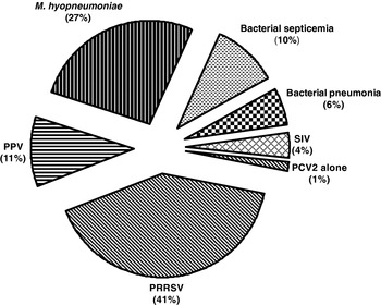
PCVAD rarely occurs in pigs infected with PCV2 alone. The rates of occurrence of co-infecting pathogens in PCVAD as consolidated values (Fig. 3) based on four independent studies include PRRSV (41%), M. hyopneumoniae (27%), bacterial septicemia (10.0%), bacterial pneumonia (6%) and SIV (4%). PCV2 alone caused disease in only 1% of the cases (Ellis et al., Reference Ellis, Bratanich, Clark, Allan, Meehan, Haines, Harding, West, Krakowka, Konoby, Hassard, Martin and McNeilly2000; Pallares et al., Reference Pallares, Halbur, Opriessnig, Sorden, Villar, Janke, Yaeger, Larson, Schwartz, Yoon and Hoffman2002; Drolet et al., Reference Drolet, Larochelle, Morin, Delisle and Magar2003; Grau-Romaet al., Reference Grau-Roma, Crisci, Sibila, Lopez-Soria, Nofrarias, Cortey, Fraile, Olvera and Segales2008) (Fig. 3). Previous experimental and field studies that included retrospective analyses have emphasized the importance of co-infections in pathogenesis of PMWS (Ellis et al., Reference Ellis, Clark, Haines, West, Krakowka, Kennedy and Allan2004) so much so that it was believed that another agent was required for PCV2 to cause disease until it was categorically shown that PCV2 alone could independently cause PMWS (Magar et al., Reference Magar, Larochelle, Thibault and Lamontagne2000a).
Pie chart showing the average percentages of co-infections between PCV2 and various other pathogens in PCVAD cases. Data were compiled from Pallares et al. (Reference Pallares, Halbur, Opriessnig, Sorden, Villar, Janke, Yaeger, Larson, Schwartz, Yoon and Hoffman2002), Ellis et al. (Reference Ellis, Bratanich, Clark, Allan, Meehan, Haines, Harding, West, Krakowka, Konoby, Hassard, Martin and McNeilly2000), Drolet et al. (Reference Drolet, Larochelle, Morin, Delisle and Magar2003) and Grau-Romaet al. (Reference Grau-Roma, Crisci, Sibila, Lopez-Soria, Nofrarias, Cortey, Fraile, Olvera and Segales2008).

PRRSV is most frequently associated with PCV2 in PMWS cases. It is believed that they act synergistically to induce PMWS. PRRSV has been detected in 83% of the PMWS cases in the Netherlands (Wellenberg et al., Reference Wellenberg, Stockhofe-Zurwieden, Boersma, De Jong and Elbers2004a) and about 52% of the cases in the USA. It has been shown that while PCV2 had the strongest statistical association with PMWS, co-infection with PRRSV increased the risk of PCVAD by several folds (Pogranichniy et al., Reference Pogranichniy, Yoon, Harms, Sorden and Daniels2002; Rose et al., Reference Rose, Larour, Le Diguerher, Eveno, Jolly, Blanchard, Oger, Le Dimna, Jestin and Madec2003). PRRSV also has a strong association with PDNS and necrotizing pneumonia, with a high with co-infection rates of 42 and 45%, respectively (Drolet et al., Reference Drolet, Larochelle, Morin, Delisle and Magar2003; Grau-Roma et al., Reference Grau-Roma, Crisci, Sibila, Lopez-Soria, Nofrarias, Cortey, Fraile, Olvera and Segales2007). PCV2 infection has been implicated in the reduction of PRRSV vaccine efficacy (Opriessnig et al., Reference Opriessnig, McKeown, Harmon, Meng and Halbur2006c). It has been shown that there is a dramatic decrease in leukocyte counts in experimental dual infections of PRRSV and PCV2 (D'Allaire et al., Reference D'Allaire, Moore and Cote2007).
Porcine parvovirus (PPV) is a common pathogen of pigs that causes sporadic reproductive failure. Under natural conditions, PPV and PCV2 have been co-isolated in about 15% of PMWS cases (Ellis et al., Reference Ellis, Bratanich, Clark, Allan, Meehan, Haines, Harding, West, Krakowka, Konoby, Hassard, Martin and McNeilly2000). Experimental co-infection of PPV and PCV2 resulted in reproduction of PMWS in gnotobiotic and colostrum-deprived pigs (Ellis et al., Reference Ellis, Krakowka, Lairmore, Haines, Bratanich, Clark, Allan, Konoby, Hassard, Meehan, Martin, Harding, Kennedy and McNeilly1999; Allan et al., Reference Allan, McNeilly, Ellis, Krakowka, Meehan, McNair, Walker and Kennedy2000a). Recently, it was determined that high levels of TNF-α are induced by co-infection of PPV and PCV2. It is speculated that the strong pro-inflammatory environment may lead to precipitation of PMWS (Kim et al., Reference Kim, Ha and Chae2006). PPV vaccination, however, did not reduce the severity of PMWS in co-infected pigs (Opriessnig et al., Reference Opriessnig, Fenaux, Yu, Evans, Cavanaugh, Gallup, Pallares, Thacker, Lager, Meng and Halbur2004a) but a combined PPV and swine erysipelas vaccine appeared to protect against PCV2-induced reproductive failure (Rose et al., Reference Rose, Blanchard, Cariolet, Grasland, Amenna, Oger, Durand, Balasch, Jestin and Madec2007).
M. hyopneumoniae is another important pathogen that has been found to co-infect with PCV2 in a significant percentage of PMWS cases. The observation that it was easier to experimentally reproduce PMWS in immune-stimulated pigs (Allan et al., Reference Allan, McNeilly, Ellis, Krakowka, Meehan, McNair, Walker and Kennedy2000a, Reference Allan, McNeilly, Ellis, Krakowka, Botner, McCullough, Nauwynck, Kennedy, Meehan and Charreyre2004; Krakowka et al., Reference Krakowka, Ellis, McNeilly, Ringler, Rings and Allan2001; Kyriakis et al., Reference Kyriakis, Saoulidis, Lekkas, Miliotis Ch, Papoutsis and Kennedy2002) led to the speculation that vaccination of piglets with M. hyopneumoniae vaccine or other vaccines between 3 and 5 weeks according to the recommended protocols may potentiate PMWS. It has been shown that immune stimulation that occurs due to Mycoplasma vaccination or infection does increase the incidence of PMWS (Opriessnig et al., Reference Opriessnig, Thacker, Yu, Fenaux, Meng and Halbur2004b, Reference Opriessnig, Halbur, Yu, Thacker, Fenaux and Meng2006b; Krakowka et al., Reference Krakowka, Ellis, McNeilly, Waldner, Rings and Allan2007) and it has been suggested that the timing of vaccination to 2 weeks before anticipated exposure to PCV2 could prevent such an occurrence (Opriessnig et al., Reference Opriessnig, Halbur, Yu, Thacker, Fenaux and Meng2006b).
Some of the other pathogens that have been associated with PCVAD include Cryptosporidium in enteritis cases (Nunez et al., Reference Nunez, McNeilly, Perea, Sanchez-Cordon, Huerta, Allan and Carrasco2003), Aujeszky's disease (Quintana et al., Reference Quintana, Segales, Rosell, Calsamiglia, Rodriguez-Arrioja, Chianini, Folch, Maldonado, Canal, Plana-Duran and Domingo2001; Maldonado et al., Reference Maldonado, Segales, Martinez-Puig, Calsamiglia, Riera, Domingo and Artigas2005), swine influenza and bacterial pneumonia (Pogranichniy et al., Reference Pogranichniy, Yoon, Harms, Sorden and Daniels2002; Kim et al., Reference Kim, Chung and Chae2003; Dorr et al., Reference Dorr, Baker, Almond, Wayne and Gebreyes2007; Gagnon et al., Reference Gagnon, Tremblay, Tijssen, Venne, Houde and Elahi2007; Grau-Roma and Segales, Reference Grau-Roma, Crisci, Sibila, Lopez-Soria, Nofrarias, Cortey, Fraile, Olvera and Segales2007).
Based on these studies, no single co-infecting disease agent can be considered indispensable for development of PCVAD. It is possible that an unknown agent or agents might be playing a role in the development of PCVAD. It is also not known whether the immunosuppressive effect of PCV2 infection facilitates co-infection or whether the co-infecting pathogen aids in inducing PMWS. Clearly, more research is required to understand the specific interactions between these insidious swine pathogens.
Molecular pathogenesis
The molecular pathogenesis of PCV2 is still largely unexplored, probably because of the difficulty in correlating viral genetic information with pathogenicity. The absence of a virally encoded DNA polymerase dictates a heavy dependency of PCV on host cellular enzymes. It has been determined that PCV replicates best in cells that are in the S phase of the cell cycle (Tischer et al., Reference Tischer, Peters, Rasch and Pociuli1987). Indeed in PMWS cases, PCV2 antigen localizes to most actively replicating cells. A conserved heparin-binding motif has been identified in the Cap protein of PCV2, and the glycosaminoglycans (GAGs), heparan sulfate and chondroitin sulfate have been identified as molecules that facilitate attachment of PCV2 to host cells (Misinzo et al., Reference Misinzo, Delputte, Meerts, Lefebvre and Nauwynck2006). GAGs are ubiquitously distributed in animal tissues and while they may serve as the first point of attachment, other fusion and internalization receptors are likely to be involved in the internalization of viral particles. However, considering the fact that PCV2 can infect both immune and epithelial cells in a variety of tissues, it is probable that PCV2 may not require a unique receptor that is indispensable for viral entry.
Recently, a new protein encoded by the ORF3 has been reported to be involved in host cell apoptosis (Liu et al., Reference Liu, Chen and Kwang2005, Reference Liu, Chen, Du, Chua and Kwang2006, Reference Liu, Zhu, Chen, Lau, He, Lau, Wang, Karuppannan and Kwang2007). However, independent confirmation of the existence of the ORF3 protein is lacking to date, and unpublished data from our laboratory contradict this claim (Juhan, Reference Juhan2007).
It has been shown that inhibition of the endolysosomal acidification, actin organization and clathrin-mediated endocytosis in monocytes reduces PCV2 infection (Misinzo et al., Reference Misinzo, Meerts, Bublot, Mast, Weingartl and Nauwynck2005, 2008a), whereas in epithelial cells, inhibition of the endolysosomal acidification increases PCV2 replication. It was also shown that a serine protease mediated-event, probably uncoating of the capsid in the endosome, was required for replication in epithelial cells (Misinzo et al., Reference Misinzo, Delputte and Nauwynck2008b). Recently, analyses of differentially expressed lymph node transcripts in healthy and PMWS-affected pigs showed that genes encoding an RNA helicase, RNA splicing factor and a hyaluronan-mediated motility receptor (RHAMM) were up-regulated in PMWS-affected pigs. CD44 and RHAMM are receptors of hyaluronan, an extracellular matrix protein, which is involved in cell motility, adhesion and proliferation. RHAMM is found to be up-regulated in disease processes such as tissue injury, cancer cells and transformed cells. It is speculated that PCV2 up-regulates RHAMM to arrest the cell cycle at the G2/M phase to prevent host cell apoptosis (Bratanich and Blanchetot, Reference Bratanich and Blanchetot2006).
Immunity to PCV2
The interaction of PCV2 with the host immune system is probably the single most important factor in PCV2 pathogenesis. Unfortunately, the study of host immune responses to infection and determination of responses that are critical for protection are complicated by several factors such as the limitations of the pig model in terms of lack of knock-outs, cytokine panels, commercial antibodies for swine cell surface markers or cytokines as well as complications induced by coinfections with other pathogens.
Immunostimulation by co-infections
While it is recognized that PCV2 is immunosuppressive, an opposing school of thought is that extraneous immunostimulation is required to trigger PCVAD. Initial studies to reproduce PMWS in pigs did not result in full-spectrum PMWS unless a co-infecting agent such as PPV, PRRSV or adjuvants like Freund's adjuvant and keyhole limpet hemocyanin or immune stimulation by vaccination were included in the protocol (Balasch et al., Reference Balasch, Segales, Rosell, Domingo, Mankertz, Urniza and Plana-Duran1999; Allan et al., Reference Allan, McNeilly, Kennedy, Meehan, Ellis and Krakowka2000b, Reference Allan, McNeilly, Ellis, Krakowka, Botner, McCullough, Nauwynck, Kennedy, Meehan and Charreyre2004; Harms et al., Reference Harms, Sorden, Halbur, Bolin, Lager, Morozov and Paul2001; Ladekjaer-Mikkelsen et al., Reference Ladekjaer-Mikkelsen, Nielsen, Stadejek, Storgaard, Krakowka, Ellis, McNeilly, Allan and Botner2002; Opriessnig et al., Reference Opriessnig, Thacker, Yu, Fenaux, Meng and Halbur2004b; Krakowka et al., Reference Krakowka, Ellis, McNeilly, Waldner, Rings and Allan2007). However, it was later shown that PCV2 alone can induce PMWS, thus confounding the hypothesis (Magar et al., Reference Magar, Larochelle, Thibault and Lamontagne2000a). This situation is not unlike other multifactorial disease syndromes such as shipping fever and kennel cough that affect domestic animals that are under stressful conditions. In these syndromes, any one of the participating organisms can also cause disease by itself, but stressors and co-infections exacerbate disease. In the case of PCV2 infections, no such unique stressor or molecular mechanism of immune suppression or stimulation has been identified so far.
Role of lymphoid depletion and immunosuppression
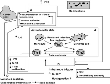
Infection with PCV2 is known to produce an immunosuppressive effect on the host. This finding is not surprising, given the characteristic lesions of PCV2 that include lymphoid depletion and leucopenia. Typically, destruction of lymphoid follicles, depletion of lymphocytes associated with a reduction in the numbers of interfollicular dendritic cells, interdigitating cells, B cells, NK cells, γ/δ T cells, CD4+ and CD8+ T lymphocytes and reduced expression of high endothelial venules along with an increase in the number of monocytes and granulocytes are seen in affected piglets. The degree of depletion had a direct correlation with the amount of PCV2 antigen in tissues (Sarli et al., Reference Sarli, Mandrioli, Laurenti, Sidoli, Cerati, Rolla and Marcato2001; Segales et al., Reference Segales, Alonso, Rosell, Pastor, Chianini, Campos, Lopez-Fuertes, Quintana, Rodriguez-Arrioja, Calsamiglia, Pujols, Dominguez and Domingo2001; Darwich et al., Reference Darwich, Segales, Domingo and Mateu2002; Grierson et al., Reference Grierson, King, Tucker, Donadeu, Mellencamp, Haverson, Banks and Bailey2007). Co-infection with PRRSV has been shown to further exaggerate immune cell depletion (Shi et al., Reference Shi, Li, Guo, Ge, Jia, Zheng and Yang2007). PCV2 is known to persist in dendritic cells without altering their immune function. However, in natural interferon (IFN)-producing cells it is known to impair IFN-α and TNF-α production, thereby interfering with immune priming (Vincent et al., Reference Vincent, Carrasco, Herrmann, Meehan, Allan, Summerfield and McCullough2003; Vincent et al., Reference Vincent, Carrasco, Guzylack-Piriou, Carrasco, Guzylack-Piriou, Herrmann, McNeilly, Allan, Summerfield and McCullough2005). Examination of the T lymphocyte surface markers in neonatal pigs with and without PMWS revealed a state of activation with higher level and earlier expression of MHC-II on T and B cells and higher level of CD25 [interleukin-2 (IL-2) receptor] expression in diseased pigs (Grierson et al., Reference Grierson, King, Tucker, Donadeu, Mellencamp, Haverson, Banks and Bailey2007) (Fig. 4).
A hypothesis for the possible mechanisms of immunopathogenesis of PCV2. (A) In the normal state, PCV2 may coexist with the host with minimal replication and a limiting but balanced Th1/Th2 response. (B) A disturbance in the cytokine balance with a probable influence of host genetics may trigger the onset of PCVAD. (C) Pathogenesis of PCV2 in the lymph nodes and other organs. (D) An out-of-control inflammatory response and lymphoid depletion may lead to clinical manifestations of PCVAD.

Lymphoid depletion can be a direct result of viral replication in lymphoid tissues or the indirect consequence of infection such as host cell apoptosis in response to infection. Attempts to identify cells that support PCV2 replication showed that, in vitro, PCV2 antigen was detected in cultured cells of the monocytic lineage but not lymphocytes. However, monocytic cells did not contain replicative intermediates, indicating that they were not the primary sites of viral replication (Gilpin et al., Reference Gilpin, McCullough, Meehan, McNeilly, McNair, Stevenson, Foster, Ellis, Krakowka, Adair and Allan2003). In contrast, a recent examination of cells supporting viral replication by detection of cap gene mRNA showed that viral replication occurred in tissues derived from bronchial lymph nodes, inguinal lymph nodes, tonsils, lungs, liver, kidneys, spleen and thymus from infected pigs from day 14 post-infection. T and B lymphocyte and monocyte populations from peripheral blood mononuclear cells, and bronchial lymph nodes also supported replication. However, replication was greater in B cells of the bronchial lymph node than in peripheral blood mononuclear cells (Yu et al., Reference Yu, Opriessnig, Kitikoon, Nilubol, Halbur and Thacker2007a) and in activated T cells than in the monocytes and macrophages in peripheral blood mononuclear cells (Yu et al., Reference Yu, Vincent, Opriessnig, Carpenter, Kitikoon Halbur and Thacker2007b) Therefore, it would appear that both B and T cells are important targets for PCV2.
The role of apoptosis in PCV2-induced lymphoid depletion has been controversial. Some studies report an increase of pro-apoptotic gene expression and markers (Shibahara et al., Reference Shibahara, Sato, Ishikawa and Kadota2000; Bassaganya-Riera et al., Reference Bassaganya-Riera, Pogranichniy, Jobgen, Halbur, Yoon, O'Shea, Mohede and Hontecillas2003; Krakowka et al., Reference Krakowka, Ellis, McNeilly, Meehan, Oglesbee, Alldinger and Allan2004; Chang et al., Reference Chang, Jeng, Lin, Liu, Chang, Tsai, Chia and Pang2007; Liu et al., Reference Liu, Zhu, Chen, Lau, He, Lau, Wang, Karuppannan and Kwang2007; Seeliger et al., Reference Seeliger, Brugmann, Kruger, Greiser-Wilke, Verspohl, Segales and Baumgartner2007), whereas others contradict the role of apoptosis in PCV2 pathogenesis (Krakowka et al., Reference Krakowka, Ellis, McNeilly, Meehan, Oglesbee, Alldinger and Allan2004; Mandrioli et al., Reference Mandrioli, Sarli, Panarese, Baldoni and Marcato2004; Resendes et al., Reference Resendes, Majo, Segales, Mateu, Calsamiglia and Domingo2004). Therefore, the mechanism by which PCV2 causes lymphadenopathy in PMWS-affected pigs has yet to be identified.
Antibodies and PCVAD
Despite the pathology associated with the B cell population, pigs infected with PCV2 appear to mount strong PCV2-specific antibody responses. A typical response in PCV2 seropositive pigs in the field is characterized by a decrease in maternal antibody titers from 3 until 11 weeks of age, increase in titers at 15 weeks and persisting PCV2 antibody titers thereafter. Experimental inoculation of colostrum-deprived pigs with PCV2 elicited PCV2-specific antibody responses within 14 days postinfection, and neutralizing antibodies at about 21 days post-infection (Pogranichniy et al., Reference Pogranichnyy, Yoon, Harms, Swenson, Zimmerman and Sorden2000; Rodriguez-Arrioja et al., Reference Rodriguez-Arrioja, Segales, Calsamiglia, Resendes, Balasch, Plana-Duran, Casal and Domingo2002; McKeown et al., Reference McKeown, Opriessnig, Thomas, Guenette, Elvinger, Fenaux, Halbur and Meng2005). Initial studies did not identify significant differences in the antibody responses between PMWS and non-PMWS pigs (Larochelle et al., Reference Larochelle, Magar and D'Allaire2003; Sibila et al., Reference Sibila, Calsamiglia, Segales, Blanchard, Badiella, Le Dimna, Jestin and Domingo2004). More recently, it has been shown that the levels of neutralizing antibodies and IgM isotype antibodies were lower in pigs with PMWS, while the total antibody levels remained the same in both PMWS and non-PMWS pigs. It is believed that while IgM is not the neutralizing isotype, it is indicative of an active infection and functions by stearic interference with viral attachment to host cells (Meerts et al., Reference Meerts, Van Gucht, Cox, Vandebosch and Nauwynck2005b, Reference Meerts, Misinzo, Lefebvre, Nielsen, Botner, Kristensen and Nauwynck2006; Fort et al., Reference Fort, Olvera, Sibila, Segales and Mateu2007). In subclinically infected pigs, it has been shown that increased IL-10 levels lead to a high ratio of IgG to IgM (Darwich et al., Reference Darwich, Segales, Resendes, Balasch, Plana-Duran and Mateu2008).
Vaccination and in vitro neutralization studies have shown that there is a direct correlation between antibody titers and protection (Fan et al., Reference Fan, Ju, Tong, Huang, Lv and Chen2007; Song et al., Reference Song, Jin, Zhang, Xu, Xiao, Cao and Chen2007). The immunogenic epitopes of PCV2 capsid protein have been identified (Mahe et al., Reference Mahe, Blanchard, Truong, Arnauld, Le Cann, Cariolet, Madec, Albina and Jestin2000; Troung et al., Reference Truong, Mahe, Blanchard, Le Dimna, Madec, Jestin and Albina2001; Lekcharoensuk et al., Reference Lekcharoensuk, Morozov, Paul, Thangthumniyom, Wajjawalku and Meng2004), and it has been shown that amino acid sequences 47–57 and 165–200 contain neutralizing epitopes. Whether antibodies to these two epitopes are necessary and sufficient for protection against PCV2 is not known. However, considering that the strong antibody response that is mounted is not always effective in viral neutralization, it is possible that PCV2 may employ a decoy mechanism to evade the host immunity. Such an evasion is usually achieved by directing a strong antibody response to immunogenic epitopes that are not protective while masking the protective epitopes (Ostrowski et al., Reference Ostrowski, Galeota, Jar, Platt, Osorio and Lopez2002). Therefore, further work in characterizing the neutralizing antibodies and their cognate antigens is warranted.
Cytokines and PCVAD
Examinations of cytokine mRNA profiles in serum and lymphoid organs of PMWS-affected and healthy pigs lend further credence to the theory that PCV2 is immunosuppressive. Increased levels of IL-10 mRNA in thymus were associated with the thymic depletion and atrophy that is observed in PMWS pigs. IFN-γ mRNA in tonsils was up-regulated, and decreased mRNA expression of several cytokines such as IL-2 and IL-12p40 in the spleen, IL-4 in tonsils and IL-10, IL-12p40 and IL-4 in inguinal lymph nodes were detected. Comparison of subclinically infected and uninfected pigs showed that serum IL-10 levels peaked transiently with peak viremia. A shift of isotype from IgM to IgG occurred at the same time, following which normalcy was restored (Darwich et al., Reference Darwich, Pie, Rovira, Segales, Domingo, Oswald and Mateu2003b; Darwich et al., Reference Darwich, Segales, Resendes, Balasch, Plana-Duran and Mateu2008). Recently, it has been shown that increased levels of IL-10 in PCV2-infected pigs are responsible for suppression of Th1 responses in the PBMCs of the infected pigs. PCV2-induced IL-10 leads to impaired IFN and antigen recall responses in psuedorabies virus-immunized animals, thus lending a possible explanation for the role of PCV2 in the pathogenesis by co-infecting agents (Kekarainen et al., Reference Kekarainen, Montoya, Mateu and Segales2008a). It has also been shown that modulation of host IL-10 levels is not influenced by CpG oligodeoxynucleotides (ODNs) present in the viral DNA and therefore are likely to be due to viral proteins (Kekarainen et al., Reference Kekarainen, Montoya, Dominguez, Mateu and Segales2008b).
In vitro results obtained with PBMCs and macrophages from PMWS pigs showed a reduced antiviral activity and an increase in pro-inflammatory cytokines. In one study, proliferation in response to mitogens in PBMCs from PMWS-affected pigs was reduced (Darwich et al., Reference Darwich, Balasch, Plana-Duran, Segales, Domingo and Mateu2003a). However, their memory functions and ability to produce IL-10 and IFN-γ in response to stimulation by PCV2 antigens remained intact. PCV2 suppressed IL-4 and IL-2 production in PBMCs from both healthy and PMWS-affected animals while increasing the pro-inflammatory IL-1β and IL-8 expressions. Similarly, swine alveolar macrophages that were experimentally infected with PCV2 showed a reduced microbicidal activity with a decrease in the production of O2 free radicals and H2O2, and an increased production of TNF-α, IL-8, alveolar macrophage-derived neutrophil chemotactic factors-II (AMCF-II), granulocyte colony-stimulating factor (G-CSF) and monocyte chemotactic protein-1 (MCP-1) (Chang et al., Reference Chang, Jeng, Lin, Liu, Chia, Tsai, Chia and Pang2006). Other studies have confirmed that pro- inflammatory cytokines like TNF-α, macrophage chemotactic proteins, macrophage inflammatory protein and C reactive proteins are up-regulated during PCV2 infection (Kim and Chae, Reference Chae2004; Stevenson et al., Reference Stevenson, McCullough, Vincent, Gilpin, Summerfield, Nielsen, McNeilly, Adair and Allan2006). Co-infection with PPV was also found to substantially increase the pro-inflammatory cytokine TNF-α (Kim et al., Reference Kim, Ha and Chae2006). However, cytokine profiles may vary based on the detection method used. In a study by Sipos et al. (Reference Sipos, Duvigneau, Willheim, Schilcher, Hartl, Hofbauer, Exel, Pietschmann and Schmoll2004), mRNA measurements confirmed the findings of others in that TNF-α mRNA levels were increased and IL-2 mRNA was down-regulated. However, when intracellular cytokine levels were measured by flow cytometry it was found that IL-2 was increased whereas TNF- α expression was not affected.
Overall, notwithstanding a temporal and spatial influence, an increase in IL-10 and pro-inflammatory cytokines and a decrease in general antiviral responses were found in pigs affected with PMWS when compared with non-PMWS pigs. Efforts to delineate those cell-mediated immune responses that are protective and those responses that result in viral-induced pathology are important in devising improved methods to strengthen the immune system against PCV2 infections.
IFNs and PCV2
ODNs containing CpG motifs which are naturally present in the DNA of pathogens are known to modulate host IFN response. Five such ODNs have been identified in PCV2 DNA. Although only 1 out of 5 ODNs had INF-α inhibitory activity, the strength of the inhibition was strong enough to suppress the stimulatory activity of the other four ODNs (Hasslung et al., Reference Hasslung, Berg, Allan, Meehan, McNeilly and Fossum2003). The immunomodulatory effect of the ODNs is believed to be dependent on secondary structure and to involve Toll-like receptor-7 (TLR-7) (Wikstrom et al., Reference Wikstrom, Meehan, Berg, Timmusk, Elving, Fuxler, Magnusson, Allan, McNeilly and Fossum2007). Most of the Cpg-ODNs with inhibitory activity are located on the rep gene and appear to induce INF-α in PBMCs, while they did not influence INF-α levels in bone-marrow-derived dendritic cells. Therefore, the immune response induced by the many PCV2 viral components vary with the cell type and may be important in host and viral gene regulation in response to infection (Kekarainen et al., Reference Kekarainen, Montoya, Dominguez, Mateu and Segales2008b).
It has recently been shown that infection of natural IFN-producing cells by PCV2 inhibits their ability to respond to ODNs, TLR-7 and TLR-9 agonists, as well as viruses such as pseudorabies virus, transmissible gastroenteritis virus and classical swine fever virus, thereby preventing maturation of dendritic cells (Vincent et al., Reference Vincent, Balmelli, Meehan, Allan, Summerfield and McCullough2007). However, stimulation of PK-15 and 3D4/31 cells with IFN-α and -γ before and during infection with PCV2 has led to increased PCV2 titers (Meerts et al., Reference Meerts, Misinzo and Nauwynck2005a; Misinzo et al., Reference Misinzo, Delputte, Lefebvre and Nauwynck2008a). The accepted paradigm in viral infections is that stimulation of a strong IFN response is required for protection. There is no definitive study on whether type 1 and 2 IFNs are protective or detrimental in PCV2 infections and more research is required to clarify the role of IFNs in the pathogenesis of PCV2.
Taken together, these findings indicate a complex and multifactorial mechanism for the immuno-pathogenesis for PCV2-induced PMWS (Fig. 4). In an infected but asymptomatic animal, PCV2 may coexist with the host by undergoing minimal replication and inducing a limiting but balanced Th1/Th2 response (Fig 4). A potential trigger or triggers, some of which could be unidentified, may set disease progression in motion. The triggers could include stress and exposure to other pathogens and resistance could be influenced by host genetics (Fig. 4). At the molecular level, the triggers may induce increased viral proliferation, leading to clinical signs and a further disturbance of the cytokine milieu. Identification of the triggers and characterization of their interactions with PCV2 at the immune and molecular levels will help in better understanding PCV2 pathogenesis.
Prevention and control of PCV
Management practices
The common risk factors that increase the odds of PCVAD in piglets have been identified as concomitant infections with other pathogens, stress, vaccination, insemination with semen from infected breeding stock, large weaning pens, lack of treatment against external parasites, mixing of batches, poor air and thermal control of pens, crowding and lack of an all-in-and-all-out policy with strict disinfection of pens (Rose et al., Reference Rose, Larour, Le Diguerher, Eveno, Jolly, Blanchard, Oger, Le Dimna, Jestin and Madec2003; Dorr et al., Reference Dorr, Baker, Almond, Wayne and Gebreyes2007; Kawashima et al., Reference Kawashima, Katsuda and Tsunemitsu2007; Woeste and Grosse Beilage, Reference Woeste and Grosse Beilage2007). A 20-point plan has been proposed (Madec et al., Reference Madec, Eveno, Morvan, Larour, Jolly, Le Diguerher, Cariolet, Blanchard and Jestin2001) to help producers identify management practices that can contain the disease. Important recommendations include following an all-in-and-all-out system with thorough disinfection, avoiding mixing of batches and cross-fostering, maintaining appropriate temperature and airflow conditions in pens and following recommended de-worming, ectoparasite treatments and vaccinations. Additionally, supplementing feed with antibiotics to reduce secondary bacterial infections and carrying out Mycoplasma vaccinations 2 weeks prior to the anticipated exposure to PCV2 may be useful in reducing the severity of PCV2 infections (Opriessnig et al., Reference Opriessnig, Halbur, Yu, Thacker, Fenaux and Meng2006b).
Vaccination
Vaccination-induced antibodies against PCV2 are known to be highly effective in controlling PCVAD. At least four commercial vaccines are available against PCV2 infection and PCVAD in piglets and sows. The Fort Dodge Suvaxyn® PCV2 One Dose™ vaccine contains inactivated whole PCV1–2 chimeric viral particles (Fenaux et al., Reference Fenaux, Opriessnig, Halbur and Meng2003, Reference Fenaux, Opriessnig, Halbur, Elvinger and Meng2004a). The Ingelvac CircoFLEX vaccine and the Intervet vaccine both consist of PCV2 capsid protein expressed in baculovirus. The Muriel Circovac vaccine contains inactivated PCV2 viral particles. The vaccines have been successful in reducing mortality caused by PCV2 in Europe, Canada and the USA (Opriessnig et al., Reference Opriessnig, Meng and Halbur2007b). Several other experimental vaccines that include an ORF2 baculovirus vaccine (Blanchard et al., Reference Blanchard, Mahe, Cariolet, Keranflec'h, Baudouard, Cordioli, Albina and Jestin2003; Fan et al., Reference Fan, Ju, Tong, Huang, Lv and Chen2007), an ORF2 DNA vaccine (Kamstrup et al., Reference Kamstrup, Barfoed, Frimann, Ladekjaer-Mikkelsen and Botner2004), a recombinant pseudorabies virus expressing an ORF1 and ORF2 fusion protein (Ju et al., Reference Ju, Fan, Tan, Liu, Xi, Cao, Wu and Chen2005; Song et al., Reference Song, Jin, Zhang, Xu, Xiao, Cao and Chen2007) and a recombinant adenovirus virus expressing the ORF2 protein (Wang et al., Reference Wang, Jiang, Jiang, Li, Feng and Xu2006) have been successful under experimental conditions in inducing protection against PCV2. Therefore, it appears that prevention and control of PCV2 infection are an achievable goal if proper management and immunization practices are adopted.
Summary
The emergence of new infectious diseases is usually associated with infectious or environmental factors such as genetic alterations leading to host switching or antigenic variation, changes in animal husbandry practices, increase in animal movement and proximity to transmission vectors. PCV appears to have coexisted with their hosts for a long time without any apparent ill effects. Several studies have identified risk factors that are associated with increased incidence of PCVAD. However, the establishment of a definitive genetic or environmental cause for the emergence of PCVAD may help in further controlling the disease. The fact that PCV2 is one of the smallest viruses in existence indicates that the virus is very heavily dependent on the host cell machinery for survival. Some of the viral and host factors that make this complex association possible, such as the proteins associated with replication, the capsid protein, viral transcripts that are produced during viral infection, the kinetics of viral replication, influence of acidification of endolysosomes on viral replication, host antibody and cytokine responses, have been studied so far. It also appears that PCV2 has evolved sophisticated mechanisms that allow it to evade host immunity and coexist with the host and that a probable imbalance in such mechanisms may culminate in the disease condition (Fig. 4). As future research sheds more light on these areas, an improved understanding and control of PCVAD will be possible. A highly significant recent achievement in this direction has been the development of effective vaccines against PCV2. With a combination of good management practices and adequate prophylaxis, substantial reduction of economic losses due to PCV2 could be a very achievable goal in the near future. At this juncture, it is important to determine the optimal levels of herd immunity required to avoid co-evolution of the virus in response to selection pressure that may be exerted by suboptimal immunization and to make a co-ordinated, global effort to achieve such levels of protection.
Acknowledgments
We acknowledge Dr Manoharan Seeralan for help withproofreading the citations and Dr Elankumaran Subbiah for help with construction of the phylogenetic tree.