I. INTRODUCTION
Mo–Si intermetallics compound (e.g., MoSi2 and Mo5Si3) has been investigated as a potential material for high temperature structural and heating element applications. Reference Gnesin, Gnesin and Nekrasov1,Reference Chen, Shao, Wang, Pu, Ma and Huang2 MoSi2 has a desirable combination of high melting (2293 K), high Young's modulus (440 GPa), relatively low density (6.24 g/cm3), high oxidation resistance at high temperatures (>1473 K), and good thermal and electric conductivity. Reference Xu, Hu, Yan, Lu, Munroe and Xie3 However, it has low toughness at room temperature (2–3 MPa·m1/2), low strength at high temperature (>brittle–ductile transition temperature), and poor oxidation resistance at about 500 °C (pesting). Reference Zamani, Bakhsheshi-Rad, Shokuhfar, Vaezi, Kadir and Shafiee4,Reference Seong-Ho, Kyosuke, Kouichi, Rong and Takashi5 Multi-component is one of the most efficient means to improve the toughness of intermetallics alloys. Reference Lu and Wang6 Compared to the Mo–Si binary metal silicide, the ternary metal silicide Mo2Ni3Si possesses better toughness. Reference Lu and Wang7 Meanwhile, the high melting point, inherent high hardness, and strong atomic bonds make Mo2Ni3Si excellent candidate for high temperature structural applications. Reference Xu and Wang8–Reference Lu, Wang and Zhou14 However, like most of the intermetallics, Mo2Ni3Si also exhibits a poor strength and creep resistance at elevated temperature. Introduction of a second reinforcement phase is an important way to improve mechanical properties of Mo2Ni3Si. Reference Ignacio, Maria and David15 For instance, the addition of SiC, Si3N4, Mo5Si3, ZrO2, or Al2O3 leads to strengthening of MoSi2-based composites at elevated temperature. Reference Manukyan, Kharatyan, Blugan, Kocher and Kuebler16–Reference Hu, Luo and Yan19 Among these compounds, Al2O3 is an oxide known for its low density, high strength, and oxidation resistance even at temperatures higher than 1400 °C. Reference Ramezanalizadeh and Heshmati-Manesh20 The network microstructure of Al2O3 was supposed to block the paths of oxygen diffusion that caused pest oxidation (Mo–Si system compound) at the low temperature (400–800 °C). Meanwhile, Al2O3 has a thermal expansion coefficient similar to that of Mo2Ni3Si, which suppresses the related absence of residual thermal stresses during sintering and cooling. Therefore, its use as reinforcement phase in Mo2Ni3Si matrix can reduce the thermal tension and improve the thermal shock resistance and creep resistance.
Mechanical alloying is basically a dry and high energy ball milling process and used to synthesize alloys, amorphous alloys, and various intermetallics compounds. Reference Zakeri, Yazdani-Rad, Enayati and Rahimipoor21,Reference Chen, Ma, Shao, Ma, Wang and Huang22 Meanwhile, mechanical alloying is an important preparation method of nanostructured powders. Reference Krakhmalev23 Unfortunately, conventional methods of consolidation at high temperature usually lead to the degradation of nanostructures obtained during the mechanical alloying. Medium frequency induction hot-pressing method has a high-energy heat source for sintering simultaneously with a uniaxial pressure to sinter powders, which can produce dense materials within 5 min. Thus, this method gives an opportunity to carry out the rapid sintering of nanostructured materials from elemental or composite powders. Some intermetallics, silicides and ceramics prepared by medium frequency induction hot-pressing sintering method have a high density and fine microstructure.
In the present work, Mo2Ni3Si–Al2O3 nanocomposite powders were synthesized use MoO3, Ni, Si, and Al as starting materials by mechanically alloying. This synthesis method does not require pure Mo as starting material, and the preparation cost can be reduced. Moreover, the reaction between MoO3 and Al is highly exothermic. Thus, a self-propagating combustion reaction can be ignited by mechanical activation at a much lower temperature. The combustion synthesis represents an in situ processing technique for the preparation of composite materials with interpenetrating phase microstructures and improved properties. The consolidation of dense nanocrystalline Mo2Ni3Si–Al2O3 composite was fabricated by medium frequency induction hot-pressing sintering method. The morphology, particle size, and phase constitution of composite powders were investigated using SEM and XRD methods. Finally, the microstructure and mechanical properties of the consolidated products were also investigated in detail.
II. EXPERIMENTAL PROCEDURE
MoO3 (99.5% purity and particle size of 3–5 μm), Ni (99.9% purity and particle size of <8 μm), Al (99.7% purity and particle size of 10–35 μm), and Si (99.9% purity and particle size of <3 μm) powders were used as starting materials. The powders were mixed to give the desired stoichiometric composition according to following reactions:
 $${\rm{Mo}}{{\rm{O}}_3} + 2{\rm{Al}} = {\rm{Mo}} + {\rm{A}}{{\rm{l}}_2}{{\rm{O}}_3}\left( {\Delta G_{25}^o = - 914.783{\rm{\ kJ/mol\ and\ }}\Delta H_{25}^{\rm{o}} = - 931.092{\rm{\ kJ/mol}}} \right)$$
 $$2{\rm{Mo}} + 3{\rm{Ni}} + {\rm{Si}} = {\rm{M}}{{\rm{o}}_2}{\rm{N}}{{\rm{i}}_3}{\rm{Si}}$$
 $${\rm{or:\ }}2{\rm{Mo}}{{\rm{O}}_3} + 3{\rm{Ni}} + {\rm{Si}} + 4{\rm{Al}} = {\rm{M}}{{\rm{o}}_2}{\rm{N}}{{\rm{i}}_3}{\rm{Si}} + 2{\rm{A}}{{\rm{l}}_2}{{\rm{O}}_3}\quad .$$
Mechanical alloying was performed in a planetary ball mill (QM-3SP2, Nanjing Nanda Instrument Plant, Nanjing, China) at rotation speed of 300 rpm under air atmosphere in room temperature. The mixture of powders and the steel balls of 8 mm in diameter were charged into a steel vial (120 mL). The weight ratio of ball-to-powder was 10:1. To prevent a significant temperature rise in the mechanically alloyed powders, 30 min milling and 10 min stop (natural cooling) milling cycle was used. The milling process was interrupted and small amounts of powder samples were taken out for analysis at various intervals. Subsequently, heat treatment of as-milled powders at 1000 °C for 2 h was conducted in a tube furnace under an Ar atmosphere.
After milling, the composite powders were placed in a graphite die with an inner diameter of 40 mm, and introduced into a medium-frequency induction-heated sintering system. The sintering was conducted at 1450 °C for 0.5 h under Ar atmosphere, mechanical pressure of 50 MPa, and heating rate was 75 °C/min. The final hot pressed samples were in a disc shape with a diameter of 40 mm and a thickness of 6–8 mm.
The relative density of the sintered sample was measured using the Archimedes method. Vickers hardness was tested in a HV-1000 Vickers hardness tester (Shanghai Shuangxu Electronics Co. Ltd., Shanghai, China) at a load of 500 g with the dwell time of 10 s. Average hardness values were obtained from five points. The samples were machined into 3 × 4 × 35 mm bars. The flexural strength was measured at room temperature using the three-point bending test with a span length of 30 mm and a cross-head speed of 0.05 mm/min. Fracture toughness tests were carried out by a single edge notch bending (SENB) specimens with 30 mm span length, 4 mm width, 3 mm thickness, and a straight notch of about 2 mm depth. High temperature flexural strength was measured in a THERMORESTOR-W thermal simulator with a compression speed of 0.5 mm/min. The specimens (ϕ 5 × 8 mm) were put in the thermal simulator and heated to 400, 800, 1000, 1200, and 1400 °C.
The phase compositions of the composites powders and sintered samples were analyzed by x-ray diffraction (XRD; D/Dmax-2400, Rigaku Corporation, Tokyo, Japan) with Cu Kα radiation (λ = 0.15418 nm). Microstructures, morphologies of the samples, and chemical compositions were investigated by scanning electron microscopy (SEM: JSM-6700F, JEOL Ltd., Tokyo, Japan) and energy dispersive x-ray analysis (EDS).
III. RESULTS AND DISCUSSION
A. Mechanical alloying
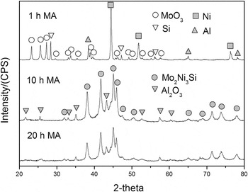
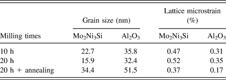
Figure 1 shows the XRD patterns of powder mixture after various milling times. The as-received powder mixture consisted of MoO3, Ni, Si, and Al that had been mixed on the stoichiometry of the reaction (3). As seen in Fig. 1, after 10 h of mechanical alloying, the XRD peaks of the raw material disappeared due to the reaction of MoO3, Ni, Si, and Al to give Mo2Ni3Si and Al2O3 phases according to the reaction (3). Those phases in Fig. 1 are based on the JCPDS file numbers 05–0508(MoO3), 65–2865(Ni), 40–0932(Si), 65–2869(Al), 15–0489(Mo2Ni3Si), and 46–1212(Al2O3). With increasing the milling time to 20 h, no further reaction took place and only the XRD peaks became broader and weaker. The peak broadening indicates the reduction of grain size and higher microstrain induced in powder particles. The grain size and microstrain of Mo2Ni3Si and Al2O3 were calculated from the broadening of XRD peaks using the Williamson–Hall method, as shown in Table I. After 20 h of milling, the grain size and lattice microstrain of Mo2Ni3Si and Al2O3 were (15.9 nm, 0.48%) and (32.4 nm, 0.25%), respectively.
The XRD patterns of powders after various milling times.

Grain size and lattice microstrain of powder particles at different conditions.

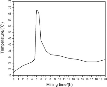
In general, the adiabatic temperature T ad > 1800 K is required for the propagation of self-sustaining reaction. Reference Zamani, BakhsheshiRad, Kadir and Shafiee24 This value for the reaction (1) is 4285 K. Reference Zakeri, Yazdani-Rad, Enayati, Rahimipour and Mobasherpour25 It is therefore expected that a combustion reaction takes place during milling of the powder mixture. The change of vial temperature was monitored during milling, as shown in Fig. 2. It can be seen that the temperature of the vail showed a sudden increase after 5 h of milling, which indicates that the reaction of MoO3, Ni, Si, and Al took place in an explosive mode, analogous to self-propagating high temperature synthesis reaction. The process begins with an activation period, during which size reduction and defect formation take place, and then the self-propagating synthesis reaction is ignited when the powders reaches a well defined critical state.
Variation of vial temperature versus time during mechanical milling of MoO3–Ni–Si–Al powder mixture.

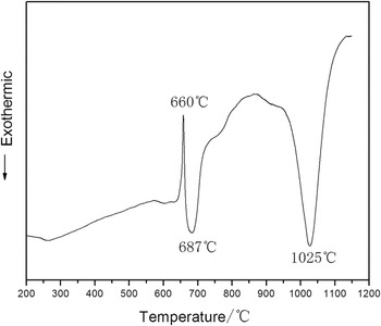
To elucidate the mechanism of reaction of MoO3–Ni–Si–Al powder mixture, thermal analysis of the mixed powders were carried out, as shown in Fig. 3. It can be seen that three peaks appear in DTA curve. A sharp endothermic peak at about 660 °C is due to the melting of aluminum in the starting material. Meanwhile, the sample has exothermic peaks at 687 °C and 1025 °C, respectively. The sharp exothermic peak at 687 °C is mainly attributed to the reaction between Al and MoO3 powders in the mixture, which is a high exothermic reaction. According to the XRD results, the sample mainly consists of Mo2Ni3Si and Al2O3 after 20 h of milling. Therefore, the exothermic peak at 1025 °C should be attributed to the reaction of Mo, Ni, and Si to give Mo2Ni3Si.
The DTA analysis of as-blended MoO3–Ni–Si–Al powder mixture under argon atmosphere.

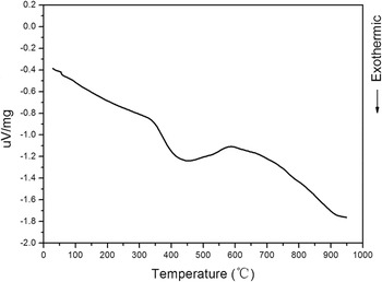
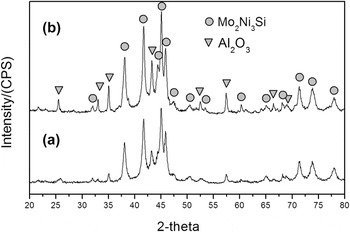
To make sure that the thermal stability of Mo2Ni3Si–Al2O3 composite structure, DTA analysis was performed for this mixture powder after 20 h of milling, as shown in Fig. 4. It can be seen that there is only a broad exothermic peak at temperature ranging from 327 to 581 °C. Meanwhile, as shown in Fig. 5, before and after annealing at 1000 °C for 2 h, Mo2Ni3Si–Al2O3 composite powders with 20 h milling has same phase composition of Mo2Ni3Si and Al2O3 phases. According to the thermal analysis and XRD analysis results, the Mo2Ni3Si–Al2O3 composite is stable during an annealing at 1000 °C. Moreover, as shown in Table I, the width of XRD peaks decreased and their intensity increased after annealing due to reduction of lattice strain as well as grain growth. But this temperature (327–581 °C) is not high enough for grain growth. Therefore, the broad exothermic peak may mainly originate from strain relaxation in the lattice due to the decrease of faults and dislocations.
The DTA analysis of powder mixture after milling for 20 h.

XRD patterns of Mo2Ni3Si–Al2O3 composite powders (a) as-milled for 20 h and (b) after subsequent annealing for 2 h at 1000 °C.

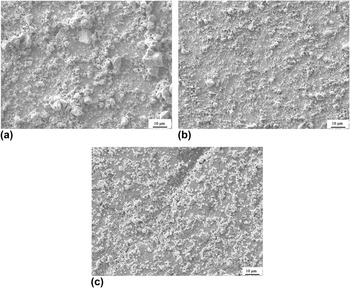
SEM images of powder particles after 1, 10, and 20 h of milling are shown in Fig. 6. The powder particle size is uneven and irregular with a distribution of very small particles and large agglomerates (10 μm) after 1 h of milling [Fig. 6(a)]. With the increasing milling time to 10 h, the refining rate of powders became quicker and the average size of particles reduced to about 3 μm. After 20 h milling, the particles became much finer and more homogeneous with an average size of about 1 μm.
SEM micrograph of milled powders after: (a) 1 h, (b) 10 h, and (c) 20 h.

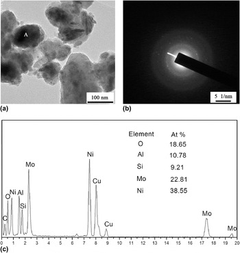
The microstructure and selected area diffraction pattern (SADP) of the 20 h milled composite powder observed in TEM is shown in Fig. 7. As can be seen in TEM image, highly agglomerated crystalline nanoparticles about 50–100 nm in diameter was observed. The agglomerated nanoparticles is usually explained as a common way to minimize their surface free energy. Reference Mousavi, Karimzadeh and Abbasi26 The selected area diffraction rings clearly demonstrate that the particles are nanocrystalline. Reference Bodaghi, Zolfonoon, Tahriri and Karimi27 Figure 7(c) shows the energy dispersive analysis of point A in Fig. 7(a). Grain A is composed of Mo, Ni, Si, Al, and O elements and the atomic ratio of these elements is close to the stoichiometric composition of Mo2Ni3Si–Al2O3. The XRD results (Table I) show that the mean crystallite size of both phases is <50 nm. Thus, the mean crystallite size estimated using XRD method is approximately consistent with TEM results.
TEM observation of powder mixtures milled for 20 h (a) TEM micrograph, (b) associated SADP, and (c) EDS spectrum.

B. Consolidated product
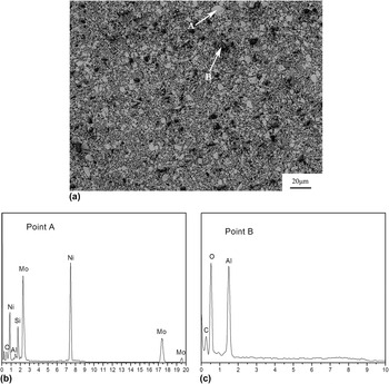
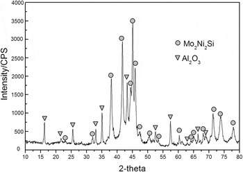
The composite powders of Mo2Ni3Si–Al2O3 were consolidated by hot pressing as shown in Fig. 8. After the hot pressing, the Mo2Ni3Si–Al2O3 composite has a uniform microstructure consisting of the dual-phase structure. Further, it may be noted that the microstructural features are very fine in the microns and submicrometer range. Figure 9 shows the XRD patterns of Mo2Ni3Si–Al2O3 composite by hot pressing at 1450 °C. According to the reports of Zamani and BakhsheshiRad, Reference Zamani, Bakhsheshi-Rad, Shokuhfar, Vaezi, Kadir and Shafiee4,Reference Zamani, BakhsheshiRad, Kadir and Shafiee24,Reference Khalajabadi, Abdul Kadir, Izman and Mohd Yusop28 the area under the diffraction peaks is proportional to the volume fraction of concentration, which can be obtained from the following formula:
 $${R_{\rm{x}}} = {{{a_{\rm{x}}}} \mathord{\left/ {\vphantom {{{a_{\rm{x}}}} {\sum\limits_i {{a_i}} }}} \right. \kern-\nulldelimiterspace} {\sum\limits_i {{a_i}} }}\quad ,$$
where R x is the volume fraction, x is Mo2Ni3Si, and Al2O3, a x is the area of the principal peak, and a i is the area of the total peaks. The volume fractions of the Mo2Ni3Si and Al2O3 in the sample are 78 and 22%, respectively. Results of x-ray and EDS analyses indicate that the brighter areas correspond to Mo2Ni3Si grains and darker grains are Al2O3. Average hardness of the Mo2Ni3Si–Al2O3 composite is about 13 GPa, harder than the single Mo2Ni3Si (about 11 GPa Reference Xu and Wang29 ) due to the reinforcing effect of the amount of Al2O3. The relative density of the Mo2Ni3Si–Al2O3 composite is approximately 96.3%.
SEM micrographs (a) of the Mo2Ni3Si–Al2O3 composite and EDS patterns of the composite corresponding to (a): (b) point B and (c) point C.

XRD patterns of Mo2Ni3Si–Al2O3 composite by hot pressing at 1450 °C.

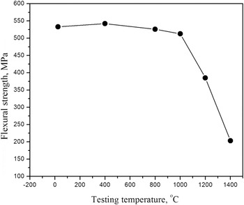
The flexural strength of Mo2Ni3Si–Al2O3 composite as a function of temperature is shown in Fig. 10. The values obtained are the average value of 3–4 bend tests per material. It can be seen that the flexural strength of the composite was quite high. At room temperature, the strength of the composite was 533 MPa. The strength remained stable up to 1000 °C. It has been shown that an interphase barrier mechanism, as a generalization of the Hall–Petch strengthening in polycrystalline structures, is the main high-temperature strengthening mechanism in intermetallic composites. Reference Manukyan, Kharatyan, Blugan, Kocher and Kuebler16 Similarly, grain refinement and second phase strengthening were suggested to be the main reason for the high strength and hardness. The flexural strength for composite degraded significantly at higher temperature (>1000 °C) due to the low high-temperature creep resistance of the Mo2Ni3Si phase.
The flexural strength of the Mo2Ni3Si–Al2O3 composite as a function of temperature.

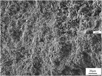
The fracture toughness of Mo2Ni3Si alloy is quite poor (about 2.13 MPa·m1/2) and similar to most of other intermetallics. However, the addition of Al2O3 and the formation of Mo2Ni3Si–Al2O3 composite result in the improvement of toughness, and the average fracture toughness of Mo2Ni3Si–Al2O3 composite is 6.29 MPa·m1/2. The fracture toughness is in general dependent on the microstructural features of the composite. The nature and morphology of the reinforcement seem to be important in achieving improved fracture toughness value. It was reported that addition of up to 0.9 wt% La2O3 reinforcement increased the fracture toughness of MoSi2 to about 6.5 MPa·m1/2. Reference Wang, Wang, Liu, Zhu and Zan30 In the present study, the fracture toughness value increase with the addition of Al2O3 reinforcement phase due to formation of fine equiaxed crystals (the grain size of the composite is about 15.9 nm–32.4 nm). The fracture morphology of the composite after three-point bend test is shown in Fig. 11. It can be seen that the fracture mode of the composite is intergranular fracture, transgranular fracture, and a small amount of dimple structure characteristic of a ductile fracture. Generally, the existence of second particles in composite may change the orientation of crack growth, which make the crack tip blunted. Reference Zan, Wang, Shi, Sun and Xu31 The change of crack morphology and the formation of new fracture surface will both absorb more energy. Thereby, the as-obtained Mo2Ni3Si–Al2O3 composite exhibits better strength and toughness than single Mo2Ni3Si.
Surface morphology of the Mo2Ni3Si–Al2O3 composite fractured during fracture toughness testing. The inset is a SEM image showing the enlarged microstructure of Mo2Ni3Si–Al2O3 composite.

IV. CONCLUSIONS
Mo2Ni3Si–Al2O3 nanocomposite was successfully synthesized by mechanical milling mixture of MoO3, Ni, Si, and Al powders. The composite was obtained after 10 h of milling. At the end of milling, the grain size and lattice microstrain of Mo2Ni3Si and Al2O3 were (15.9 nm, 0.48%) and (32.4 nm, 0.25%), respectively. The reaction mechanism was investigated. The results indicated that the reaction of MoO3, Ni, Si, and Al takes place in an explosive mode, analogous to self-propagating high temperature synthesis reaction. According to the thermal analysis and XRD analysis results, the Mo2Ni3Si–Al2O3 composite is stable and no new phase formation during an annealing at 1000 °C. After the hot pressing, the Mo2Ni3Si–Al2O3 composite has a uniform microstructure consisting of the dual-phase structure (Mo2Ni3Si and Al2O3). The relative density of the Mo2Ni3Si–Al2O3 composite is approximately 96.3%. The hardness and fracture toughness of the Mo2Ni3Si–Al2O3 composite is harder than the single Mo2Ni3Si due to the reinforcing effect of the amount of Al2O3. The flexural strength of the composite was quite high at room temperature: 533 MPa. The strength remained stable up to 1000 °C. The flexural strength for composite degraded significantly when the temperature was raised above 1000 °C due to the low high-temperature creep resistance of the Mo2Ni3Si phase.
ACKNOWLEDGMENTS
This research was supported by the Natural Science Foundation of Shandong Province of China (Grant Nos BS2015CL007 and ZR2012EMM007), China Postdoctoral Science Foundation funded project (Grant No. 2015M572026), Doctor Scientific Research Startup Funding of Liaocheng University (Grant No. 31805). The authors acknowledge Mr Ke Xu and Ms Xue Zhao for their assistance on experiments of hot press sintering.