A hallmark of inflammation is the movement of leukocytes out of the circulation and into diseased tissues. Leukocytes receive antigen specificity and activation cues in secondary lymphoid tissues, which sensitise them to receive additional cues that can direct their infiltration into specific target tissues. Leukocyte trafficking from the circulation into various tissues is a multistep process involving selectins, integrins and chemokines (Ref. Reference Butcher and Picker1). Selectins (which bind sialylated carbohydrates) mediate the ‘capture and rolling’ of lymphocytes by the activated endothelium, which is followed by firm adhesion to the vessel wall by the interaction of lymphocyte integrins and their ligands (members of the immunoglobulin superfamily) on endothelial cells; endothelial-cell-derived chemokines (which bind G-protein-coupled receptors on lymphocytes) then direct lymphocyte extravasation into the tissue (Refs Reference Springer2, Reference Johnston and Butcher3). Various combinations of these molecules appear to provide direct cues enabling tissue-specific homing. For example, trafficking to the skin occurs via a specific combination of selectin, integrin and chemokine molecules and their respective receptors including: E-selectin and cutaneous lymphocyte antigen (CLA); very late antigen (VLA)-4 (also known as α4β1 integrin) and vascular cell adhesion molecule (VCAM)-1; and the chemokine ligand CCL17 and chemokine receptor CCR4 (Ref. Reference Zollner, Asadullah and Schon4). The precise combination of molecules directing cellular trafficking to the CNS is still yet to be defined.
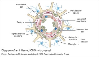
The CNS is protected from immune cell surveillance under normal conditions by a highly specialised system of microvasculature known as the blood–brain barrier (BBB) (Fig. 1) (Ref. Reference Lucas, Rothwell and Gibson5). A variety of alterations distinguish the BBB from the peripheral vasculature, including specialised endothelial cells, the presence of additional basement membranes and the addition of astrocytes. These specialisations contribute to the formation of a formidable barrier between the blood and CNS parenchyma, with the interaction of endothelial cells and astrocytes providing the fundamental basis of BBB formation and maintenance. CNS endothelial cells are securely held together by tight-junction proteins and are encased by astrocyte end-feet, which enhances barrier properties. The endothelial cells also possess specialised transport systems that limit the penetration of molecules. A basement membrane, which includes embedded pericytes, together with the astrocyte foot processes provide the structural basis for the perivascular space. The astrocyte end-feet with associated microglia form the glial limitans, which is thought to provide an additional mechanical barrier to cells entering the CNS (Ref. Reference Ballabh, Braun and Nedergaard6).
Nevertheless, despite this formidable barrier, immune cells are capable of BBB penetration during neuroinflammation resulting from a variety of pathologies, including CNS infection and putative autoimmune diseases such as multiple sclerosis (MS) (Ref. Reference Frohman, Racke and Raine7). Infiltrating leukocytes accumulate in the perivascular space, leading to the ‘perivascular infiltrate’ – the cardinal lesion associated with all neuroinflammatory diseases (Fig. 1). While these inflammatory infiltrates are required to clear pathogen during bacterial, viral and parasitic infections, they induce severe tissue destruction within the CNS in diseases like MS. The ubiquitous nature of this lesion suggests there are generalised mechanisms involved in its formation. What is unclear, however, is whether infiltration is a nonspecific process in which the mechanical barrier is overwhelmed by the bulk of the infiltrate or if there are specific mechanisms for directing leukocyte movements into the CNS parenchyma.
Diagram of an inflamed CNS microvessel. Depicted is a cross-section of a capillary in the central nervous system (CNS) with interendothelial-cell tight and adherens junctions, and periendothelial accessory structures including the endothelial-cell basement membrane with embedded pericytes and the glial limitans, composed of astrocyte end-feet and associated microglia. During neuroinflammatory diseases, mononuclear cells traverse the endothelial cells and congregate within the perivascular space between the basement membrane and glial limitans.

Leukocyte trafficking in multiple sclerosis
MS is second only to trauma as a leading cause of neurological disability in young adults in North America (Ref. Reference Sospedra and Martin8). During MS exacerbations, leukocytes enter the CNS parenchyma leading to damage of oligodendrocytes, the myelinating cells of the CNS, by destruction, apoptosis and/or necrotic cell death. This results in demyelinating, inflammatory lesions that can be detected by magnetic resonance imaging (MRI) (Ref. Reference Prat and Antel9). The loss of myelin manifests with a variety of motor deficits, depending on the site of the damage. While clinical courses of MS vary greatly, there are four main defined subtypes: relapsing–remitting (RRMS), primary progressive (PPMS), progressive–relapsing (PRMS), and secondary progressive (SPMS) (Ref. Reference Siva10). The RRMS and SPMS subtypes are characterised by exacerbations or ‘attacks,’ between which the patient is clinically stable or progressing downhill, respectively. The less common PPMS and PRMS subtypes begin with downhill progression. Although studies suggest that all forms of MS are associated with autoimmune attack, there is little understanding of the mechanisms responsible for the differences in the rates of and recoveries from exacerbations that define the MS subtypes.
Although the cause of MS is unknown, it is considered an autoimmune disease mediated by the activities of myelin-specific CD4+ T cells (Ref. Reference Frohman, Racke and Raine7). While there is some evidence that CD8+ T cells are able to transfer CNS autoimmune disease in rodent models (Ref. Reference Huseby11), the vast majority of research centres around CD4+ T cells, and these are the focus of this review. CD4+ T cells, also known as T helper (Th) cells, differentiate into separate and distinct subsets. Cytokine production by CD4+ cells defines their subset and leads to direct inhibition of differentiation into an alternative subset. To date, there are four known subsets of T helper cells: Th1, Th2, Th3 and Th17 cells, which produce interferon γ (IFN-γ), interleukin 4 (IL-4), transforming growth factor (TGF)-β and IL-17, respectively (Refs Reference Weaver12, Reference Harrington13, Reference Akbar14). It is generally accepted that Th2 cells are not the primary subset causing experimental autoimmune encephalomyelitis (EAE; an animal model of brain inflammation) and MS, because Th2 cells have been shown to play a protective role during EAE (Ref. Reference Chen15). Th1 cells are the mediators of viral and bacterial clearance and were traditionally characterised as the subset involved in both MS and EAE. Th3 cells are a type of regulatory T cell thought to be involved in recovery from autoimmune inflammation. Data analysing the role of the recently defined Th17 subset during EAE and MS are challenging the theory that Th1 cells are the primary mediators of EAE and MS (Refs Reference Cua16, Reference Langrish17). Owing to the very recent emergence of Th17 cells, it is unknown if much of the information provided in this review applies to Th1, Th17 or both subsets.
While T cells are often the focus of research and discussion surrounding leukocyte trafficking into the CNS during neuroinflammatory disease, a role for mononuclear cells (lymphocytes and monocytes/macrophages) that aid T-cell trafficking and function should be acknowledged. Circulating monocytes that move into the CNS parenchyma play an active role in exacerbating disease as tissue macrophages by providing a source of inflammatory cytokines. The presence of monocytes and macrophages early in MS lesions has been correlated with demyelination (Ref. Reference Bruck18) and their elimination has been shown to ameliorate EAE (Ref. Reference Huitinga19). Blocking effector functions of macrophages via depleting antibodies and genetic deletion of macrophage migration inhibitory factor significantly decreases EAE (Ref. Reference Powell20) and interferes with T-cell trafficking into the CNS during EAE (Ref. Reference Denkinger21). Additional supportive roles for CNS-infiltrating macrophages include major histocompatibility complex (MHC) class II expression and antigen presentation, although these roles are far less well understood (Ref. Reference Juedes and Ruddle22). Nevertheless, this review concentrates on the role of CD4+ T cells in CNS autoimmunity.
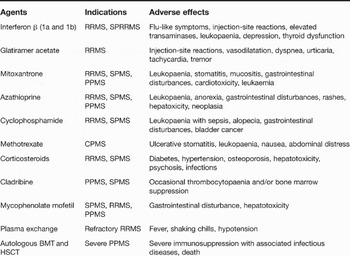
Because MS has been defined as a cell-mediated autoimmune disease, much of the focus on pharmacological treatments has been on immunosuppressants, particularly to target peripheral inflammatory responses. These approaches have met with varying success, depending on the type of MS and on the clinical criteria used to judge efficacy. In general, various cytolytic immunosuppressive therapies are used in patients with refractory, progressive forms of MS. These agents induce the destruction of cell-mediated immunity and are thus fraught with serious, adverse side effects (Table 1). However, the ability simply to prevent the trafficking of autoreactive lymphocytes into the CNS would best interrupt the cycle of inflammatory demyelination that repeatedly occurs in MS. Identification of some of the molecular events responsible for interactions between infiltrating mononuclear cells and the CNS microvasculature, resulting in movement of leukocytes into the CNS, has revealed new putative therapeutic targets. In this review, we highlight putative molecular targets on lymphocytes, emphasising those that appear to impart some specificity to the tissue-specific homing of lymphocytes into the CNS. In addition, we discuss new biologically based strategies that target the CNS microvasculature itself as a specific target for the treatment of MS exacerbations.
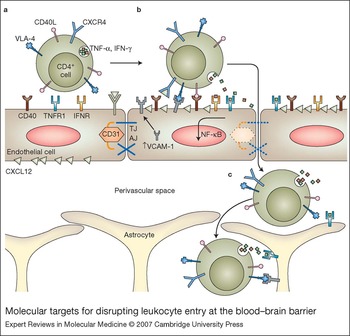
Molecular targets for disrupting leukocyte entry at the blood–brain barrier. The trafficking of CD4+ T cells across central nervous system (CNS) endothelial-cell barriers and then into and out of the perivascular space is accomplished via signalling cascades that lead to interactions between multiple cell types. Depicted are CD4+ T-cell and endothelial-cell receptor–ligand pairs recently determined to play significant roles in these migratory pathways. (a) Activated CD4+ T cells express very late antigen 4 (VLA-4), CD40 ligand (CD40L), the chemokine receptor CXCR4 and secrete the Th1 cytokines tumour necrosis factor α (TNF-α) and interferon γ (IFN-γ). Normal CNS endothelial cells express CD40 and cytokine receptors, exhibit CD31-associated tight and adherens junctions (TJ and AJ), and display polarised expression of the chemokine CXCL12. (b) Interactions between CD4+ T cells and CNS endothelium leads to cytokine- and CD40L-mediated signalling pathways that activate transcription factors, such as nuclear factor κB (NF-κB), which up-regulate endothelial-cell surface expression of adhesion molecules, such as vascular cell adhesion molecule 1 (VCAM-1). Activated CNS endothelial cells display dissociation of CD31 and loss of junctional integrity. In the case of autoimmunity, polarised expression of CXCL12 is lost, which may lead to CXCR4 signalling within lumenal leukocytes. (c) Perivascular accumulation of CD4+ T cells is associated with cytokine-mediated up-regulation of adhesion molecules on astrocytes within the glial limitans. Loss of CXCL12 expression at the basolateral surfaces of endothelial cells is associated with exit of CD4+ T cells out of the perivascular space, which continue to associate with astrocytes via interactions of adhesion molecules.

Immunosuppressive agents used for the treatment of multiple sclerosisa

aFor additional information, see Refs Reference Kremenchutzky, Morrow and Rush144, Reference Perini145, Reference Perumal146, Reference Zaffaroni, Ghezzi and Comi147.
Abbreviations: BMT, bone-marrow transplantation; CPMS, chronic progressive multiple sclerosis; HSCT, haematopoietic stem-cell transplantation; RRMS, relapsing–remitting multiple sclerosis; SPMS, secondary progressive multiple sclerosis; SPRRMS, secondary progressive relapsing–remitting multiple sclerosis; PPMS, primary progressive multiple sclerosis.
Molecular basis of leukocyte trafficking into the CNS
Much of the work elucidating the molecular events required for T-cell trafficking across the BBB during MS has been accomplished via the use of the animal model EAE. EAE can be induced in mice by immunisation with myelin peptides or via adoptive transfer of myelin-specific CD4+ T cells (Refs Reference Sospedra and Martin8, Reference Laschinger and Engelhardt23). As in patients with MS, the hallmark features of mice with EAE are paralysis and CNS-demyelinating lesions containing myelin-specific T cells and tissue macrophages. The combination of research utilising both MS patient samples and the EAE model has revealed a number of important molecular players involved in directing the entry of leukocytes into the CNS. These include the intercellular adhesion molecule (ICAM)-1 and VCAM-1 (Ref. Reference Laschinger and Engelhardt23) and P-selectin, which are expressed on activated endothelium (Ref. Reference Piccio24). It is important to note that even within the CNS there may be differences in the mechanisms of cellular trafficking. This is exemplified in the debate over the role of P-selectin for which data suggest that it may be important in trafficking to the brain (Ref. Reference Piccio25) but not to the spinal cord (Ref. Reference Engelhardt26).
Expression of these adhesion molecules is detected on endothelial cells activated by treatment with tumour necrosis factor α (TNF-α), CD40 ligand (CD40L), lipopolysaccharide (LPS) and IL-1β or during onset of EAE (Ref. Reference Ubogu, Cossoy and Ransohoff27). Autoreactive T cells present in the circulation are initially captured on the activated BBB endothelium via interactions of P-selectin with P-selectin glycolipid (PSGL)-1, and of ICAM-1 with lymphocyte-function-associated antigen (LFA)-1 (integrin αLβ2) (Refs Reference Piccio24, Reference Engelhardt28, Reference Biernacki29, Reference Bullard30). The interaction between VLA-4 and adhesion molecule ligands is more ambiguous and depends on the mononuclear cell type in question: VCAM-1 appears to direct monocyte movements while ICAM-1 guides CD4+ T-cell migration (Refs Reference Biernacki29, Reference Bullard30, Reference Floris31). In vitro and in vivo experiments using human brain endothelial cells have implicated the chemokines CCL2, CCL4 and CCL5 (Refs Reference Mahad32, Reference Prat33, Reference Quandt and Dorovini-Zis34) as providing the molecular cues for extravasation of CCR2- and CCR5-expressing T cells into the perivascular space. In addition, we have recently shown that the basolateral expression of the chemokine CXCL12 on BBB endothelial cells retains infiltrating CXCR4-expressing lymphocytes within the perivascular space (Ref. Reference McCandless35).
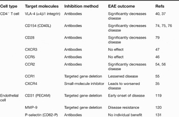
A variety of these and probably other receptor–ligand interactions between leukocytes and endothelial cells determine whether immune cells gain entry into the CNS (Fig. 2). Thus, there exists the potential to target leukocyte and/or endothelial-cell constituents that participate in these interactions as a therapeutic strategy to treat diseases of neuroinflammation such as MS. The best targets would interfere with lymphocyte trafficking to the CNS without altering the ability of the immune system to carry out normal surveillance and pathogen clearance in peripheral tissues. Because the inhibition of leukocyte-based molecules might potentially disturb their trafficking patterns in a universal fashion, the specialisations of the CNS microvasculature might present more unique and therefore CNS-specific targets for the prevention of leukocyte entry during MS. Using tools such as targeted gene deletion and antibody inhibitors, investigators have tested some of the molecules known to be involved in directing lymphocyte trafficking across the BBB. Although it is not yet clear whether the important goal of CNS specificity can be achieved, some of the most promising targets tested to date are discussed below (Table 2).
T-cell and endothelial-cell molecular targets for the treatment of CNS autoimmunity

Abbreviations: CCR1, -2 and 5, chemokine (C-C motif) receptor 1, 2 and 5; CD40L, CD40 ligand; CNS, central nervous system; CXCR3, chemokine (C-X-C motif) receptor 3; MMP-9, matrix metalloproteinase 9; VLA-4, very late antigen-4.
Targeting leukocyte molecules for the treatment of MS
Various drugs directed against T-cell surface ligands have been developed and include monoclonal antibodies (mAbs), fusion proteins and oligonucleotides. Most of these inhibitors target cell-surface molecules that participate in the movement of immune cells through the BBB. However, several interfere with the development of lymphocyte effector functions, which may also occur as lymphocytes move into the CNS. It is anticipated that the identification of CNS-specific molecules for the migration of lymphocytes will lead to therapies with minimal interference of peripheral lymphocyte trafficking. Unfortunately, inhibition of lymphocyte trafficking into the CNS will continue to pose issues regarding CNS immune surveillance, including decreased control of CNS infection with opportunistic pathogens.
Adhesion molecules
VLA-4 mediates adhesion and migration of T cells via interaction with its ligand, VCAM-1. Anti-VLA-4 antibodies have proved efficacious in the prevention of autoimmune disease in both animal models and in patients with MS (Refs Reference Brocke36, Reference Kent37, Reference Miller38, Reference Tubridy39, Reference Yednock40). Natalizumab is a humanised monoclonal antibody against the α4 chain of the α4β1 integrin that markedly reduced the relapse rate in clinical trials performed on patients with the RRMS (Ref. Reference Miller38). Although the US Food and Drug Administration (FDA) and the European Agency for the Evaluation of Medicinal Products (EMEA) have lifted the clinical hold on natalizumab use for the treatment of RRMS, its safety for the treatment of MS and other autoimmune diseases is unclear because of the emergence of several cases of progressive multifocal leukoencephalopathy (PML), a neuroinfectious disease caused by the JC virus (Ref. Reference Adelman, Sandrock and Panzara41). Approximately 80% of the population is seropositive for exposure to JC virus, and PML has occurred in patients with either MS or Crohn's disease treated with natalizumab alone, or in combination with either IFN-β or azathioprine, respectively (Ref. Reference Adelman, Sandrock and Panzara41). Thus, it is likely that α4 integrin is important for directing the immune response that suppresses the dissemination of JC virus from tissue sites where the virus normally resides. Thus, despite the apparent CNS specificity associated with anti-α4-integrin therapeutics, they do not selectively inhibit pathological T-cell trafficking, leaving patients susceptible to CNS infections. Nevertheless, the success of natalizumab in brief clinical trials and after chronic treatment suggests that targeting mononuclear cell trafficking for the treatment of neuroinflammatory diseases will be a viable therapeutic approach, once issues of specificity are determined through continued research into the molecular mechanisms.
Chemokine receptors
The expression of a variety of chemokines, including CCL2–5 and CXCL9–11, within CNS tissues from animals and patients with autoimmune disease correlate with the appearance of pathogenic T cells bearing chemokine receptors that bind these ligands (CCR1, CCR2, CCR5 and CXCR3) (Refs Reference Giunti42, Reference Misu43, Reference Sorensen and Sellebjerg44, Reference Sorensen45). Neutralising antibodies and targeted deletion studies of CXCR3 and CCR5 did not demonstrate essential roles for these receptors in the pathogenesis of EAE (Refs Reference Tran, Kuziel and Owens46, Reference Liu47). In addition, patients with Delta32CCR5, a mutated allele of the chemokine receptor CCR5, are not protected from developing MS (Refs Reference Pulkkinen48, Reference Bennetts49); indeed, the mutation may worsen the course and/or severity of disease (Refs Reference Favorova50, Reference Gade-Andavolu51, Reference Kantarci52). However, antagonism or deletion of CCR2 and its ligand CCL2 is completely protective and of CCR1 appears to be partially protective (Refs Reference Huang53, Reference Izikson54, Reference Rottman55, Reference Fife56). These results have provided the impetus for the design of CCR1 and CCR2 antagonists for the treatment of MS. In one study, BX 471, a CCR1 antagonist, was tested for the prevention of new CNS inflammatory lesions in patients with RRMS and observed to be safe but with no efficacy (Ref. Reference Zipp57). A small-molecule antagonist of CCR2 has also been developed, INCB3344, which was shown to reduce disease significantly in mice with EAE but has not been tested for its efficacy in the treatment of MS (Ref. Reference Brodmerkel58).
Other lymphocyte targets
Other members of the greater chemoattractant network have also been targeted for the prevention of mononuclear cell trafficking. In a Phase II controlled clinical trial involving 255 patients with active, relapsing MS, oral fingolimod (also known as FTY720, by Novartis Pharmaceuticals Corp) significantly reduced signs of inflammation on MRI scans (Ref. Reference Kappos59). Fingolimod binds to the sphingosine-1-phosphate (S1P) receptor on lymphocytes, which plays an essential role in the emigration of lymphocytes from lymphoid tissues (Ref. Reference Rosen and Goetzl60). Thus, targeting S1P may have limits in its chronic administration as it is likely to interfere with the trafficking and effector functions of antigen-specific T cells.
Although not a chemoattractant, 3-hydroxy-3-methylglutaryl coenzyme A (HMG-CoA) reductase has been recently implicated in the migration of leukocytes into the CNS and its inhibition, via the administration of statins, has been shown to reduce neuroinflammatory infiltrates and inhibit the development of disease in animal models of MS (Refs Reference Youssef61, Reference Nath62, Reference Greenwood63). Statins reduce circulating cholesterol by inhibiting HMG-CoA reductase, a rate-limiting enzyme in the cholesterol synthesis pathway. Recently, a variety of anti-inflammatory effects of statins have been described, including decreases in T-cell activation and proliferation, down-regulation of expression of matrix metalloproteinases (MMPs), chemokines, cytokines and MHC II expression and, most importantly, blockade of ICAM signalling (Refs Reference Mach64, Reference Neuhaus65, Reference Weber66, Reference Bernot67, Reference Takeuchi68). While the mechanism of statin-mediated immune modulation is complicated, it primarily involves inhibition of isoprenoid lipid production and protein prenylation of small GTPase signalling proteins within immune cells (Refs Reference Greenwood63, Reference Greenwood, Steinman and Zamvil69). Prior studies have demonstrated that these are essential molecules in the ICAM signalling pathway that lead to phosphorylation of cytoskeleton-associated proteins and leukocyte movement (Refs Reference Greenwood, Steinman and Zamvil69, Reference Etienne70). A small, open-label clinical trial administering simvastatin for RRMS demonstrated a decrease in the numbers and sizes of new lesions as assessed by MRI (Ref. Reference Neuhaus71). In addition, combinations of statins appears to have additive immunomodulatory effects, suggesting that therapies may be tailored to address the severity of different forms of MS. Several larger trials are now under way to examine the efficacy of various statins in different combinations for the treatment of MS.
Lymphocyte receptors
Among the myriad of surface receptors involved in the functions of T cells are those that are involved in their activation and differentiation into effector cells. These include the T-cell receptor (TCR) and several costimulatory molecules including CD28, CD40L (CD154), CD134 and CD137. Disruption of TCR activity with TCR antagonists or altered peptide ligands in theory would promote tolerance or T-cell unresponsiveness. Clinical trials evaluating the efficacy of such compounds revealed that many caused immediate-type hypersensitivity reactions in a significant number of patients (Ref. Reference Kappos72). In general, the immunogenicity of these protein-based therapies limits their usage. However, a recent study in which MS patients were vaccinated with a trivalent self-peptide contained within the TCR chains reported the induction of regulatory T-cell responses and a reduction in MRI lesions (Ref. Reference Vandenbark73). Continued studies employing this approach are needed to evaluate its long-term efficacy in controlling clinical symptoms and preventing exacerbations. Anti-CD40L monoclonal antibodies reduced the severity of EAE by limiting the expansion of encephalitogenic T cells (Refs Reference Howard74, Reference Howard75, Reference Howard and Miller76), and IDEC-131, a humanised version, proved to be safe in a clinical trial for the treatment of systemic lupus erythematosus (SLE) (Ref. Reference Kalunian77). Although IDEC-131 does not appear to be efficacious for the treatment of SLE, clinical trials administering this agent to MS patients are ongoing (Ref. Reference Dumont78).
Recently, anti-CD28 antibodies have been observed to promote interactions between regulatory T cells and encephalitogenic T-cell clones within secondary lymphoid organs during the induction of EAE (Ref. Reference Tischner79). Although promising in these preclinical studies, these antibodies have yet to be tested for the treatment of ongoing disease or for their tolerability in patients. Two new TNFR family members – OX40 (CD134) and 4-BB (CD137) – are potential new targets for the treatment of MS. CD134 and CD137 are both costimulatory molecules expressed by activated CD4+ T cells that promote T-cell survival and effector functions such as cytokine expression, and CD134 has been implicated in the pathological cytokine storm that occurs during T-cell responses to viral infections (Ref. Reference Humphreys80). CD134 is up-regulated within the CNS of MS patients, and CD134 deficiency is associated with attenuation of EAE (Ref. Reference Carboni81). Elevated cerebrospinal fluid (CSF) levels of CD137 have also been observed in MS patients and administration of anti-CD137 monoclonal antibodies ameliorates EAE (Refs Reference Sharief82, Reference Sun83).
Interestingly, arvanil, a synthetic analogue of the endocannabinoid anandamide, down-regulated the expression of these and other activation markers and ameliorated EAE in preclinical studies (Ref. Reference Malfitano84). The connection between CNS autoimmune disease and endocannabinoids had already been identified; the cannabinoid receptor agonist R (+) WIN55,212 thwarts the progression of virus-induced demyelinating disease induced by infection with Theiler's virus by down-regulating T-cell effector functions (Ref. Reference Croxford and Miller85). More recently, this agonist was found to decrease the interactions between leukocytes and CNS endothelial cells during EAE (Ref. Reference Ni86), and mice with loss of endocannabinoids experienced more severe damage than those with this system intact (Ref. Reference Witting87). Thus, in addition to the known role of these compounds for the treatment of pain, tremor and spasticity in MS patients, cannabinoids might also exert counter-regulatory effects on the immune system. Studies of immune function in patients enrolled in clinical trials evaluating cannabinoid-mediated amelioration of neurological symptoms are needed to determine these effects.
Rituximab, a humanised mAb that targets CD20 antigen and depletes B cells, was observed to decrease exacerbations in an open-label study on a small group of patients with neuromyelitis optica, a demyelinating disease of the CNS characterised by transverse myelitis and optic neuritis (Ref. Reference Cree88). Given this result, several placebo-controlled trials examining the efficacy of rituximab for the treatment of RRMS are currently under way. Preliminary results indicate that both B and T cells are depleted from the CSF of MS patients after six months of rituximab treatment (Ref. Reference Cross89). These cellular alterations were not accompanied by a significant decrease in CSF or serum antibodies against myelin peptides and suggest that loss of B cells affects T-cell function and/or trafficking in the CNS. Further studies evaluating the affects of B-cell depletion on clinical disease and MRI findings are currently pending.
The CNS microvasculature in neuroinflammation
Most of the strategies aimed at preventing leukocyte entry have focused on cell membrane receptors present on leukocytes; however, various studies indicate that the migration of these cells into the CNS during MS is related to permeability changes of the BBB. Therefore, it is possible that the defective proteins within patients that develop MS reside in the microvasculature itself. Recent studies indicate that even highly activated, myelin-specific T cells require alterations in the patterns and conformations of proteins expressed by endothelial cells in order to engage in trafficking events (Ref. Reference Piccio24). Thus, in this section, we highlight data that suggests that CNS autoimmune disease may occur as a result of inappropriate activation of CNS endothelial cells or deficiencies in the molecules that maintain BBB impermeability.
As MRI indicates that exacerbations in patients with CNS autoimmune disease are associated with increased BBB permeability (Refs Reference Broom90, Reference Dousset91, Reference Larsson92, Reference Wuerfel93), studies in MS patients and in animals with EAE have examined the molecules involved in CNS endothelial-cell activation. TNF-α, IFN-γ and IL-1β are elevated within the CNS in MS patients during periods of increased symptoms and in mice during acute EAE (Refs Reference Gimenez, Sim and Russell94, Reference Probert95, Reference Kassiotis96, Reference Eugster97, Reference Cannella and Raine98, Reference Minagar and Alexander99) and their signalling is associated with alterations in the expression of adhesion molecules within the CNS microvasculature (Refs Reference Gimenez, Sim and Russell94, Reference Cannella and Raine98, Reference Duran100). In patients, elevations in serum levels of TNF-α and various adhesion molecules correlate with higher numbers of gadolinium-enhancing lesions (Refs Reference Khoury101, Reference Rieckmann102, Reference Rieckmann103). TNF-α and other proinflammatory cytokines induce endothelial-cell activation characterised by increased expression of CD40, dispersion of CD31 and the expression of MMP-7 and MMP-9 within CNS endothelium (Refs Reference Lin104, Reference Williams105, Reference Omari and Dorovini-Zis106, Reference Kieseier107, Reference Chiodoni108, Reference Melter109). MMPs, which target cellular matrix proteins such as fibronectin and laminin, have thus been implicated in the neuropathogenesis of MS and EAE. MMP-9 levels are elevated in patients with MS and correlate with enhanced BBB permeability detected by gadolinium-enhanced MRI (Refs Reference Fainardi110, Reference Avolio111, Reference Sellebjerg and Sorensen112). Similarly, MMP-7 and MMP-9 levels are increased in mice with EAE and correlate with clinical disease severity scores (Ref. Reference Clements113). Interaction of CD40 with its ligand CD40L (expressed by T cells) leads to increased expression of MMPs as well as adhesion molecules such as ICAM-1, VCAM-1 and E-selectin, and chemokines such as CCL2–5 (Refs Reference Omari and Dorovini-Zis106, Reference Omari, Chui and Dorovini-Zis114). These alterations in protein expression are all further associated with abnormalities in the appearance of the junction proteins zonula occludens 1 (ZO-1) and occludin, loss of claudin-3 and the disruption of junctional integrity (Refs Reference Harkness115, Reference Hawkins116, Reference Mori117, Reference Reijerkerk118).
Use of mice with targeted deletion of genes encoding some of these proteins indicate that endothelial-cell biology plays a significant role in leukocyte trafficking during CNS autoimmune attack. Mice with CD31 deficiency exhibit enhanced trafficking of T cells across endothelial-cell barriers, increased vascular permeability and an earlier onset of EAE (Ref. Reference Graesser119). By contrast, loss of MMP-9 or CD40 induces resistance to EAE (Refs Reference Agrawal120, Reference Becher121, Reference Samoilova122). In the case of CD40, adoptive transfer of wild-type, encephalitogenic T cells into chimaeric mice with CD40 deficiency within the CNS led only to an attenuation of EAE (Ref. Reference Ponomarev, Shriver and Dittel123). These animals displayed decreased trafficking of mononuclear cells, decreased activation of microglia and decreased proliferation of encephalitogenic T cells. The authors of this study concluded that CD40 is required for the activation of microglia during autoimmune inflammation. However, expression of CD40 by activated endothelial cells could also contribute to these results, leading to alterations in T-cell migration, antigen encounter and interactions with microglia. Thus, pathological responses of CNS endothelial cells could underlie alterations in leukocyte restriction at the BBB.
As mentioned above, recent data suggest that the chemokine CXCL12 acts to localise mononuclear cells to the perivascular space surrounding CNS microvessels, thereby limiting their entry into the CNS parenchyma (Ref. Reference McCandless35). Expression of CXCL12 at the BBB normally displays a basolateral polarity within endothelial cells. This pattern of expression is lost during CNS autoimmune disease, leading to extensive mononuclear cell trafficking. Although the regulation of CXCL12 expression is incompletely understood, several studies have demonstrated that interactions between members of the TNF ligand–receptor superfamilies, including TNF-α–TNFR1 and CD40–CD40L, may up-regulate CXCL12 in various cell types (Refs Reference Nanki124, Reference Jung125). In the normal CNS, microvessel endothelial cells express high levels of CXCL12 only along their parenchymal surfaces, suggesting CXCL12 might participate in leukocyte interactions with the BBB prior to leukocyte entry into the CNS parenchyma (Ref. Reference Stumm126). Activation of endothelial cells and parenchymal invasion by T cells has been shown to require TNFR1 (Ref. 127). Under circumstances of immune surveillance, T cells might thus increase endothelial cell expression of CXCL12 via either TNF-α or CD40L, which would promote the perivascular association of T and other effector cells. During CNS autoimmune disease, anomalous expression of CXCL12 by a dysfunctional BBB might interfere with this process, allowing inappropriate leukocyte entry. The association between leukocyte entry and loss of CXCL12 polarity suggests this molecule participates in the maintenance of immune privilege within the CNS.
Targeting dysfunctional CNS microvasculature during MS
The molecular alterations in activated endothelial cells that occur during MS exacerbations might be specifically targeted for the prevention of leukocyte extravasation and extension of inflammatory lesions. Consistent with this, corticosteroids have been used successfully to treat MS flares because, in part, they regulate the expression of adhesion molecules (E-selectin, P-selectin, ICAM-1 and VCAM-1) and MMPs in endothelial cells through glucocorticoid receptor modulation of nuclear factor κB (NF-κB) (Refs Reference Harkness115, Reference Engelhardt128, Reference Trojano129). Newer strategies have emerged that interfere with the activities of endothelial-cell molecules without targeting immune cells and eliciting the adverse effects associated with steroid usage.
P-selectin, an adhesion molecule expressed by most activated endothelium, is an attractive therapeutic target to prevent the CNS entry of T cells, which express its ligand PSGL-1. Although initial experiments using intravital microscopy determined that antibodies directed against P-selectin blocked lymphocyte rolling and capture along TNF-α- or LPS-activated brain vasculature (Ref. Reference Piccio24), studies administering anti-PSGL-1 antibodies did not induce alterations in lymphocyte trafficking across CNS endothelium either in vitro or in vivo (Ref. Reference Engelhardt130). Additionally, loss of PSGL-1 signalling, via antibody administration or targeted deletion, does not affect the clinical course of EAE (Refs Reference Engelhardt130, Reference Osmers, Bullard and Barnum131). Despite these data, targeting P-selectin might prove beneficial in certain contexts as more-recent studies combining anti-P-selectin and anti-VLA-4 antibodies induced lower levels of T-cell rolling and adhesion to inflamed microvessels as well as more significant amelioration of EAE than the use of either antibody alone (Ref. Reference Kerfoot and Kubes132). Thus, combinational targeting of adhesion molecules present on both T and endothelial cells may prevent the infiltration of autoreactive inflammatory cells. Unfortunately, these targets do not provide high levels of CNS specificity because P-selectin is found on vasculature throughout the body (Refs Reference Auchampach133, Reference Sanders134).
Based on years of research into the molecular cascades that lead to BBB disruption, small-molecule inhibitors that modulate NF-κB activation, which leads to MMP expression, are currently under development and, in preclinical studies, attenuate the development of CNS autoimmune disease by maintaining BBB integrity (Refs Reference Prasad135, Reference Prasad136). Similarly, administration of ch5D12, a humanised antagonist antibody against human CD40, successfully inhibits both the induction of EAE and suppresses enlargement of pre-existing brain lesions in MOG-immunised marmosets by inhibiting endothelial-cell activation (Ref. Reference 't Hart137). In addition to monoclonal antibodies, additional molecular approaches to CD40 inhibition are currently under exploration including Fab fragments, chimaeric and/or fusion proteins and peptidomimetics (Ref. Reference Allen138). Finally, oestrogens, which potently suppress EAE, down-regulate adhesion molecules on CNS endothelial cells, alter cytokine and chemokine expression patterns and enhance the activation of regulatory T cells (Refs Reference Subramanian139, Reference Matejuk140, Reference Polanczyk141, Reference Morales142, Reference Matejuk143). A controlled clinical trial of oestriol involving 130 women with RRMS is currently under way (http://www.nationalmssociety.org/research). Because endothelial-cell activation occurs only within active lesions, these newer treatments might need to be administered only during exacerbations, similar to the way β-agonist inhalers are used only at the occurrence of asthmatic symptoms. The arrest of leukocyte entry via pharmacological closure of the BBB may serve to thwart an evolving MS attack and prevent the initiation of an inflammatory cascade that leads to permanent deficits.
Concluding remarks
Although we have outlined numerous potential strategies for the prevention of leukocyte trafficking during CNS autoimmune disease, data that prove the overall specificity of these molecules for the CNS are still lacking. Thus, many of the clinical trials discussed above are likely to reveal additional peripheral effects of new drugs attempting to specifically target CNS-infiltrating leukocytes. However, the use of intermittent therapies in RRMS and other nonprogressive forms of MS may minimise these immunosuppressive effects and lower the risks of opportunistic infections that occur with chronic inhibition of leukocyte trafficking. Because of the specialisation of the CNS microvasculature, efforts at completely elucidating the mechanisms of BBB disruption may ultimately prove the most fruitful in the development of therapies that prevent unwanted leukocyte extravasation. In addition, the ability to manipulate the integrity of the BBB may prove to be extremely useful in promoting the entry of virus-specific lymphocytes and of drugs that treat other CNS diseases, such as brain tumours.
Acknowledgements and funding
We thank Drs Anne Cross, Joshua Rubin and John Russell for their critical reviews of the manuscript. We are also greatly appreciative of the insightful comments and helpful suggestions provided by reviewers of the manuscript. This work is supported by NIH/NINDS (awards K02NS045607 and R01NS052632) and by grants from the National Multiple Sclerosis Society, the Dana Foundation and the Midwest Regional Center for Excellence in Emerging Infectious Diseases (all funding to R.S.K.).