Introduction
Edible clays are obtained from naturally occurring kaolinite (Wilson Reference Wilson2003). In Africa, the two major types of edible clay are distinguished by their colors: gray and white. These clays comprise a group of 1:1 aluminosilicates (Al2O32SiO2H2O) which result from the breakdown of Al-rich silicate rocks either through weathering or hypodermal activity and the weathering of pegmatites containing mica. Pegmatite has been noted to contain kaolinized alkali-feldspar with quartz and abundant mica (Franco et al. Reference Franco, Cecilia, Perez–Marqueda, Perez-Rodriguez and Gomez2007). The pegmatite, quartz, and mica have the ability to disperse in water, but are mostly insoluble in it. Impurities may cause various colors or tints (Badmus & Olatinsu Reference Badmus and Olatinsu2009), making them ideal as a pigment (Aja & Gbohinor Reference Aja and Gbohinor2013).
Edible clays have been consumed by humans since antiquity; they are also the preferred material for use in pharmaceutical preparations for the treatment and management of diarrhea (Adelabu Reference Adelabu2012; Tabletwise 2019) e.g. ‘Mist Kaolin,’ a mixture of proprietary drugs with kaolin, from Moko® (referred to below as “Moko”). The finely divided kaolinite particles yield a comparatively large surface area which adsorbs a wide variety of compounds. These clay minerals have a range of physical, chemical, and physicochemical properties (Burdock et al. Reference Burdock, Carabin and Crincoli2009). As excipients, they are also used widely in pharmaceutical preparations, to improve the physicochemical properties such as viscosity of the active ingredients (emulsifying, thickening, and anti-caking agents), to facilitate their elaboration (lubricants, diluents, binders, isotonic agents), or conservation (desiccants, opacifiers), and to facilitate liberation of the active ingredient within an organism. Though clayey soils can be medicinal, they can also be hazardous to human health if ingested (Ali et al. Reference Ali, Idrees, Ali, Hussain, Rehman, Ali, Iqbal and Kamel2014; Macheka et al. Reference Macheka, Olowoyo, Matsela and Khine2016; Misyak et al. Reference Misyak, Burlaka, Golotiuk and Lukin2016). Edible clay deposits are widespread throughout Nigeria, notably the Ozubulu, Darazo, Akpene-Obom, Kankara, and Porter deposits in the States of Anambra, Bauchi, Cross River, Kaduna, and Plateau, respectively. The three most extensively studied deposits are the Ozubulu, Kankara, and Porter (Badmus & Olatinsu Reference Badmus and Olatinsu2009).
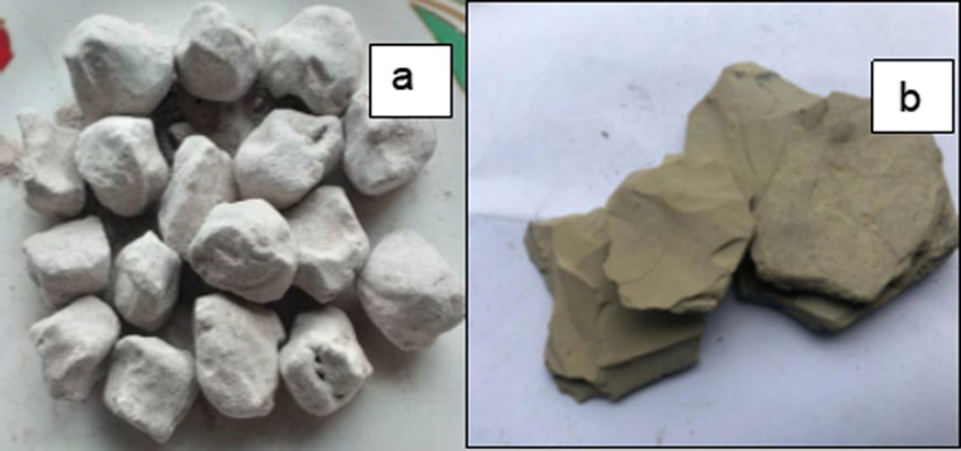
Clay consumption occurs in cultural groups on every inhabited continent (Pessoa Reference Pessoa2005). It is sold in several markets and mined from various sources both within and outside Nigeria. Clay consumption is observed widely, mostly in pregnant women, probably because of its anti-nausea effect (Bonglaisin et al. Reference Bonglaisin, Mbofung and Lantum2011; Pessoa Reference Pessoa2005), but may also be a symptom of nutritional disorders observed frequently in pregnant women (Ekosse Reference Ekosse2010; Gueslin Reference Gueslin2005). Clay consumption also exists in the animal world (Bonglaisin et al. Reference Bonglaisin, Mbofung and Lantum2011). Culturally, consumption of clay by children seems to occur because they witness their mothers or close relatives doing so (Bisi-Johnson et al. Reference Bisi-Johnson, Obi and Ekosse2010). Edible clays known locally as ‘kalaba,’ ‘nzu,’ or ‘ulo’ are the preferred types of clay for geophagy (Bisi-Johnson et al. Reference Bisi-Johnson, Obi and Ekosse2010). African studies on geophagy and pregnancy show its use by 65% of pregnant women in Kenya, 28% in Tanzania, and 50% in Nigeria (Kmeic et al. Reference Kmeic, N'Guyen, Rouger, Beger, Lambert and Hentzein2017). White edible clay (WEC) (Fig. 1a) is prepared by powdering the clay sample, mixing the powder with salt, molding it into spherical shapes, and then drying on an open fire. Gray edible clay (GEC) is mined, broken into pieces, and sold without further processing (Fig. 1b).
a WEC and b GEC

The objectives of the present study were to ascertain the chemical composition of WEC and GEC, to determine the toxicity profiles using analytical chemical methods, to test the acute and sub-acute toxicity of edible clays in their natural form as consumed, and to compare the raw (GEC) and processed (WEC) clays with each other and to compare the latter with the proprietary Moko formulation, all with an eye to determining their toxicity to humans when consumed.
Materials and Methods
The GEC and WEC were obtained from Yaba market, Lagos, Nigeria. Moko was purchased from a pharmacy at Idi-Araba, Mushin, Lagos, Nigeria. The clays were ground with an agate mortar and pestle until a fine powder was obtained. Samples were labeled as GEC or WEC and stored in foil at room temperature until needed.
Determination of Mineral Constituents
The clay mineralogy of the samples was determined by wet chemical analysis and by Fourier-transform infrared spectroscopy (FTIR) (Appendices 1–3). The chemical composition of each clay sample with respect to Ca, K, Na, Mg, Fe, Cd, and Pb was determined by atomic absorption spectrophotometry (AAS) (AA model 2004, ThermoScientific, Germany) using the triple-acid digestion method described by AOAC (2011). Each clay sample was also prepared as a KBr pellet and analyzed by FTIR (Agilent Cary 630 spectrophotometer, Agilent Technologies, Waldbronn, Germany) with a resolution of 8 cm–1 over the range 400–6500 cm–1. The dried powder was mixed thoroughly with spectrophotometric-grade KBr (0.0020.01 clay:KBr ratio) in an agate mortar, then pressed into a 3-mm pellet with a hand press. Mixing was limited to 3 min so as to achieve minimal grinding. Peaks were reported based on percentage transmittance over the range 400–6500 cm−1.
Gas Chromatography/Mass Spectrometry (GC/MS)
Each clay sample was prepared for GC/MS analysis by weighing ground samples into a conical flask and suspending in distilled water, then filtered with a cotton cloth as a sieve. The filtrate was passed through a glass column packed with pre-heated and cooled silica gel. Anhydrous sodium sulfate was added to absorb any moisture in the system. The eluate resulting from the chromatography column was concentrated and stored in a glass vial for subsequent injection into the GC/MS instrument, an Agilent 7820A gas chromatograph coupled to an Agilent Mass Spectrometer 5977E (Agilent Technologies, Waldbronn, Germany), equipped with an Equity-5 fused silica capillary column (60 m × 0.32 mm inner diameter, film thickness of 0.25 nm, oven temperature ranging from 70 to 250°C programmed to increase at a rate of 3°C/min, with initial and final hold times of 2 min). The carrier gas was helium at a constant pressure of 10 psi; a split ratio of 1:40; the temperature in the injector, transfer line, and source was kept at 250°C. An ionization energy of 70 eV was used over a mass scan range of 40–450 amu. Characterization was achieved on the basis of retention time.
Animal Studies
The animals used for the study were obtained from the Laboratory Animals Center, College of Medicine of the University of Lagos, Idi-Araba, Lagos, and allowed to acclimatize for 7 days prior to the study. The animals were maintained on standard animal feeds (Pfizer Nigeria Limited, Lagos. Nigeria) and provided with water ad libitum. Each clay sample was prepared by grinding and mixing in distilled water; no sieving was done; animals were fed only the clay suspended in the water.
Acute Toxicity Study
Six female Wistar mice were used, three for each dose of GEC and WEC. The animals were fed orally via oral gavage, 5000 mg/kg of GEC or WEC suspended in distilled water. The mice were allowed to move around freely and were observed for 72 h for signs of toxicity such as inability to move, sickness, or death.
Sub-acute Toxicity Study
Healthy female Wistar rats (120 ± 20 g) were used for the sub-toxicity study. The animals were divided randomly into eight groups of five animals each. Sample administration was by oral gavage and they were fed the samples daily for 28 days. At the end of the study, the animals were fasted overnight and were anesthetized with inhaled diethyl ether and sacrificed humanely to harvest vital organs for histo-pathological studies.
Grouping
Groups with GEC: A, low dose of 250 mg/kg (LDGEC); B, medium dose of 500 mg/kg (MDGEC); C, high dose of 1000 mg/kg (HDGEC); and D: GEC control, distilled water only (G). Groups with WEC: E, low dose of 250 mg/kg (LDWEC); F, medium dose of 500 mg/kg (MDWEC): G, high dose of 1000 mg/kg (HDWEC); and H, WEC control, distilled water only (W).
Histo-pathology
Post-mortem examination was performed on the selected rats from each treatment and control group after the animals had been sacrificed; their vital organs were recovered and dissected carefully en bloc for histopathological examinations. After rinsing in normal saline, the organs were fixed in 10% formosaline before they were dehydrated completely in absolute (100%) ethanol. The organs were treated with acetone and cleared in xylene. They were embedded in paraffin blocks and 4–5 μm-thick sections were prepared and stained with hematoxylin–eosin. The slides were examined at high magnification for any associated histopathological lesions and photomicrographs were taken (Mbaka et al. Reference Mbaka, Anunobi, Ogunsina and Osiagwu2017).
Results
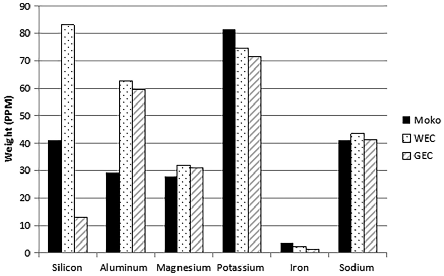
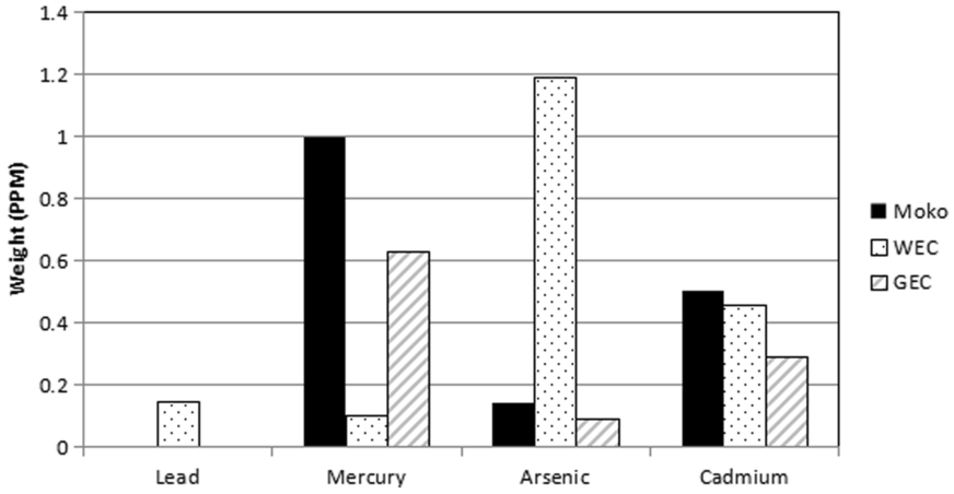
Elemental analysis (Figs 2,3) of GEC, WEC, and Moko revealed that heavy metals Pb, Hg, Cd, and As were present in all the samples to varying degrees, except that samples GEC and Moko contained no Pb. The limits for food set by the Food and Agricultural Organization (World Health Organization, Food and Agricultural Organization of the United Nations 1999) are 0.2 ppm for Pb, 0.1 ppm for Cd, 0.3 ppm for Hg, and 0.01 ppm for As. WEC contained 0.149 ppm of Pb. Lead targets multiple organs in the body due to its systemic toxicity and causes cardiovascular, renal, gastrointestinal, and hematological damage (Nkansah et al. Reference Nkansah, Korankye, Darko and Dodd2017), but none of the observed levels reached the recommended maximum value. Further, the United States Pharmacopoeia (USP 2013) recommends permissible daily exposure limits of Pb in pharmaceutical products of 5 µg/day.
Amounts of Si, Al, Mg, K, Fe, and Na (ppm) in GEC, WEC, and Moko

Amounts of Pb, Hg, As, and Cd (ppm) in GEC, WEC, and Moko

Moko, WEC, and GEC contained 1.000 ppm, 0.101 ppm, and 0.631 ppm of Hg, respectively. The values for Moko and GEC were much greater than the WHO limit of 0.3 ppm.
Moko, WEC, and GEC contained 0.503 ppm, 0.458 ppm, and 0.289 ppm of Cd, respectively; all of which are greater than the recommended limit of 0.1 ppm. Elevated concentrations of Cd in foodstuffs are toxic and have been associated with kidney disorders (United States Pharmacopeia 2013; Deloris et al. Reference Deloris, Desire, Lakisha, Kara, DeJuana, Micoya, Eddy, Rangari, Vijaya, Ramble, Kokoasse and Curtis2015).
With regard to As, the levels found in Moko, WEC, and GEC samples were 0.142 ppm, 1.191 ppm, and 0.092 ppm, respectively, all also well above the recommended limit for ingesting arsenic.
As far as other metals are concerned, Fe levels in Moko, WEC, and GEC were 3.634, 2.364, and 1.39 ppm; Mg, 86, 31.86, and 31.01 ppm; K, 81.52, 74.66, and 71.49 ppm; and Na, 41.07, 43.37, and 41.34 ppm, respectively.
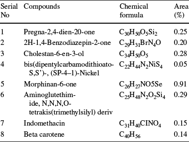
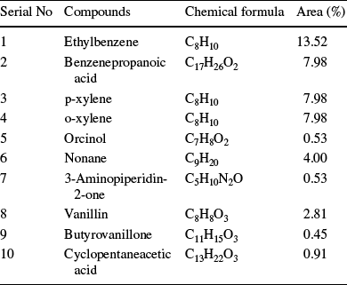
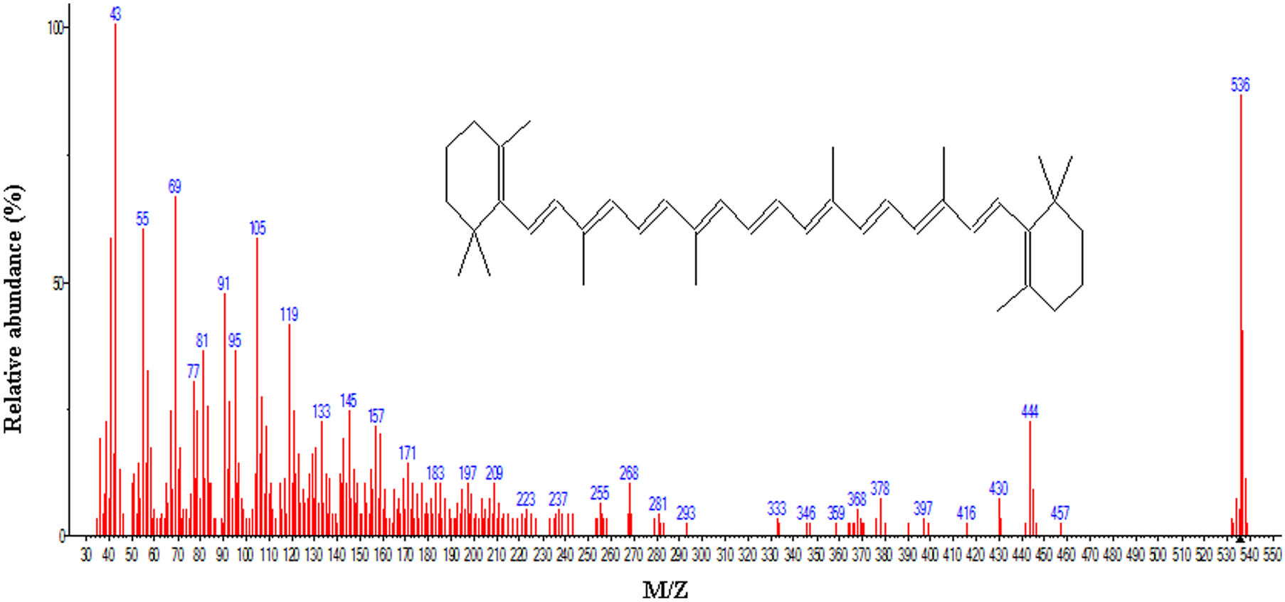
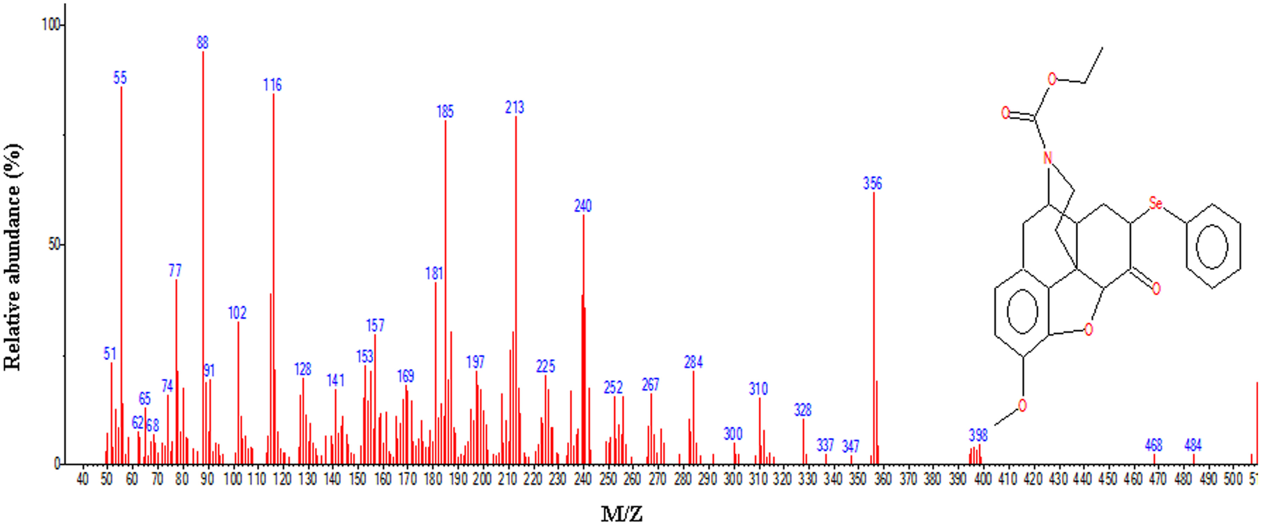
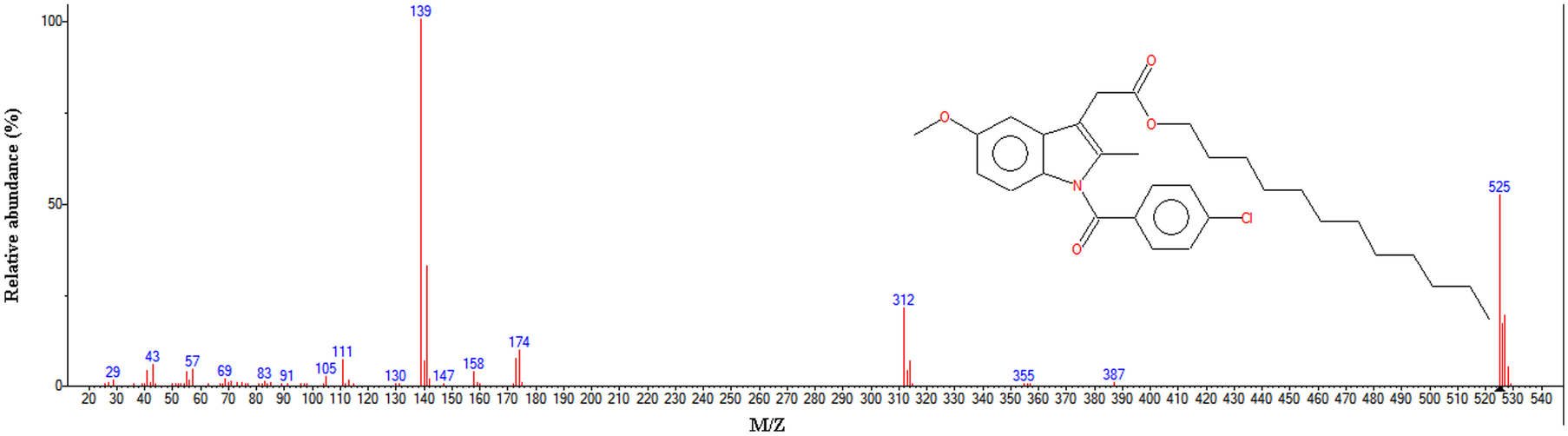
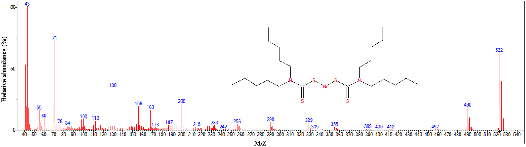
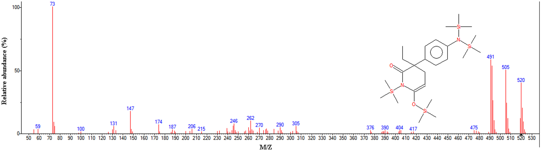
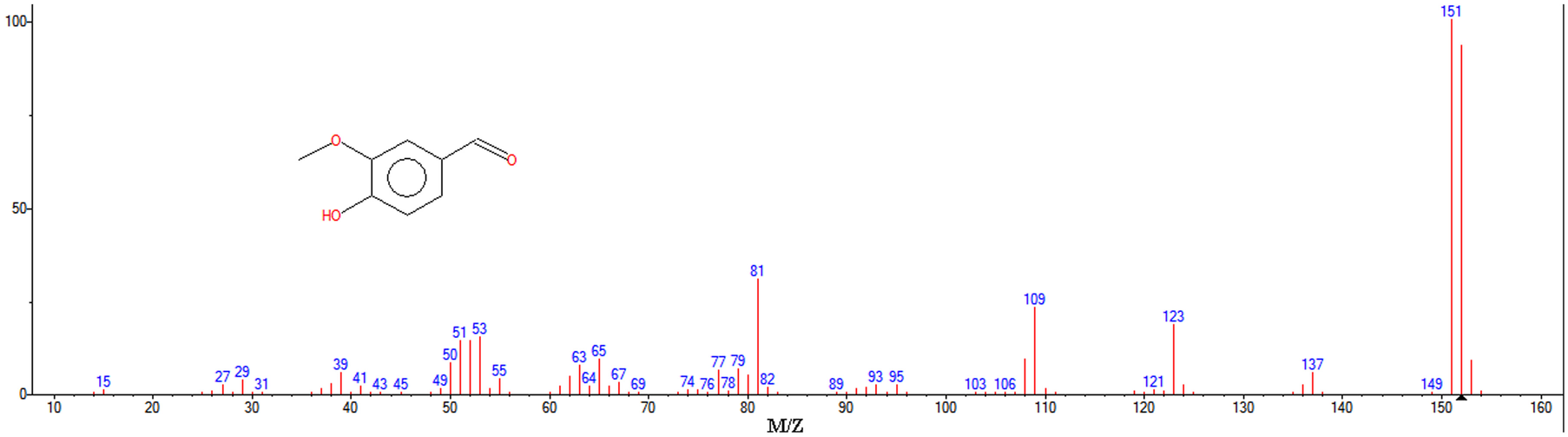
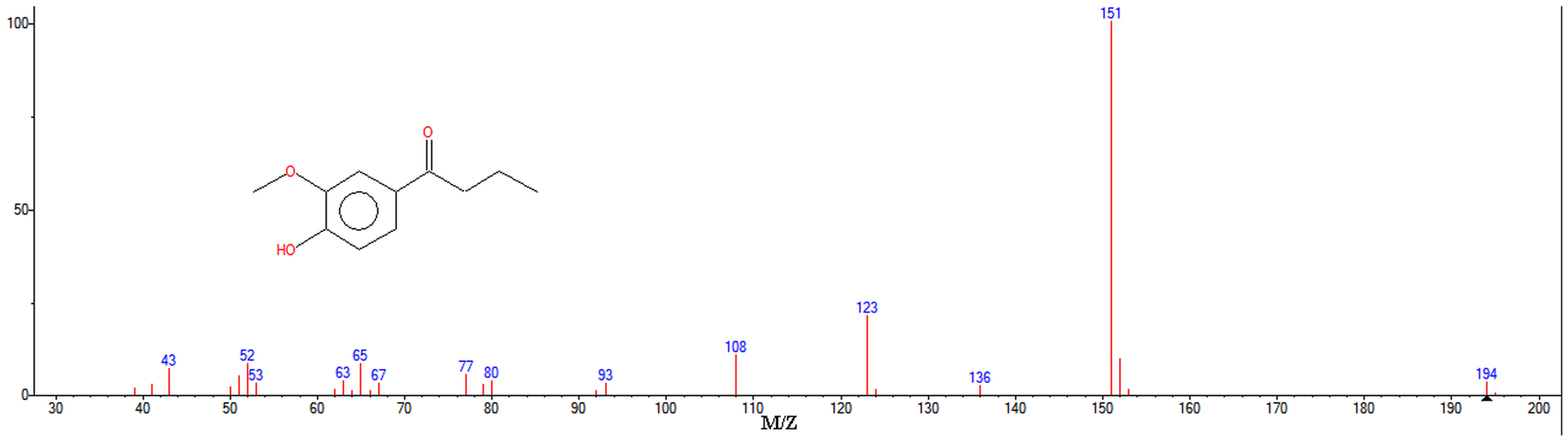
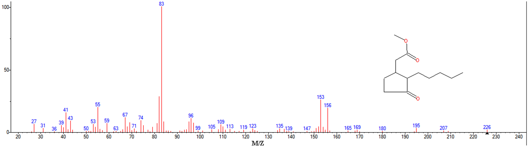
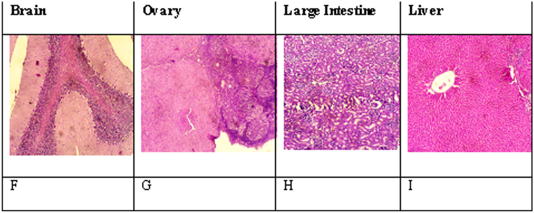
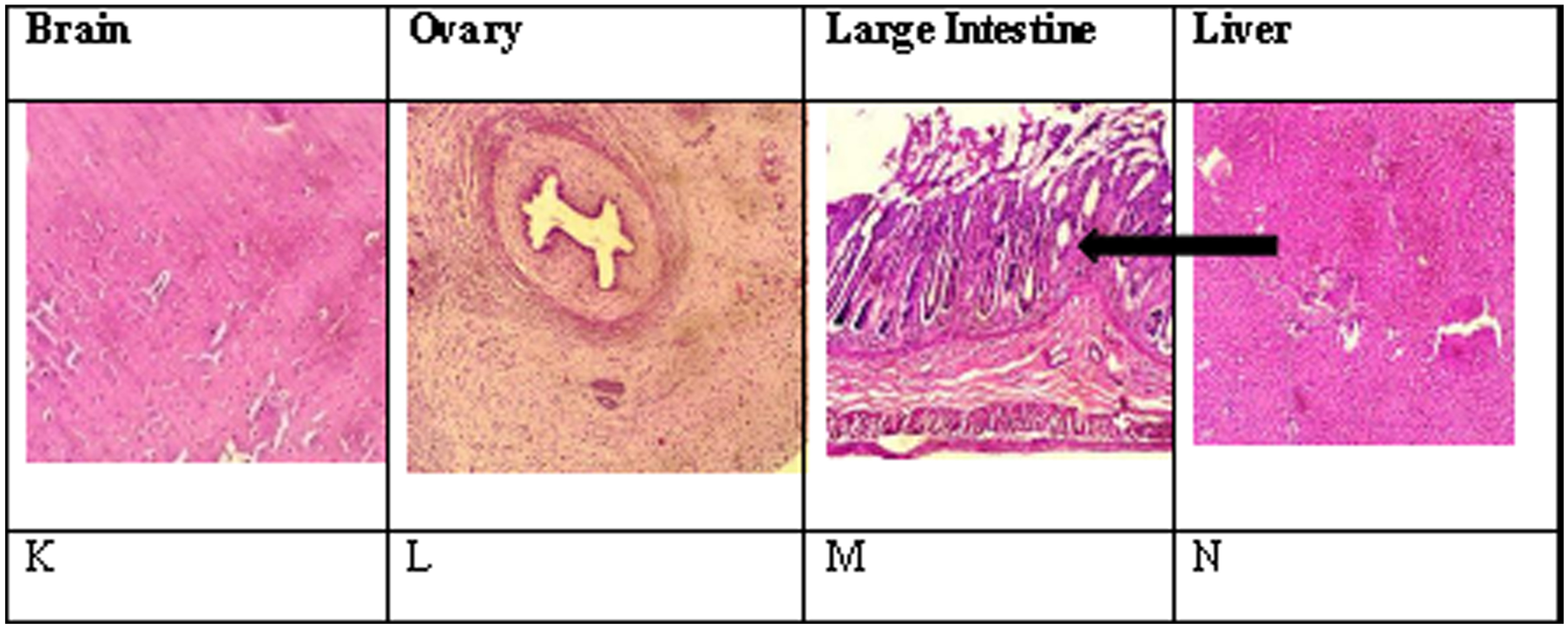
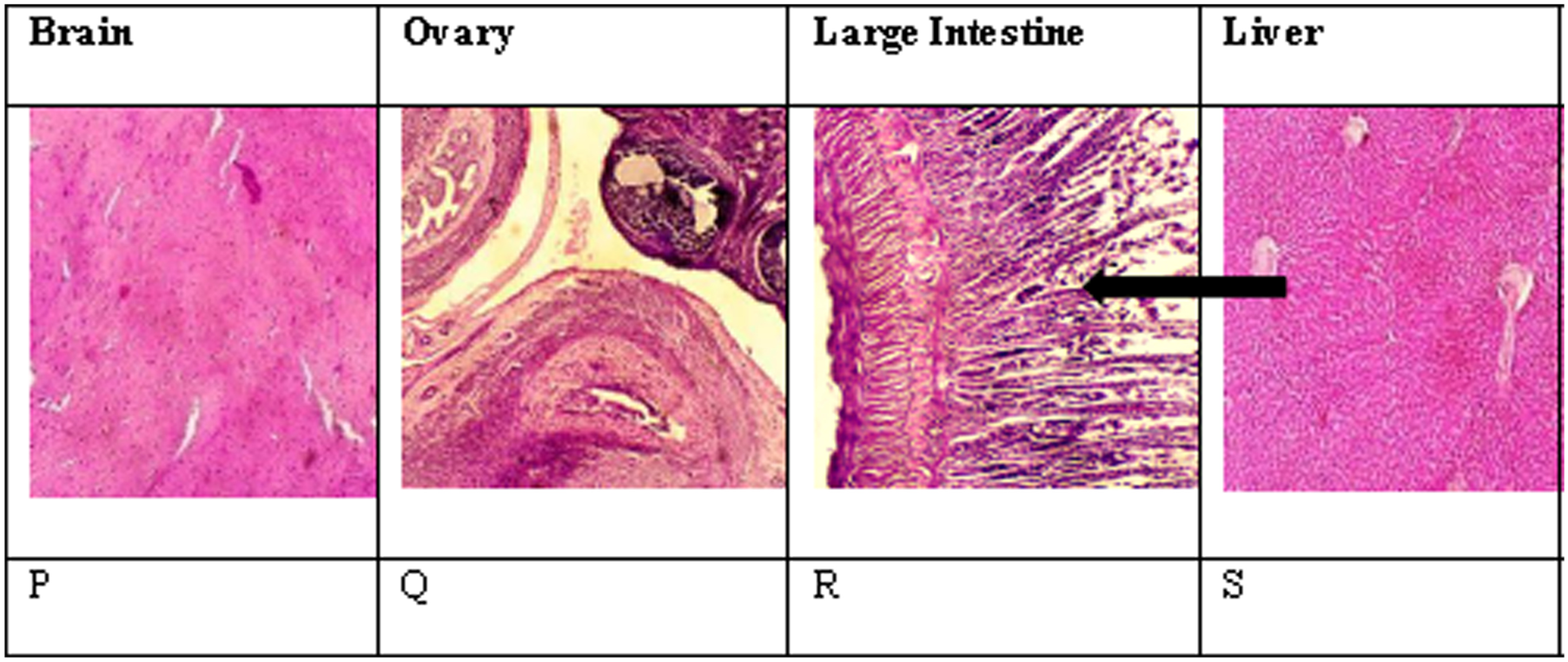
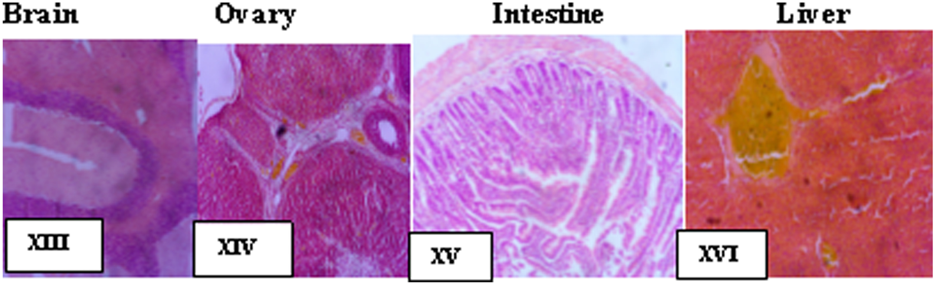
Results from GC/MS analysis confirmed the presence of steroids in both GEC and WEC (Tables 1, 2); this may be attributed to adsorption of organic compounds by the clays, possibly from runoff water. The GEC contained steroids such as cortisone metabolites, however, which were absent from WEC. A number of compounds were identified that were unique to either GEC and WEC. In GEC, beta carotene, morphinan-6-one, aminoglutethimide, bis(dipentylcarbamodithioato-S,S')-, (SP-4–1)-nickel, and indomethacin were found (Table 1, Figs 4–10). The compound with the greatest concentration was morphinan-6-one with an area of 0.91% observed in the chromatogram. In WEC, toluene was observed in addition to various alkanes (Table 2). Other compounds observed in WEC were vanillin, butyrovanillone, and cyclopentaneacetic (Table 2, Figs 11–13). Histopathological results from the organs of animals fed with the various samples revealed normal tissue for the initial 250 mg/kg in the kidney, liver, ovary, and brain (Fig. 14). The intermediate sample of 500 mg/kg showed an inflamed intestinal mucosa, normal brain cells, normal kidney tissue, normal ovaries, and no abnormalities in the liver (Fig. 15). The high dosage of 1000 mg/kg resulted in an inflamed intestinal mucosa with mild erosion, normal brain cells, normal ovaries, normal kidney cells, and severe vascular congestion in the liver structures (Fig. 16). The acute toxicity study revealed no lethal effects nor behavioral signs of toxicity at the tested doses, indicating that the LD50 is > 5000 mg/kg.
Compounds identified in GEC from GC/MS analysis

Compounds identified in WEC using GC/MS analysis

Mass spectrum of beta carotene in GEC

Mass spectrum of orcinol in GEC

Discussion
The present study aimed to determine the constituents in both GEC and WEC to check whether consumption would be toxic to humans. Organic and inorganic compounds and drugs such as indomethacin and morphinone and metals such as Ni were discovered and their consumption was determined to be threatening to human health, especially in pregnant women, but not life threatening. Chronic oral exposure of humans to inorganic As at doses as low as 0.05–0.1 mg/kg/day is commonly associated with As in soils ranging from 5 to 20 ppm. Nevertheless, results from the present study indicated that levels elevated above these had no apparent harmful effects.
The consumption of large amounts of edible clay, however, can lead to severe vomiting, disturbances in blood circulation, damage to the nervous system, and eventually to death (Nkansah et al. Reference Nkansah, Korankye, Darko and Dodd2017). Edible clays, therefore, should not be consumed in large quantities.
The GC/MS analyses revealed compounds unique to each sample of the edible clays (Tables 1, 2). In GEC, surprising organic compounds such as beta-carotene (Fig. 4) and orcinol (Fig. 5) were recorded. The GC/MS also revealed drugs such as indomethacin (Fig. 6), morphinan-6-one (Fig. 7), and aminoglutethimide (Fig. 8) in the GEC. They could be there as contaminants having been adsorbed by the clay from contaminated run-off water (Massaro et al. Reference Massaro, Colletti, Bommarito, Giordano, Milioto, Noto, Poma and Lazzara2015). Edible clays are structured in such a way that the 1 nm-thick clay aluminosilicate layers are stacked (Gobel et al. Reference Gobel, Thomsen, McArdell, Ternes and Siegrist2005; Massaro et al. Reference Massaro, Colletti, Bommarito, Giordano, Milioto, Noto, Poma and Lazzara2015; Stackelberg et al. Reference Stackelberg, Furlong, Meyer, Zaugg, Henderson and Reissman2004). The clay is able to expand a little (due to the strong hydrogen bond in the interlayer space) allowing molecules to penetrate the structure. The soil environment is thus susceptible to the active pharmaceutical ingredients and their metabolites when sludge from the water-treatment process is applied to land either as an agricultural fertilizer or when the soil is flushed with waste-water effluents from industries (Tristan et al. Reference Tristan, Michael, Bruce, John and Milena2003).
Mass spectrum of indomethacin in GEC

Mass spectrum of morphinan-6-one in GEC

Mass spectrum of aminoglutethimide in GEC

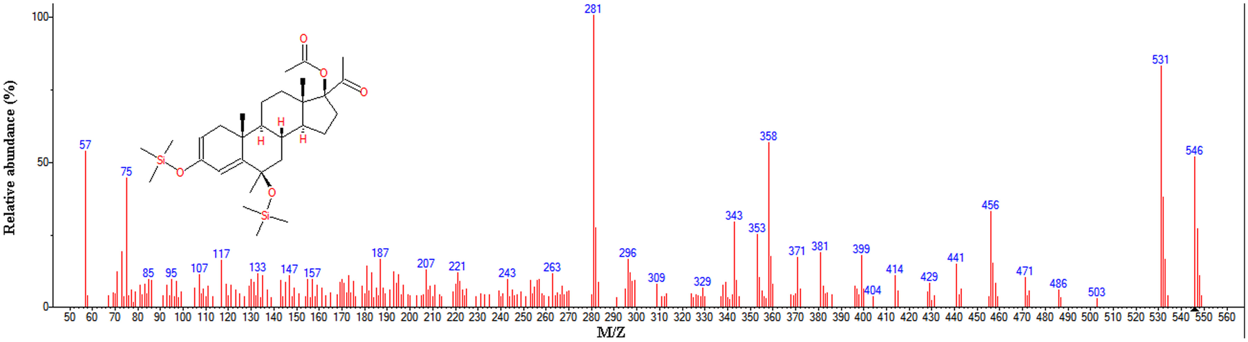
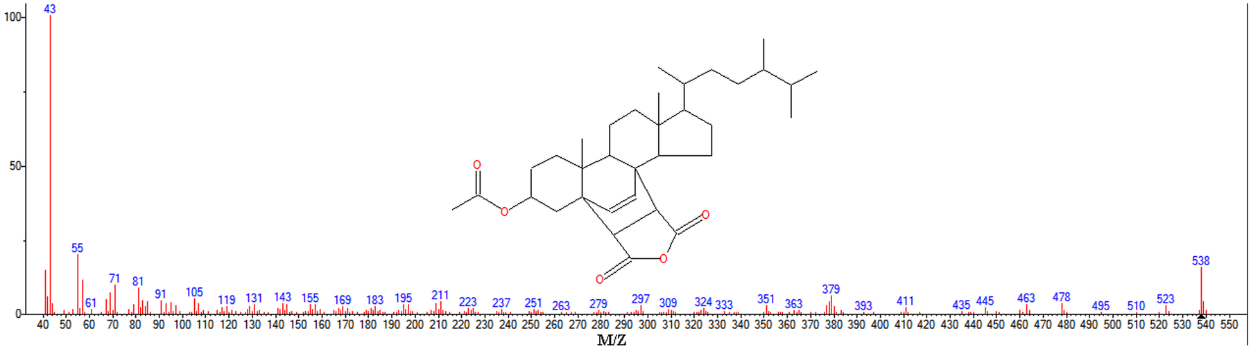
While only a few studies have discussed the occurrence of active pharmaceutical ingredients in soil, the available data reveal that a range of active pharmaceutical ingredients, including non-steroidal trace metals such as Ni, were also found to be present in the GEC. This is due to contamination of the soil with heavy metals (Yu et al. Reference Yu, Xiao, Huang and Peng2004; Zhang et al. Reference Zhang, Xie, Li, Wang, Yang and Nie2015). Pregna-2,4-dien-20-one (Fig. 9) was also found; this is a pregnane derivative which serves as an example of the sterones which are seen in clay compounds with the same chemical composition as edible clay (Dongen et al. Reference van Dongen, Rijpstra, Philippart, de Leeuw and Sinninghe Damste2000). Cholestan-6-en-3-ol (Fig. 10), a derivative of cholesterol, was discovered in the GEC sample as previously observed and reported by Dongen et al. (Reference van Dongen, Rijpstra, Philippart, de Leeuw and Sinninghe Damste2000). In the WEC, vanillin (Fig. 11), butyrovanillone (Fig. 12), and cyclopentaneacetic acid (Fig. 13) were observed. Toluene and other alkanes were also found.
Mass spectrum of pregna-2,4-dien-20-one in GEC

Mass spectrum of cholestan-6-en-3-ol in GEC

Mass spectrum of vanillin in WEC

Mass spectrum of butyrovanillone in WEC

Mass spectrum of cyclopentaneacetic acid in WEC

The origin of the homologous series of n-alkanes is still being debated (Vancampenhout et al. Reference Vancampenhout, De Vos, Wouters, Van Calster, Swennen, Buurman and Deckers2010) but possible sources include microbial polymers (Lichtfouse Reference Lichtfouse, Chenu, Baudin, Leblond, Da Silva, Behar, Derenne, Largeau, Wehrung and Albrecht1998). Other aromatic alkanes observed within the WEC include ethyl benzene, o-xylene, p-xylene, benzene propanoic acid, nonane, cyclopentenacetic acid, benzaldehyde, and ethanone. To the best of the authors’ knowledge, this is the first time in vivo toxicity assays have been carried out on these samples. A study by World Health Organization/International Programme (2005) reported that in a series of 100 necropsies among 11 mammalian and eight avian species in the San Diego Zoo, California, USA, interstitial fibrosis was observed in 20% of the mammals studied, including rats and mice that consume clayey soils from that environment. The study found that clay is toxic to a variety of mammalian cells, and it produces transient inflammation in the lungs of experimental animals after intra-tracheal instillation. The absence of lethal effects from the acute toxicity study indicates the relative safety of GEC and WEC, though caution should still be applied to the quantities consumed.
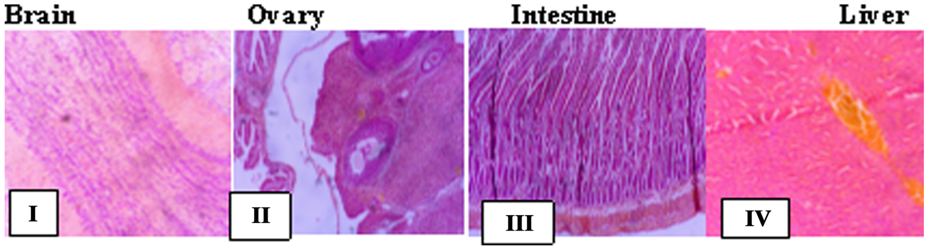
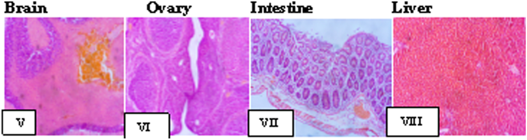
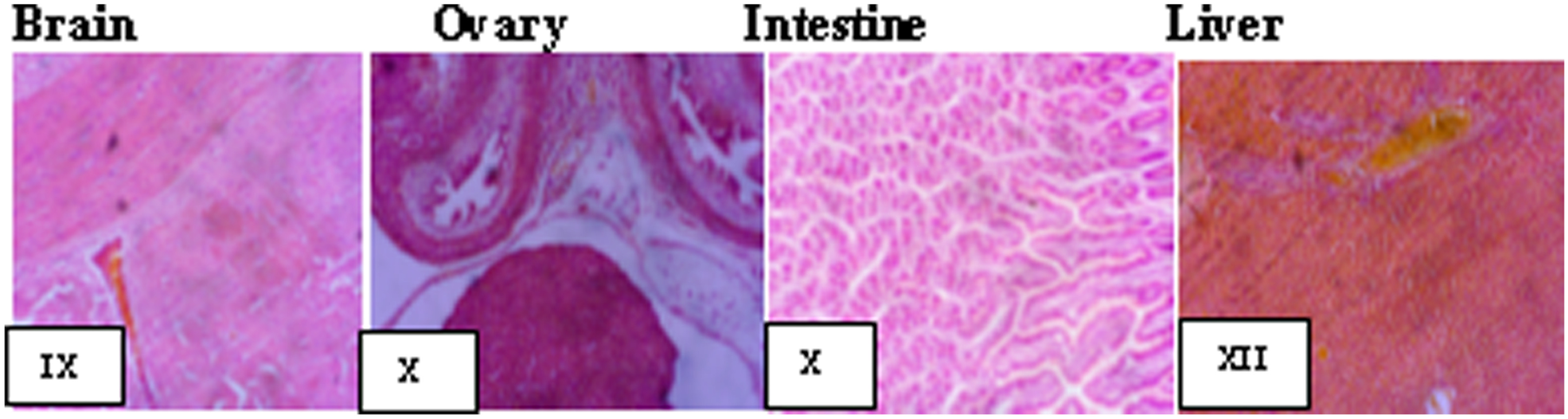
The results of the toxicity study were very similar for GEC and WEC; histological sections of all the control organs (W) showed them to be normal (Fig. 17). No abnormalities were recorded in the organs for LDGEC (Fig. 14), but intestinal erosion was observed from LDWEC (Fig. 18: VII). After the MDGEC exposure (Fig. 15), the ovary (K), liver, (N), and brains (L) were normal but the large intestines (M) (Fig. 15) showed that the mucosa was infiltrated by dense aggregates of inflammatory cells (black arrows in M). This is consistent with the findings of WHO (2005). After MDWEC ingestion, the brain showed vascular congestion (Fig. 19: X); the liver (Fig. 19: XII) also showed congested blood vessels.
Histological sections from animals fed low-dose 250 mg/kg Gray Edible Clay (LDGEC). F – Section of brain: tissue shows neuronal cell bodies disposed on a background of neuropil. No abnormalities seen. G – Section of ovary: sections of ovarian tissue show show follicles at varying stages of development and corpora lutea (indicating ovulation). No abnormalities seen. H – Section of large intestine: section of tissue shows viable muscularis propria wall and mucosal lining cells. No abnormalities seen. I – Section of liver: liver tissue shows parallel radially arranged plates of hepatocytes. No abnormalities seen

Histological sections from animals fed medium-dose 500 mg/kg Gray Edible Clay (MDGEC). K – Section of brain: tissue shows neuronal cell bodies disposed on a background of neuropil. No abnormalities seen. L – Section of ovary: sections of ovarian tissue show follicles at varying stages of development and corpora lutea (indicating ovulation). No abnormalities seen. M – Section of large intestine: section of tissue shows viable muscularis propria wall. The mucosa is infiltrated by dense aggregates of inflammatory cells. Inflamed intestinal mucosa. N – Section of liver: liver tissue shows parallel radially arranged plates of hepatocytes. No abnormalities seen

Histological sections from animals fed high-dose 1000 mg/kg Gray Edible Clay (HDGEC). P – Section of brain: tissue showing neuronal cell bodies disposed on a background of neuropil. No abnormalities seen. Q – Section of ovary: sections of ovarian tissue show follicles at varying stages of development and corpora lutea (indicating ovulation). No abnormalities seen. R – Section of large intestine: section of tissue shows viable muscularis propria wall. The mucosa is infiltrated by dense aggregates of inflammatory cells. Inflamed intestinal mucosa with mild erosion. S – Section of liver: liver tissue shows general structure, and the basophilic portion with nucleus and the acidophilic cytoplasm of the acinar cells. Congested blood vessels also seen. Severe vascular congestion

Histological sections from animals fed a control diet (W). I – Brain tissue showing neuronal cell bodies disposed on a background of neuropil. No abnormalities seen. II – Ovarian tissue showing follicles at varying stages of development and corpora lutea. III – Tissue showing viable muscularis propria wall and mucosal lining cells. IV – Liver tissue showing general structure, and the basophilic portion with nucleus and the acidophilic cytoplasm of the acinar cells

Histological sections from animals fed low-dose 250 mg/kg White Edible Clay (LDWEC). V – Brain tissue showing neuronal cell bodies disposed on a background of neuropil. Congested blood vessels are seen with aggregates of inflammatory red cells. VI – Ovarian tissue showing follicles at varying stages of development and corpora lutea (indicating ovulation). No abnormalities seen. VII – Tissue showing viable muscularis propria wall and mucosal lining cells. Areas of erosion are also seen. Intestinal erosion. VIII – Liver tissue showing parallel, radially arranged plates of hepatocytes with central vein, portal vein, and the basophilic portion with nucleus and the acidophilic cytoplasm of the acinar cells

Histological sections from animals fed medium-dose 500 mg/kg White Edible Clay (MDWEC). IX – Brain tissue showing neuronal cell bodies disposed on a background of neuropil. Congested blood vessels are also seen. Vascular congestion. X – Ovarian tissue sections showing follicles at varying stages of development and corpora lutea (indicating ovulation). No abnormalities seen. XI – Tissue showing viable muscularis propria wall and mucosal lining cells. No abnormalities seen. XII – Liver tissue showing general structures. Congested blood vessels are also seen. Vascular congestion

Sections from animals fed HDGEC (Fig. 16) showed normal brains and ovaries but the large intestine was infiltrated by dense aggregates of inflammatory cells (R) with mild erosion also. The liver was also affected adversely with congested blood vessels (Fig. 20 S) and recorded as severe vascular congestion.
Histological sections from animals fed high-dose, 1000 mg/kg White Edible Clay (HDWEC). XIII – Brain tissue showing neuronal cell bodies disposed on a background of neuropil. No abnormalities observed. XIV – Ovarian tissue showing follicles at varying stages of development. No abnormalities seen. XV – Tissue showing viable muscularis propria wall and mucosal lining cells. No abnormalities seen. XVI – Liver tissue showing general structure. Congested blood vessels are also seen. Vascular congestion

Conclusions
These findings confirm that edible clays, WEC and GEC, are not toxic to mammals despite the presence of some heavy metals; extensive consumption, however, could harm bodily organs. Both GEC and WEC, the ingestion of which is locally regarded as a “dirty” habit, are interesting mineral resources for use in medicines and excipients in the pharmaceutical industry because of their capacity to retain organic and inorganic compounds as found in the present study. For comparison, a proprietary drug, known as “Mist Kaolin” or Moko, was investigated and results revealed that it contains kaolinite; it also contains levels of As that far exceed recommended limits.
The study protocol was approved by the Ethics and Grants Committee, College of Medicine of the University of Lagos, Idi-Araba and granted an approval number CMULACUREC/02/20/724.
Declarations
Conflict of Interest
The authors declare that they have no conflict of interest.
Appendix 1 FTIR spectra of GEC

Appendix 2 FTIR spectra of WEC

Appendix 3 FTIR spectra of Mist Kaolin Moko