Child maltreatment is associated with posttraumatic stress disorder (PTSD; De Bellis, Reference De Bellis2001), impairing subthreshold PTSD (Carrion, Weems, Ray, & Reiss, Reference Carrion, Weems, Ray and Reiss2002), and other mental illness later in life (Anda et al., Reference Anda, Felitti, Bremner, Walker, Whitfield and Perry2006). While there are suggestions that maltreated males may be less resilient to emotional dysregulation and antisocial outcomes compared with maltreated females (Bergen, Martin, Richardson, Allison, & Roseger, Reference Bergen, Martin, Richardson, Allison and Roeger2004; De Bellis & Keshavan, Reference De Bellis and Keshavan2003; Garnefski & Diekstra, Reference Garnefski and Diekstra1997; McGloin & Widom, Reference McGloin and Widom2001), studies of Gender × Maltreatment interactions where sufficient numbers of males and females were included are lacking (Maas, Herrenkohl, & Sousa, Reference Maas, Herrenkohl and Sousa2008). Sexual dimorphism is present in the developing human brain (De Bellis, Keshavan, et al., Reference De Bellis, Broussard, Herring, Wexler, Moritz and Benitez2001; Lenroot et al., Reference Lenroot, Gogtay, Greenstein, Wells, Wallace and Clasen2007; Neufang et al., Reference Neufang, Specht, Hausmann, Gunturkun, Herpertz-Dahlmann and Fink2009) and has been demonstrated in anatomical magnetic resonance imaging (MRI) studies as early as infancy (Gilmore et al., Reference Gilmore, Lin, Prastawa, Looney, Vetsa and Knickmeyer2007). Furthermore, the presence of testosterone early in fetal life not only determines physical gender but also is involved in sexual dimorphism of brain structures and neural connections involved in reproductive and non-gender-related networks (e.g., mood and cognition; McEwen, Reference McEwen2006). Prospective studies show that maltreated boys have poorer outcomes in adolescence (De Bellis & Keshavan, Reference De Bellis and Keshavan2003) and adulthood (McGloin & Widom, Reference McGloin and Widom2001). McGloin and Widom (Reference McGloin and Widom2001) prospectively studied resilience defined across a variety of domains (psychiatric, emotional, and behavioral) in a large group of adults with histories of substantiated cases of child abuse and neglect prior to age 11 years and a control group closely matched for age, sex, race, and social class background. In this study, resilience was comprehensively operationalized across eight domains (i.e., employment, homelessness, education, social function, presence of psychiatric disorders and substance abuse, and two measures of antisocial behaviors) and included multiple assessment waves of their data. They found that overall, adults maltreated as youth were less resilient than nonmaltreated youth; however, cases of maltreated males were lowest and nonmaltreated females highest on their constructed measure of resilience (McGloin & Widom, Reference McGloin and Widom2001), suggesting increased vulnerability in maltreated males. In a relatively large cross-sectional anatomical MRI study (De Bellis & Keshavan, Reference De Bellis and Keshavan2003), maltreated boys with PTSD showed more evidence of adverse brain development (smaller cerebral volumes and larger lateral ventricular volumes) than did maltreated girls with PTSD, suggesting sex differences in brain maturation in traumatized youth even though both boys and girls showed similar psychopathology and trauma histories. A follow-up of a subsample from the original study (De Bellis, Keshavan, et al., Reference De Bellis, Baum, Birmaher, Keshavan, Eccard and Boring1999) demonstrated that 32% of the maltreated males with PTSD and 5% of the maltreated females with PTSD studied, but none of the controls, developed serious antisocial behaviors within 3 years of initial brain scan, suggesting less resilience in maltreated males (De Bellis & Keshavan, Reference De Bellis and Keshavan2003). However, investigations of gender differences on maltreated girls' and boys' developing neural networks are understudied.
The phenotype of PTSD resembles both depression and generalized anxiety disorder, for which two neural networks play key roles (Phillips, Drevets, Rauch, & Lane, Reference Phillips, Drevets, Rauch and Lane2003): an executive network that supports effortful regulation of behavior, attention, and emotion (Duncan & Owen, Reference Duncan and Owen2000; Yamasaki, LaBar, & McCarthy, Reference Yamasaki, LaBar and McCarthy2002); and an affective network that processes emotional information and vigilance, including stress and fear responses (Phelps, Reference Phelps2004). The executive network comprises the lateral and dorsomedial prefrontal cortex (dmPFC), anterior cingulate cortex, and posterior parietal cortex. The affective network comprises the ventral medial PFC and subcortical regions (e.g., hippocampus and amygdala; Phelps, Delgado, Nearing, & LeDoux, Reference Phelps, Delgado, Nearing and LeDoux2004). Dysfunction in these networks and in their interactions with brain regions involved in social cognition (Gilboa et al., Reference Gilboa, Shalev, Laor, Lester, Louzoun and Chisin2004; Mitchell, Reference Mitchell2006) are hypothesized to contribute to distress disorders, particularly PTSD (Charney, Deutch, Krystal, Southwick, & Davis, Reference Charney, Deutch, Krystal, Southwick and Davis1993; Lang, Davis, & Ohman, Reference Lang, Davis and Ohman2000; LeDoux, Reference LeDoux1998; Mayberg, Reference Mayberg1997).
The pathophysiology of adult PTSD involves hypoactivation of the executive and hyperactivation of the affective emotional networks (Rauch, Shin, & Phelps, Reference Rauch, Shin and Phelps2006). In adults, PTSD is associated with medial PFC hypoactivation in response to aversive stimuli (Bremner et al., Reference Bremner, Narayan, Staib, Southwick, McGlashan and Charney1999, Reference Bremner, Vythilingam, Vermetten, Southwick, McGlashan and Staib2003, Reference Bremner, Vermetten, Vythilingam, Afzal, Schmahl and Elzinga2004, Reference Bremner, Vermetten, Schmahl, Vaccarino, Vythilingam and Afzal2005; Britton, Phan, Taylor, Fig, & Liberzon, Reference Britton, Phan, Taylor, Fig and Liberzon2005; Lanius et al., Reference Lanius, Williamson, Densmore, Boksman, Gupta and Neufeld2001, Reference Lanius, Williamson, Hopper, Densmore, Boksman and Gupta2003; Lindauer et al., Reference Lindauer, Booij, Habraken, Uylings, Olff and Carlier2004; Shin et al., Reference Shin, McNally, Kosslyn, Thompson, Rauch and Alpert1999, Reference Shin, Whalen, Pitman, Bush, Macklin and Lasko2001, Reference Shin, Wright, Cannistraro, Wedig, McMullin and Martis2005; Shin, Orr, et al., Reference Shin, Shin, Heckers, Krangel, Macklin and Orr2004). The degree of medial PFC hypoactivation is associated with PTSD severity (Britton et al., Reference Britton, Phan, Taylor, Fig and Liberzon2005; Hopper, Frewen, van der Kolk, & Lanius, Reference Hopper, Frewen, van der Kolk and Lanius2007; Shin, Orr, et al., Reference Shin, Shin, Heckers, Krangel, Macklin and Orr2004). PTSD is associated with amygdala hyperresponsivity to traumatic reminders, fear faces, and during acquisition of conditioned fear responses (Bremner et al., Reference Bremner, Vermetten, Schmahl, Vaccarino, Vythilingam and Afzal2005; Driessen et al., Reference Driessen, Beblo, Mertens, Piefke, Rullkoetter and Silva-Saavedra2004; Francati, Vermetter, & Bremner, Reference Francati, Vermetten and Bremner2007; Hendler et al., Reference Hendler, Rotshtein, Yeshurun, Weizmann, Kahn and Ben-Bashat2003; Liberzon, Abelson, Flagel, Raz, & Young, Reference Liberzon, Abelson, Flagel, Raz and Young1999; Pissiota et al., Reference Pissiota, Frans, Fernandez, von Knorring, Fischer and Fredrikson2002; Protopopescu et al., Reference Protopopescu, Pan, Tuescher, Cloitre, Goldstein and Engelien2005; Rauch et al., Reference Rauch, Whalen, Shin, McInerney, Macklin and Lasko2000; Shin, Orr, et al., Reference Shin, Shin, Heckers, Krangel, Macklin and Orr2004; Shin et al., Reference Shin, Wright, Cannistraro, Wedig, McMullin and Martis2005).
Limited studies on maltreated youth also suggest dysregulation in executive, affective, and social cognition networks (De Bellis & Hooper, Reference De Bellis and Hooper2012; De Bellis et al., Reference De Bellis, Keshavan, Frustaci, Shifflett, Iyengar and Beers2002). Preliminary studies of neglected children and adolescents showed impaired function in the dorsal executive regions (Mueller et al., Reference Mueller, Maheu, Dozier, Peloso, Mandell and Leibenluft2010) and hyperactivation in the amygdala and left anterior hippocampus to fearful and angry faces (Maheu et al., Reference Maheu, Dozier, Guyer, Mandell, Peloso and Poeth2010). Previously institutionalized international adoptees demonstrated hyperactivation in affective and social cognition networks (e.g., the bilateral amygdala and medial temporal gyrus) to fearful faces but hypoactivation to response cues in executive areas during an emotion-face go/no-go task compared to controls (Tottenham et al., Reference Tottenham, Hare, Millner, Gilhooly, Zevin and Casey2011). Youth with PTSD symptoms also exhibited decreased activation in the middle frontal gyrus and increased medial frontal activation in a similar task, suggesting response inhibition dysfunction in traumatized youth (Carrion, Garrett, Menon, Weems, & Reiss, Reference Carrion, Garrett, Menon, Weems and Reiss2008). However, these neuroimaging studies in maltreated youth lacked sufficient sample size and statistical power to examine group by gender differences.
In this investigation, we used a variant of the emotional oddball task to examine executive and affective processing in maltreated and nonmaltreated youth. The sample size was adequate to examine group by gender differences. The emotional oddball task was originally designed as an event-related task, which demonstrated in adults that the executive and affective neural networks are dissociable and can be examined separately in one task (Wang, Huettel, & DeBellis, Reference Wang, Huettel and De Bellis2008; Wang, LaBar, & McCarthy, Reference Wang, LaBar and McCarthy2006; Wang, McCarthy, Song, & LaBar, Reference Wang, McCarthy, Song and LaBar2005). The emotional oddball task contained four types of stimuli: targets, sad faces or photographs, neutral faces or photographs, and phase-scrambled photographs (as standards). Subjects detected infrequent circles (targets) within a continual stream of phase-scrambled images (standards). Sad and neutral images were intermittently presented instead of phase-scrambled photographs as task-irrelevant distracters. Healthy adults activate executive networks to targets and affective neural networks (i.e., the amygdala and ventral PFC) to sad images during this task (Wang et al., Reference Wang, McCarthy, Song and LaBar2005, Reference Wang, LaBar and McCarthy2006). Healthy youth also activate executive networks to targets and affective neural networks to sad images or sad distracters during this task (Wang, Huettel, et al., Reference Wang, Huettel and De Bellis2008). Adults with distress disorders show attenuated activation in executive networks (Wang, LaBar, et al., Reference Wang, LaBar, Smoski, Rosenthal, Dolcos and Lynch2008) and accentuated activity in affective neural networks (Drevets, Reference Drevets2000; Mayberg, Reference Mayberg1997; Nitschke et al., Reference Nitschke, Sarinopoulos, Oathes, Johnstone, Whalen and Davidson2009). In an exploratory study using the emotional oddball task, we found that the maltreated youth revealed significantly decreased activation in the left middle frontal gyrus and right precentral gyrus to target stimuli and significantly increased activation to sad stimuli in the bilateral amygdala, left subgenual cingulate, left inferior frontal gyrus, and right middle temporal cortex compared to nonmaltreated participants, suggesting that maltreated youth with distress disorders demonstrated dysfunction of neural networks related to executive and affective processing (De Bellis & Hooper, Reference De Bellis and Hooper2012).
To investigate the impact of the interaction of maltreatment and gender in the executive and affective neural network in youth, we conducted a functional MRI study in maltreated youth with PTSD symptoms compared with nonmaltreated controls. Participants performed an emotional oddball task that involved detection of targets presented alongside task-irrelevant fearful face distracters. We hypothesized that maltreated youth compared to controls would show increased activation in the affective emotional network during passive viewing of fearful faces and decreased executive network activation during target detection when presented with task-irrelevant fearful face distracters. Given that gender influences emotional regulation in adults (Koch et al., Reference Koch, Pauly, Kellermann, Seiferth, Reske and Backes2007; McRae, Ochsner, Mauss, Gabrieli, & Gross, Reference McRae, Ochsner, Mauss, Gabrieli and Gross2008; Schienle, Schafer, Stark, Walter, & Vaitl, Reference Schienle, Schafer, Stark, Walter and Vaitl2005), a planned investigation examining the relationship of neural correlates in maltreated males and females compared to control males and females was undertaken. We hypothesized that maltreated males would demonstrate greater executive and affective dysregulation than would maltreated females. Planned comparisons were undertaken to determine the relationship between functional activation in brain structures involved in affective emotional and executive networks, and PTSD symptoms.
Materials and Methods
Subjects
Thirty-seven maltreated and 57 healthy control youth, the latter with no history of DSM-IV Axis I disorders or Type A traumas, participated. Of these, 8 maltreated and 12 controls were eliminated owing to noncorrectable motion artifacts or gradient problems within the imaging apparatus, leaving 29 maltreated and 45 nonmaltreated participants with usable data included in this study. We recruited more controls than maltreated subjects to increase statistical power, reduce intersubject variance, and obtain a more normative comparison, given that individual developmental trajectories in adrenarchy and puberty differ during this period (Blakemore, Burnett, & Dahl, Reference Blakemore, Burnett and Dahl2010; Giedd, Keshavan, & Paus, Reference Giedd, Keshavan and Paus2008). The maltreated groups were defined by a positive forensic investigation with Child Protective Services (CPS) that indicated physical, sexual, emotional abuse, and/or neglect as defined by state criteria. Maltreated participants were recruited through statewide advertisements and recruitment presentations targeted at CPS agencies. To reduce bias, the study was advertised to CPS in the state of North Carolina on a statewide level, and participants who lived more than 75 miles from the research program were given overnight accommodations. Controls were recruited from schools and other community settings, and had a negative screen on both telephone interview for eligibility and research interview for any history of participant or participant sibling having CPS involvement.
Exclusion criteria were as follows: IQ < 70, chronic medical illness, head injury, neurological disorder, schizophrenia, anorexia nervosa, pervasive developmental disorder, birth weight under 5 lb, severe prenatal compromise with neonatal intensive care unit stay, alcohol/substance use disorder, and contraindications for safe MRI scan. The local university hospital institutional review board committee approved the study. Legal guardians gave informed consent, and youth assented prior to participation.
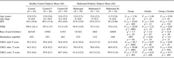
Characteristics of the maltreated and control groups are shown in Table 1. The groups were similar in age, race, handedness, and sex. The maltreated group was of lower socioeconomic status (SES) than were controls as measured by the Hollingshead Four Factor Index. Lower SES is an inherent confound and risk factor in child maltreatment (Gilman, Kawachi, Fitzmaurice, & Buka, Reference Gilman, Kawachi, Fitzmaurice and Buka2003; Lansford et al., Reference Lansford, Malone, Stevens, Dodge, Bates and Pettit2006), while higher SES or positive change in parental income reduces pediatric mental disorders (Costello, Erkanli, Copeland, & Angold, Reference Costello, Erkanli, Copeland and Angold2010). Despite attempts to control for SES between groups, lower SES children recruited as controls were more likely to meet exclusionary criteria. Two-factor IQ, measured by the Wechsler Intelligence Scale for Children (3rd ed.) and comprising vocabulary and block design (Wechsler, Reference Wechsler1991), was lower in maltreated youth versus controls. Lower IQ is a consequence of child maltreatment (De Bellis, Keshavan, et al., Reference De Bellis, Baum, Birmaher, Keshavan, Eccard and Boring1999; Perez & Widom, Reference Perez and Widom1994).
Demographic and clinical characteristics of the study participants

Note: There were no statistical differences in sex distribution between the Healthy Control and Maltreated Groups (χ2 = 1.19, p = .28). F., Females; M., males; SES, socioeconomic status; FSIQ, full scale IQ estimated from two factors; FET, Fisher's Exact Test; Caus, Caucasian; AA, African American; Other, multiracial; CBCL, Child Behavior Checklist; inter., internalizing; exter., externalizing.
Measures
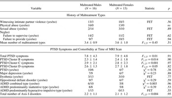
To examine psychiatric symptoms, the Kiddie Schedule for Affective Disorders and Schizophrenia—Present and Lifetime Version (Kaufman et al., Reference Kaufman, Birmaher, Brent, Rao, Flynn and Moreci1997) was administered to caregivers and youth. Because multiple sources of information are needed to gather accurate maltreatment history and related symptoms (Kaufman, Jones, Stieglitz, Vitulano, & Mannarino, Reference Kaufman, Jones, Stieglitz, Vitulano and Mannarino1994), we also used archival records (e.g., pediatric records, school attendance records, birth records, and forensics records) as sources of mental health, birth history, trauma history, and pediatric health. The Kiddie Schedule was modified to collect data on additional types of adverse life events as previously described (De Bellis, Hooper, Spratt, & Woolley, Reference De Bellis, Hooper, Spratt and Woolley2009). Child maltreatment was defined as witnessing domestic violence (which was state defined as neglect by omission or commission and/or emotional abuse), physical abuse, sexual abuse, and/or neglect. Maltreated youth experienced multiple maltreatment types that were chronic in nature. There were no significant differences in maltreatment experiences or number of maltreatment types experienced between maltreated males and females. There were no significant sex differences in PTSD symptoms or psychopathology (Table 2).
Maltreatment, PTSD symptoms, and clinical characteristics of male and female maltreated youth

Note: ADHD, Attention-deficit/hyperactivity disorder; FET, Fisher's Exact Test; MRI, magnetic resonance imaging.
PTSD was a common diagnosis in maltreated youth. In our sample, 16 had the disorder, while 13 did not meet the diagnostic criteria. As commonly seen in PTSD studies (De Bellis, Reference De Bellis2001; De Bellis, Broussard, et al., Reference De Bellis, Broussard, Herring, Wexler, Moritz and Benitez2001), there was significant comorbidity with other disorders and with impairing subthreshold PTSD (N = 8/13; Carrion, Weems, Ray, & Reiss, Reference Carrion, Weems, Ray and Reiss2002), making a comparison of maltreated subjects with and without PTSD scientifically inappropriate. Attention-deficit/hyperactivity disorder, predominantly the inattentive type, co-occurred with 77% of maltreated youth who either met PTSD criteria or had impairing subthreshold PTSD. Eight of the maltreated youth were on stable doses of medications (N = 2 stimulant and antidepressant, n = 1 female; N = 4 stimulants only, n = 2 females; N = 2 antidepressants only, n = 2 females). If significant brain differences were found between the maltreated and nonmaltreated groups, we addressed the influence of medications in secondary brain region of interest (ROI) analyses to confirm group differences by excluding all 8 maltreated subjects on medications in these secondary general linear analyses.
Experimental paradigm
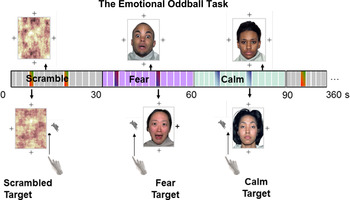
Emotional and executive control was probed using a block design variant of the emotional oddball task (Wang, Huettel, et al., Reference Wang, Huettel and De Bellis2008), consisting of fear, calm, and scrambled face stimuli mixed with target events. There were 15 trials presented sequentially of which 2 had a target (a cartoon running rabbit) on one of the four sides of the stimulus image. Participants pressed a button when they saw this target. We used randomly selected fearful and calm faces from the NimStim, a valid and reliable set of facial expression stimuli of multiracial individuals (Tottenham et al., Reference Tottenham, Tanaka, Leon, McCarry, Nurse and Hare2009), to ensure a gender and racially diverse balance that was similar to our subject demographics. The same set of faces was randomly given to all participants. The block design involved five runs, each lasting 6 min. Each run consisted of 12 blocks, or stimulus presentations, where a set of 4 of each stimulus type was presented in a pseudorandom order to ensure that 2 of the same stimulus block types were not consecutive. Images of calm expressions with relaxed facial musculature were used for the calm condition because elevated amygdala response to neutral faces was reported in children (Thomas et al., Reference Thomas, Drevets, Whalen, Eccard, Dahl and Ryan2001). Because children show heightened amygdala activations to a variety of emotional faces compared with adults (Hoehl, Brauer, van der Kolk, & Lanius, Reference Hoehl, Brauer, Brasse, Striano and Friederici2010), we planned to examine responses to both fearful and calm faces. To increase motivation, subjects could earn additional compensation for responding to targets. Fear target refers to when a target was presented with a fearful face, calm target refers to when a target was presented during a calm face, and scrambled target refers to when a target was presented during a scrambled face. The experimental task is described in further detail in Figure 1.
(Color online) Illustration of the functional magnetic resonance imaging (MRI) task. The experimental task was described to participants as the “Catch the Rabbit Game” and is described in detail here. The task was a block design consisting of fearful, calm, and scrambled face stimuli mixed with target events surrounding each image. We used fearful faces from the NimStim, a valid and reliable set of facial expression stimuli of multiracial individuals (Tottenham et al., Reference Tottenham, Tanaka, Leon, McCarry, Nurse and Hare2009), to ensure a gender and racially diverse balance that was similar to our sample demographics. Each block contained fearful faces, calm faces, or control stimuli. For control images, photographs of faces were Fourier transformed, phase scrambled, and then inverse Fourier transformed, resulting in images that were matched with the faces on average spatial frequency and luminance but that had no recognizable content. Images of calm expressions with relaxed facial musculature were used for the calm condition rather than neutral faces. The block design involved five runs, with each run consisting of 12 blocks, or stimulus presentations. For each 30-s stimulus presentation, four crosshairs were displayed on all four sides of the stimulus image (fear, calm, or scrambled). There were 15 trials presented sequentially, of which 2 had a target (a running cartoon rabbit) on one side of the stimulus image. For example, for 2 trials during each stimulus presentation, one of the crosshairs displayed with the stimulus image was randomly replaced by a running cartoon rabbit (the target). The participants' task was to press a button as soon as they saw this target. To increase motivation, subjects could earn additional compensation for responding to targets. Each picture image was presented for 1500 ms and was followed by a single crosshair during a 500-ms interstimulus interval. Scrambled target refers to when a target was presented during a scrambled face. Fear target refers to when a target was presented with a fearful face, and calm target refers to when a target was presented during a calm face. There were 12 blocks with 4 of each stimulus type, which were presented in a pseudorandom order to ensure that two of the same stimulus block types were not consecutive.

Image acquisition
Prior to scanning, subjects underwent mock scanning desensitization and task training. Anatomical and functional images were acquired using a 3.0-T General Electric Signa EXCITE HD scanner (Waukesha, WI) with 40-mT/m gradients and an eight-channel head coil. High-resolution T1-weighted anatomical images were acquired in the axial plane using spoiled gradient-recalled acquisition with repetition time = 7.5 ms, echo time = 3.0 ms, field of view = 24 cm, flip angle = 12o, matrix = 256 × 256, yielding 1 mm2 in-plane resolution with 124 contiguous images (1 mm slice thickness) per brain volume. Functional images were collected with echo-planar imaging acquisition sensitive to blood oxygen level dependent (BOLD) contrast with repetition time = 2000 ms, echo time = 28 ms, field of vision = 24 cm, flip angle = 90o, matrix = 64 × 64, yielding 4 mm isotropic voxels and 31 contiguous images per brain volume.
Image analysis
Functional images were analyzed using FMRI Expert Analysis Tool (version 5.98, Analysis Group, FMRIB, Oxford, UK). Image preprocessing included correction for slice acquisition time, motion correction with MCFLIRT (Jenkinson, Bannister, Brady, & Smith, Reference Jenkinson, Bannister, Brady and Smith2002), normalization into standard Montreal Neurological Institute stereotaxic space (MNI; Montreal, Canada), and subject to high-pass filtering (pass frequency > 1/100 Hz). FSL's Brain Extraction Tool (Smith, Reference Smith2002) was used to exclude nonbrain voxels from our analyses.
This emotional oddball paradigm was designed to characterize cognitive processing, emotional processing, and their interactions. The scrambled condition was left unmodeled as a baseline for comparison, as is conventional in the FSL analysis package. Statistical analyses were conducted using a general linear model with local autocorrelation (Woolrich, Ripley, Brady, & Smith, Reference Woolrich, Ripley, Brady and Smith2001). Events were time locked to stimulus onset and included facial stimuli and targets presented with facial stimuli. Targets were orthogonalized from corresponding face blocks. Estimated motion parameters and ventricle regressor were included as nuisance regressors.
The second-level analyses averaged results for each contrast across runs for an individual using a fixed effect model. Third-level analyses collapsed across all subjects that included an additional regressor for between-group comparisons using a random effects model (FLAME 1). Third-level analyses provided the following contrasts: fear versus calm, fear versus scrambled, calm versus scrambled, calm target versus scrambled target, fear target versus calm target, and fear target versus scrambled target. The emotional oddball task was designed to have these types of contrasts in adults to examine affective neural networks with and without executive networks. Executive neural networks were examined with attentional control to targets during the scrambled condition and represents the brain circuits for the dorsal attention–executive system task while faces (calm or emotional) during targets were distractors and measure the influence of emotion (e.g., social cues) on attention. Thus, the contrasts of interest in this study were the following: fear versus calm (fear face vs. calm face), fear versus scrambled (fear face vs. stimuli with no social cue) for examination of affective processing; and fear target versus calm target (fear face vs. social cue) and fear target versus scrambled target (fear face vs. no social cue) for examination of executive networks during emotional and nonsocial cue distractions. Because we showed in the original emotional oddball task (on which this task is based) that healthy youth activate the dorsal attention–executive system including the anterior middle frontal gyrus, dorsal anterior cingulate, posterior cingulate, insula, and supramarginal gyrus to targets like adults but, unlike adults, youth exhibited strong activation to the emotional distracter images (i.e., sad images) not only in the ventromedial PFC but also in the posterior middle frontal gyrus and in the parietal cortex (Wang, Huettel, et al., Reference Wang, Huettel and De Bellis2008); and because the limited neuroimaging studies in youth show that children show heightened amygdala activations to a variety of types of emotional faces than adults (Hoehl et al., Reference Hoehl, Brauer, Brasse, Striano and Friederici2010) including neutral faces (Thomas et al., Reference Thomas, Drevets, Whalen, Eccard, Dahl and Ryan2001), we examined two types of comparison contrasts (calm or scrambled faces) for our contrasts of interest. In the third-level analyses, group, gender, and their interactions were examined. All statistical results of whole-brain voxelwise analyses reported in figures of brain images and tables were thresholded using clusters determined by Z > 2.3 and a corrected cluster significance threshold of p = .05 (Worsley, Reference Worsley, Jezzard, Matthews and Smith2001).
To examine the relationship between brain ROI and clinical variables, we used mean ROI BOLD activation extracted from baseline (the scrambled condition) from the second-level analyses to illustrate the activation patterns during each contrast for significant clusters in third-level whole-brain analyses. Given the significant difference between maltreated and control youth in SES, IQ, and possible medication effects, these measures were included as covariates in separate ROI analyses using general linear regression models to control for the influences of these parameters. The relationship between these ROI and clinical variables (e.g., total number of PTSD symptoms) were examined with Spearman rho correlations.
Results
Task performance
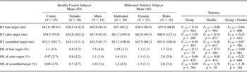
The task performance was measured by the percentage of omission errors and reaction times for target detection in each type of target event. Mixed analysis of variance did not show significant effects by group, gender, or interaction of group by gender, suggesting similar task performance between groups (Table 3).
Task performance characteristics of the study participants

Note: RT, Reaction time (ms); OE, omission error (%); fear target, target detection while viewing fearful faces; calm target, target detection while viewing neutral or “calm” faces; scrambled target, target detection while viewing scrambled faces.
Gender × Maltreated effect in brain activation during fearful face (emotional) processing
The fear versus calm contrast examined emotional processes during fearful face presentation while controlling for calm (nonemotional) faces, while the fear versus scrambled contrast examined emotional processes during a fearful face controlling for a nonsocial stimuli. The whole-brain voxelwise analyses revealed no main effects of group or gender in the fear versus calm or the fear versus scrambled contrasts.
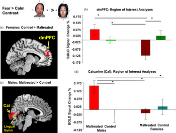
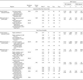
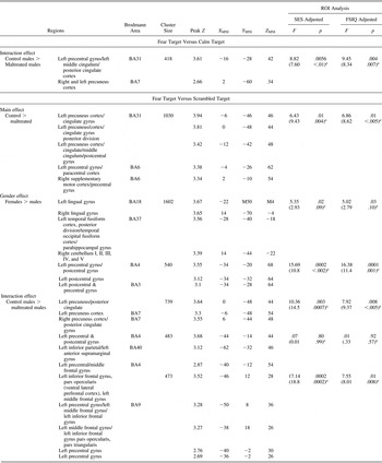
However, the whole-brain voxelwise analyses revealed significant clusters of activations during emotional processing of fear information for the Gender × Maltreated Group interaction analyses in the fear versus calm and fear versus scrambled contrasts (Table 4). Maltreated females compared to control females exhibited less BOLD signal to the fear versus calm contrast in the dmPFC (Figure 2a). Post hoc ROI analyses revealed that maltreated females showed less BOLD signal in the dmPFC than did the control female, control male, and the maltreated male groups (p < .05; Figure 2b). The maltreated females also showed less BOLD signal than did control females in the fear versus scrambled contrasts in the right cerebellum I, II, III, IV, and V, and left cerebellum I, II, III, IV, V, and VI, but more BOLD signal than control females in the left lateral occipital cortex, left middle temporal lobe, and left angular gyrus (Table 4). Post hoc ROI analyses revealed that maltreated females showed less BOLD signal in the right and left cerebellum I–V and left cerebellum VI than did the control females and control males (p < .05).
(Color online) Gender × Group effect on percentage blood oxygen level dependent (BOLD) signal in response to the fear versus calm contrast in the dorsomedial prefrontal cortex (dmPFC) and calcarine region. This contrast examined emotional processes during fearful face presentation while controlling for calm (no emotional) faces. (a) The brain image illustrates the whole-brain analysis demonstrating significantly decreased percentage BOLD signal change in the dmPFC in maltreated females than in control females (red label in brain images). (b) The region of interest (ROI) analysis (bar graph) revealed that maltreated females showed significantly decreased percentage BOLD signal when examining the individual subject's dmPFC activations extracted from the fear versus calm contrast from the scrambled baseline than did the control males, maltreated males, and control females. (c) The brain image illustrates the whole-brain analysis showing significantly increased percentage BOLD signal in maltreated males in a cluster that was composed of mainly the bilateral calcarine regions, but also the left lingual gyrus, compared with control males (red label in brain images online). (d) Post hoc ROI analyses revealed that the maltreated males showed significantly increased percentage BOLD signal in the calcarine than did control males, maltreated females, and control females. The bar graphs show the ROI analysis in the dmPFC and calcarine regions, respectively, confirming the whole-brain analysis. Post hoc analyses indicate significant ROI differences between gender groups (Dunnett method, *p < .05).

Whole brain analyses: Interaction effect in activation to fear versus calm and fear versus scramble pictures

Note: Regions were labeled in MNI coordinates with the FSL Atlases: Harvard–Oxford Cortical Structural Atlas, Harvard–Oxford Subcortical Structural Atlas, Cerebellar Atlas in MNI 152 space after normalization with FNIRT, and Talairach Daemon Labels for Brodmann areas. ROI, Region of interest; SES, socioeconomic status; FSIQ, full scale IQ.
a Under ROI adjusted analyses in parentheses throughout are further analyses controlling for medication status by excluding maltreated subjects on medications in the general linear models.
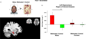
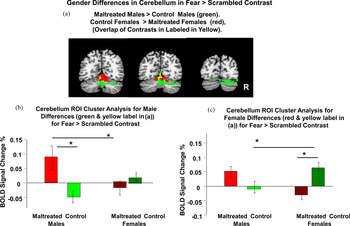
Maltreated males compared to control males showed increased BOLD signal to the fear versus calm contrast in a cluster in the calcarine cortex that included the right lingual gyrus (Figure 2b) and to the fear versus scrambled in the right cerebellum (crus I, cerebellum VI, VIIb, VIIIa, vermis VI), left middle temporal pole, left hippocampus (Figure 3a), paracentral cortex, and right supplementary motor area (Table 4). Post hoc ROI analyses revealed that maltreated males showed more BOLD signal in the calcarine cortex compared to the control male, control female, and maltreated female groups (p < .05; Figure 2d). In addition, post hoc ROI analyses revealed increased right cerebellar BOLD signal for maltreated males compared to maltreated females and control males in a large cluster that included the right cerebellum crus I (p < .05; Table 4; Figure 4b). It should be noted that these areas of cerebellar activation differences to the fear versus scrambled contrast between maltreated youth and their same-gender controls were different for males and females with little regions of overlap (Figure 4a).
(Color online) Gender × Group effect on the percentage of blood oxygen level dependent (BOLD) signal in response to fear versus scrambled in the left hippocampus. The fear versus scrambled contrast examined emotional processes during a fearful face controlling for a nonsocial stimuli. (a) The brain image illustrates the whole-brain analysis showing greater BOLD signal change in maltreated males compared with control males in response to a fear face. (b) The region of interest (ROI) analysis bar graph showed greater percentage BOLD signal in maltreated male groups in the left hippocampus to fear versus scramble compared with control males. Females did not show differences in the hippocampus. The bar graphs show the ROI analysis in the left hippocampus confirming the whole-brain analysis. Post hoc analyses indicate significant ROI differences between gender groups (Dunnett method, *p < .05).

(Color online) Gender × Group effect on percentage blood oxygen level dependent (BOLD) signal in response to fear versus scrambled in the cerebellum. (a) The brain image illustrates the whole-brain analysis demonstrating greater percentage BOLD signal in the cerebellum for maltreated males showing increased BOLD signal compared with control males (labeled in green and yellow online), and maltreated females showing decreased BOLD signal compared with control females (labeled in red and yellow online) in response to a fear face. The overlap of these two clusters is showed in yellow (online). (b) The region of interest (ROI) analysis (bar graph) showed greater percentage BOLD signal change in the cerebellum for the maltreated males compared with the control males and maltreated females to the fear versus scramble contrast for the ROI in the cerebellum from this significant cluster that involved the differences in the two male groups (i.e., the crus I and cerebellum VI, VIIb, VIIIa, vermis VI). (c) Whole-brain analysis demonstrated that control females had increased cerebellar percentage BOLD signal in response to a fear face compared with maltreated females. The bar graphs show the ROI analysis for the fear versus scramble contrast for the ROI in the cerebellum from this significant cluster that involved the differences in the two female groups (i.e., the right and left cerebellum I–V and left cerebellum VI) and showed that control females demonstrated greater activations to fear faces than did maltreated females and control males. Post hoc analyses indicate significant ROI differences between gender groups (Dunnett method, *p < .05). Note the brain regions of cerebellar activations for males and females were different and showed little areas of overlap (i.e., yellow label in (a) online).

In summary, maltreated females showed hypoactivation in the dmPFC to fearful faces compared to control females, while maltreated males showed greater BOLD signal in the visual cortex, cerebellum, and hippocampal regions compared to control males in the fear versus calm contrast, the contrast that controlled for face presentation; and the variety of gender differences seen in the fear versus scrambled contrasts most likely represented emotional processing due to both fearful face presentation and face presentation.
However, we did not find whole-brain voxelwise main effects in the maltreated versus control, gender groups, or group by gender interaction for calm versus scrambled, suggesting that the fearful face was responsible for our overall results. In order to explore these differences between the two emotional processing contrasts, we also undertook two ROI exploratory analyses to examine the relationship of dmPFC and calcarine cortex BOLD activations in the fear versus scrambled contrast. We found a significant difference for control females compared to maltreated females to show increased BOLD signal in the dmPFC for the fear versus scrambled contrast, t (1, 37) = –2.04, p < .05, which was consistent with the findings in the fear versus calm contrast. We found a trend for maltreated males compared to control males to show increased BOLD signal in the calcarine cortex, for the fear versus scrambled contrast, t (1, 33) = 1.8, p < .09. These findings were consistence with the significant Gender × Group findings seen in the fear versus calm contrast and further suggest that the fearful face was responsible for our results.
Group, Gender, and Group × Gender effects in BOLD signal during executive control processing (target detection) with fear distraction
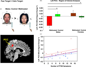
The fear targets versus calm targets contrast examined executive control processing during emotional distractors (fearful face vs. calm, nonemotional face distractors). The whole-brain voxelwise analyses revealed no main effects of group or gender in the fear targets versus calm targets contrast. However, the whole-brain voxelwise analyses revealed significant clusters of activations during executive control processing (target detection) with fear distraction for the Gender × Maltreated Group interaction analyses in the fear targets versus calm targets contrast (Table 5). Maltreated males showed decreased activations in the fear targets versus calm targets contrast in the left posterior cingulate cortex (PCC; Figure 5a). Post hoc ROI analyses revealed that maltreated males showed less BOLD signal in the left PCC compared with the control male, control female, and maltreated female groups (p < .05; Figure 5b). Greater PTSD symptoms were also correlated significantly with increased BOLD signal to fear targets versus calm targets in the PCC (Spearman ρ = 0.37, p < .05; Figure 5c). This relationship was similar in maltreated boys (Spearman ρ = 0.50, p < .05) and suggestive in maltreated girls (Spearman ρ = .52, p < .07).
(Color online) This contrast examined executive control processing during emotional distractors (fearful faces vs. calm, no emotional faces as distractors). (a) Gender × Group effect on percentage blood oxygen level dependent (BOLD) signal in response to fear target versus calm target in the posterior cingulate cortex (PCC). The brain image illustrates the whole-brain analysis, demonstrating decreased activation in maltreated males compared with control males in the PCC. (b) The bar graph illustrates decreased PCC region of interest (ROI) activations in the maltreated males compared with maltreated females, and control males and females, confirming the whole-brain analysis. Post hoc analyses indicate significant ROI differences between gender groups (Dunnett method, *p < .05). (c) Greater PTSD symptoms in maltreated youth were significantly and positively correlated with increased BOLD signal activation to fear target versus calm target in the PCC (Spearman ρ = 0.37, p < .05). This relationship was similar in maltreated boys (blue squares online, dotted line; Spearman ρ = 0.50, p < .05) and suggestive in girls (red circles online, red crossed line; Spearman ρ = 0.52, p < .07).

Whole brain analyses: Main effect or interaction effect of the analysis of variance in activation to targets

Note: Regions were labeled in MNI coordinates with the FSL Atlases: Harvard–Oxford Cortical Structural Atlas, Harvard–Oxford Subcortical Structural Atlas, Cerebellar Atlas in MNI 152 space after normalization with FNIRT, and Talairach Daemon Labels for Brodmann areas. ROI, Region of interest; SES, socioeconomic status; FSIQ, full scale IQ.
a Under ROI adjusted analyses in parentheses throughout are further analyses controlling for medication status by excluding all maltreated subjects on medications in the general linear models.
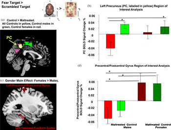
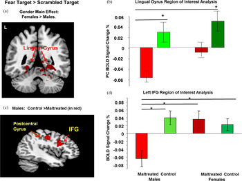
The fear targets versus scrambled targets contrast examined executive control processing during emotional distractors (fearful faces vs. nonsocial stimuli distractors). The whole-brain voxelwise analyses revealed a main effect of group and a main effect of gender in the fear targets versus scrambled targets contrast. There was a significant group difference in response to fear targets versus scrambled targets, with controls showing greater BOLD signal in the left precuneus, left middle cingulate, and right supplementary areas compared with maltreated subjects (Figure 6a; Table 5). Post hoc ROI analyses revealed that maltreated males showed less BOLD signal in the left precuneus cortex (PC) compared with control males and maltreated females (p < .05; Figure 6b), but not compared with maltreated females. Although there was a whole-brain voxelwise main group effect for controls to show greater PC activations than the maltreated groups, this finding was influenced by the lower PC activations in maltreated males. There was a main whole-brain voxelwise gender effect on the fear targets versus scrambled targets contrast in that all females showed significantly greater BOLD signal activation in the bilateral lingual gyrus, left fusiform gyrus, and right cerebellum I, II, III, IV and V, than did all males (Figure 6c and Figure 7a). The post hoc ROI analyses revealed that control females showed greater BOLD signal in the left precentral/postcentral gyrus compared with control and maltreated males, while maltreated females showed greater BOLD signal in the left precentral/postcentral gyrus compared with maltreated males (p < .05; Figure 6d). This was the only finding where gender showed a clear difference in response to executive control processing (target detection) during fear distraction that was not influenced by maltreatment status or Maltreatment × Gender interactions. Post hoc ROI analyses of the lingual gyrus revealed that whole-brain voxelwise gender effects were mainly influenced by the lower BOLD signal seen in maltreated males compared to control males and females (p < .05), but not compared with maltreated females (Figures 7a and 7b). Post hoc ROI analyses of the temporal gyrus/fusiform cortex also revealed that the main gender findings were mainly carried by the lower BOLD signal seen in maltreated males compared to control males and females (p < .05), but not compared with maltreated females (data not shown in figures).
(Color online) The fear target versus scrambled target contrast examined executive control processing during emotional distractors (fearful faces vs. nonsocial stimuli distractors). (a) Main group effect on percentage blood oxygen level dependent (BOLD) signal in response to fear target versus scrambled target in the precuneus (PC). The brain image illustrates the whole-brain analysis, demonstrating decreased percentage BOLD signal change in the maltreated group compared to the control youth; the voxels in yellow (online) indicate the main effect of group (controls greater than maltreated in the left PC). (b) The region of interest (ROI) analysis bar graph showed decreased percentage BOLD signal in maltreated males in the PC to fear target versus scrambled target compared with the control males and control females but not compared with the maltreated females. Although there was a whole-brain voxelwise main group effect for controls to show greater PC activations than the maltreated groups, this finding was influenced by the lower PC activations in maltreated males. (c) Main whole-brain voxelwise gender effect on the fear targets versus scrambled targets contrast in that all females showed significantly greater BOLD signal activation in the left precentral/postcentral gyrus, left temporal fusiform cortex, and right cerebellum I, II, III, IV, and V (data not showed), than did all males. (d) The post hoc ROI analyses revealed that control females showed greater BOLD signal in the left precentral/postcentral gyrus compared with control and maltreated males, while maltreated females showed greater BOLD signal in the left precentral/postcentral gyrus compared with maltreated males. Post hoc analyses indicate significant ROI differences between gender groups (Dunnett method, *p < .05).

(Color online) (a) Main whole-brain voxelwise gender effect on the fear targets versus scrambled targets contrast in that all females showed significantly greater blood oxygen level dependent (BOLD) signal activation in the lingual gyrus than did all males. (b) The region of interest (ROI) analysis bar graph showed decreased percentage BOLD signal in maltreated males in the lingual gyrus to fear target versus scrambled target compared with control males and control females but not compared with maltreated females, suggesting that the main gender findings were mainly carried by the lower BOLD signal seen in maltreated males compared to control males and females. (c) Gender × Group effect on a BOLD signal in response to fear target versus scrambled target in the left inferior frontal gyrus (IFG). The brain image illustrates the whole-brain analysis, demonstrating decreased percentage BOLD signal change in the maltreated males compared with the control males for the left IFG and postcentral gyrus. (d) The graph illustrates decreased percentage BOLD signal in maltreated males compared with maltreated and control female groups, and control males in the IFG (also called the ventrolateral prefrontal cortex). The bar graphs show the ROI analysis confirming the whole-brain analysis. Post hoc analyses indicate significant ROI differences between gender groups (Dunnett method, *p < .05).

In addition to the main effects of group and gender, there was a significant whole-brain voxelwise main effect of Maltreatment × Gender interaction in response to fear targets versus scrambled targets contrast. A Maltreatment × Gender interaction effect showed that maltreated males exhibited less activation to fear targets versus scrambled targets in the left PC (Figure 6b) and left inferior frontal gyrus (also known as the ventrolateral PFC [vlPFC]; Figure 7d) than did control males. Post hoc ROI analyses revealed that maltreated males showed less BOLD signal in the left PC than did the control male and females, but not the maltreated females (Figure 6b), and that maltreated males showed less BOLD signal in the left PCC than did control males and females (p < .05), but not maltreated females (data not shown in figures). Post hoc ROI analyses revealed that maltreated males showed less BOLD signal in the left vlPFC than did the control male, control female, and maltreated female groups (p < .05; Figure 7d).
We did not find main effects in the maltreated versus control, gender groups, or group by gender interaction for calm target versus scrambled target, which suggests that the fearful face was responsible for our results. The findings of greater left PC and PCC activations in control males compared with maltreated males were consistent in the fear target versus calm targets and fear target versus scrambled targets contrasts, suggesting that the fearful face distraction to targets was responsible for our results.
The influence of SES and full scale IQ on findings
In the overall sample, IQ and SES were significantly correlated (Spearman ρ = 0.34, p < .003). Therefore, we controlled for SES and full scale IQ separately in the above ROI analyses as seen in Tables 4 and 5. These analyses remained significant or suggestive, except for one finding in the fear target versus scrambled target contrast for control males to show greater BOLD response compared with maltreated males (interaction effect of control males vs. maltreated males in left precentral and postcentral gyrus cluster; Table 4). Among maltreated youth, PTSD symptoms were not significantly related to IQ (Spearman ρ = 0.17, p = .38) or SES (Spearman ρ = –0.16, p = .40). Excluding maltreated participants on medications did not influence results except for that same fear target versus scrambled target contrast (interaction effect of control males vs. maltreated males in the left precentral and postcentral gyrus cluster; Table 5). Unless otherwise reported, we did not find any other significant correlations between brain ROI reported between groups; and age, SES, IQ, or PTSD symptoms.
Discussion
Although maltreated boys and girls had similar maltreatment experiences, number of PTSD symptoms, types of Axis I mental health disorders, psychopathology, and performance on the emotional oddball task, maltreated youth significantly demonstrated gender differences during affective regulation and executive attentional control during fear distracters. During the affective processing of fearful faces controlling for calm faces, maltreated females compared to control females exhibited decreased activation in the dmPFC, while maltreated males compared to control males exhibited increased activation in the visual cortex and right lingual gyrus. When investigating executive attentional processing of oddball targets with the task-irrelevant emotional distraction of fearful faces controlling for calm faces, maltreated males compared to control males exhibited decreased activation in the left middle and PCC and the PC. Furthermore, greater PTSD symptoms were positively and significantly correlated with increased BOLD signal to fear targets versus calm targets in the PCC. This relationship remained significant in maltreated boys and was suggestive in maltreated girls. The PCC is involved in visual attention and is consistently activated during the processing of emotional stimuli and emotional memories (Maddock, Reference Maddock1999). The PC is a complex structure that is associated with multiple functions, including the posterior default mode network or resting state network (Eichele et al., Reference Eichele, Debener, Calhoun, Specht, Engel and Hugdahl2008; Fransson, Reference Fransson2005, Reference Fransson2006; Raichle et al., Reference Raichle, MacLeod, Snyder, Powers, Gusnard and Shulman2001) and integration of tasks that include visual–spatial imagery, episodic memory retrieval, and social cognition (Cavanna & Trimble, Reference Cavanna and Trimble2006). These are functions that were needed to perform this task. Only maltreated males compared to control males demonstrated differences in executive attentional processing of oddball targets with the task-irrelevant distraction of fear faces versus calm faces. Less PC activation may mean that additional deactivation of the resting state network was needed to focus more attention on the task in maltreated male youth, to integrate information and to maintain the same level of attention to the task for similar performance to the nonmaltreated youth. No differences were found in maltreated female youth, suggesting that maltreated females exhibited differences in brain regions including executive regions (e.g., dmPFC) only during the processing of affective stimuli or emotion but not during the executive component of the task. These findings suggest that maltreated male youth are more vulnerable to the influence of emotion during executive functions compared to maltreated females, while maltreated female youth may be more resilient to the influence of emotion during executive functions compared to maltreated males.
Maltreated females compared to control females exhibited decreased activation in the dmPFC during the affective processing of fearful faces versus calm faces. The dmPFC is implicated in emotion appraisal, emotion expression, and explicit threat evaluation (Etkin, Egner, & Kalisch, Reference Etkin, Egner and Kalisch2011). Decreased activation in the dmPFC in maltreated females during passive viewing of fearful faces is consistent with previous findings in both male and female adults with PTSD (Bremner et al., Reference Bremner, Narayan, Staib, Southwick, McGlashan and Charney1999, Reference Bremner, Vermetten, Vythilingam, Afzal, Schmahl and Elzinga2004, Reference Bremner, Vermetten, Schmahl, Vaccarino, Vythilingam and Afzal2005; Britton et al., Reference Britton, Phan, Taylor, Fig and Liberzon2005; Shin et al., Reference Shin, McNally, Kosslyn, Thompson, Rauch and Alpert1999, Reference Shin, Whalen, Pitman, Bush, Macklin and Lasko2001, Reference Shin, Wright, Cannistraro, Wedig, McMullin and Martis2005; Shin, Shin, et al., Reference Shin, Orr, Carson, Rauch, Macklin and Lasko2004). Previous findings in adult PTSD demonstrated decreased medial PFC activation in response to aversive stimuli including fearful faces (Francati et al., Reference Francati, Vermetten and Bremner2007; Lanius et al., Reference Lanius, Williamson, Densmore, Boksman, Gupta and Neufeld2001, Reference Lanius, Williamson, Hopper, Densmore, Boksman and Gupta2003; Lindauer et al., Reference Lindauer, Booij, Habraken, Uylings, Olff and Carlier2004; Shin, Orr, et al., Reference Shin, Shin, Heckers, Krangel, Macklin and Orr2004). Increased dmPFC activation was observed posttreatment in adults (Felmingham et al., Reference Felmingham, Kemp, Williams, Das, Hughes and Peduto2007). Thus, dmPFC hypoactivation in female youth may identify those individuals at risk for developing chronic PTSD or depression from child maltreatment. Although we did not see decreased activation in the dmPFC in maltreated females in the whole-brain voxelwise analyses for the fear versus scrambled contrast compared to control females, ROI exploratory analyses of the dmPFC showed that the maltreated females had decreased BOLD signal in the dmPFC in this contrast compared to control females, which was consist with the findings in the fear versus calm contrast and suggested that fearful face response was responsible for this finding. During the affective processing in the fear versus scrambled contrast, maltreated females compared to control females exhibited increased activation compared to control females in the left middle temporal cortex and angular gyrus. These regions are involved in face processing and social cognition, suggesting greater neural resources are needed for emotional and face processing in maltreated females than for nonmaltreated females.
We observed gender differences in maltreated male and female youth during the affective processing of fear versus scrambled faces, a contrast that reflected both response to viewing a fearful face and a face; maltreated females compared to control females exhibited decreased activation in the right cerebellum I–V and increased activation compared to control females in the left middle temporal cortex and angular gyrus. However, maltreated males compared to control males exhibited a pattern of increased activation in multiple brain regions, including the occipital cortex and fusiform gyrus, brain regions which also showed activations in the fear versus calm contrast; in addition, maltreated males demonstrated increased activations in the hippocampus, parahippocampus, left middle temporal lobe, paracentral gyrus, and right cerebellum crus I compared with control males.
Our results demonstrated gender differences during examination of executive attentional control with task-irrelevant fear distracters versus scrambled faces, a task examining executive processing during emotional distractors (fear faces vs. nonsocial stimuli distractors). Maltreated females compared to control females did not demonstrate differences in executive attentional processing of oddball targets with the task-irrelevant distraction of fear faces or scrambled faces. However, maltreated males compared to control males exhibited decreased activations in multiple brain regions including in the PC and the PCC as was seen in the fear target versus calm target contrast. Maltreated males compared to control males also exhibited decreased activations during the fear target versus scrambled target contrast in multiple brain regions (the left inferior frontal gyrus and vlPFC, bilateral precuneus, and left inferior parietal lobe). These results represent decreased brain activation to both fear faces and faces in maltreated male youth in ROI involved in visual–spatial attention and emotional regulation. The vlPFC is associated with inhibition of emotional distraction (Dolcos, Iordan, & Dolcos, Reference Dolcos, Iordan and Dolcos2011). Previous studies (Bishop, Jenkins, & Lawrence, Reference Bishop, Jenkins and Lawrence2007) found that left vlPFC activation to threat-related distracters is negatively correlated with anxiety. Hypoactivation in the PCC to fear target versus calm target and vlPFC hypoactivations to fear target versus scrambled target suggests dysfunction in the executive functions of attentional control and inhibition of emotional distraction in maltreated males compared with control males. Our ROI analyses of the inferior frontal gyrus during the fear target versus scrambled target contrast and the PCC during the fear target versus calm target contrast suggested that maltreated male youth show altered executive attentional processing during emotional and nonemotional (scrambled face) distraction compared to maltreated female, and both male and female nonmaltreated control youth.
In contrast, maltreated males compared to control males exhibited increased activation in the visual cortex (calcarine) and right lingual gyrus during affective processing in the fear versus calm contrast and exhibited a pattern of increased activation in multiple brain regions (i.e., the hippocampus, parahippocampus, left middle temporal lobe, paracentral gyrus, and right cerebellum crus I) including the occipital cortex and fusiform gyrus, as also seen in the fear versus calm contrast, during the affective processing of fear versus scrambled contrast. Maltreated males may be dedicating significant functional neural resources to processing affective and face stimuli as indicated by increased visual cortex and extended limbic system activations. The hippocampus is sensitive to stress. In early stages of stress, enlarged hippocampal volume or increased activation is seen, whereas long-term chronic stress results in hippocampal atrophy (Kitayama, Vaccarino, Kutner, Weiss, & Bremner, Reference Kitayama, Vaccarino, Kutner, Weiss and Bremner2005; Teicher, Anderson, & Polcari, Reference Teicher, Anderson and Polcari2012; Tottenham & Sheridan, Reference Tottenham and Sheridan2010; Tupler & De Bellis, Reference Tupler and De Bellis2006). Smaller hippocampi are seen in adults but not in children with PTSD (Karl et al., Reference Karl, Schaefer, Malta, Dorfel, Rohleder and Werner2006). Increased left hippocampal and parahippocampal gyrus activations were seen in adults with complex PTSD, a chronic form of PTSD that can stem from child abuse, during preferential recall of negative words (Thomaes et al., Reference Thomaes, Dorrepaal, Draijer, de Ruiter, Elzinga and van Balkom2009). Increased amygdala and left hippocampal activation to angry faces were seen in youth with PTSD symptoms, where smaller N did not permit examination of gender differences (Garrett et al., Reference Garrett, Carrion, Kletter, Karchemskiy, Weems and Reiss2012). We found increased hippocampal activation during processing fearful faces in maltreated males with PTSD symptoms. Maltreated youth did not exhibit amygdala hyperactivation in response to fearful faces. Some PTSD investigations have also failed to find exaggerated amygdala responses (Bremner et al., Reference Bremner, Narayan, Staib, Southwick, McGlashan and Charney1999, Britton et al., Reference Britton, Phan, Taylor, Fig and Liberzon2005; Lanius et al., Reference Lanius, Williamson, Densmore, Boksman, Gupta and Neufeld2001; Shin et al., Reference Shin, McNally, Kosslyn, Thompson, Rauch and Alpert1999) including a study that used fearful face stimuli in both block and event-related designs (Schäfer, Schienle, & Vaitl, Reference Schäfer, Schienle and Vaitl2005).
In the cerebellum, gender differences in maltreated youth were notable during affective processing of fearful faces versus scrambled faces, a contrast that reflected response to viewing both a fearful face and a face. It should be noted that these regions of cerebellar activations differences to the fear versus scrambled contrast between maltreated youth and their same-gender controls were not only in the opposite direction but also seen in different regions for males and females with little regions of overlap. This represents a new finding in the youth trauma literature. Maltreated males showed increased activation in the right cerebellum crus I, right cerebellum VI, VIIb, VIIIa, vermis VI, and vermis villa, whereas maltreated females exhibited decreased activation to their same-gender controls in the right and left cerebellum I–V and left cerebellum VI in response to the fear versus scramble contrasts. Thus, during both fear and face processing, maltreated females demonstrated decreased activation in cerebellar areas involved, referred to as primary sensorimotor cerebellar zones (V and VI; O'Reilly, Beckman, Tomassini, Ramnani, & Johansen-Berg, Reference O'Reilly, Beckmann, Tomassini, Ramnani and Johansen-Berg2010), and other cerebellar areas thought to be involved in higher order cognitive cerebellar regions (Schmahmann, Macmore, & Vange, Reference Schmahmann, Macmore and Vange2009). In maltreated females, decreased cerebellar activation was seen in higher order cognitive regions with corresponding decreased prefrontal activation in the dmPFC compared with control females to fearful faces. Maltreated males showed extensive increases in activations in the right cerebellum. The right cerebellum is implicated in executive functioning, language, and working memory (Habas et al., Reference Habas, Kamdar, Nguyen, Prater, Beckmann and Menon2009; Stoodley & Schmahmann, Reference Stoodley and Schmahmann2009). The crus I is involved in identifying emotional tone and cognitive function (Stoodley & Schmahmann, Reference Stoodley and Schmahmann2010). In maltreated males, increased cerebellar activation was also seen in the vermis, an area of the extended limbic system. Thus, in maltreated males greater activations were seen in cerebellar and cortical regions involved in emotional function, executive function, language, visual–spatial function, and working memory than in control males to fearful faces. These findings suggest gender differences in cerebellar–cortical activations to fear in maltreated youth. Results remained significant when controlling for SES and IQ effects. These findings are consistent with animal studies showing that stress is associated with cerebellar damage (Liu et al., Reference Liu, Wang, Shigenaga, Yeo, Mori and Ames1996) and human studies showing smaller cerebellums in youth with PTSD (De Bellis & Kuchibhatla, Reference De Bellis and Kuchibhatla2006) and previously institutionalized children (Bauer, Hanson, Pierson, Davidson, & Pollak, Reference Bauer, Hanson, Pierson, Davidson and Pollak2009). The human cerebellum is the most sexually dimorphic structure in the brain (Tiemeier et al., Reference Tiemeier, Lenroot, Greenstein, Tran, Pierson and Giedd2010). Gender differences and gender-specific responses to trauma and their relationship to the cerebellum are an area of study that requires further exploration.
We saw one main group difference between the maltreated and control groups during the executive attentional processing of oddball targets with the task-irrelevant distraction of the fear versus scrambled contrast. Maltreated youth showed less activation in a cluster that included mainly the left precuneus but also the middle cingulum, left paracentral cortex, and right supplementary motor area. However, upon examination of the ROI for these findings, post hoc ROI analyses revealed that maltreated males showed less BOLD signal in the left precuneus compared with control males and maltreated females, but not compared with maltreated females, suggesting the main group finding was influenced by these gender differences.
In the fear target versus scrambled target contrast, we saw one main gender difference. Females demonstrated increased activations in the lingual gyrus, left fusiform, and left precentral cortex as well as in the right cerebellum I–V. The post hoc ROI analyses of the findings in the lingual gyrus and temporal gyrus/fusiform cortex revealed that whole-brain voxelwise main gender effects were mainly influenced by the lower BOLD signal seen in maltreated males compared to control males and females, but not compared with maltreated females. The only contrast (fear target versus scrambled target) that indicated a clear gender effect that was not influenced by maltreatment status or Maltreatment × Gender interactions was the finding that the control females showed greater BOLD signal in the left precentral/postcentral gyrus compared with control and maltreated males, while maltreated females showed greater BOLD signal in the left precentral/postcentral gyrus compared with maltreated males. Thus, this study demonstrated gender differences during affective regulation and executive attentional control during fear distracters in maltreated youth.
To the best of our knowledge, this is the first functional imaging study of brain activation in traumatized youth that has shown gender differences during cognitive and affective information processing. Gonadal hormones influence brain development in a sexually dimorphic fashion in animals. This occurs during critical periods prenatally and in infancy when testosterone is converted to estradiol by the enzyme aromatase and then organizes neural steroid receptors (Clark, MacLusky, & Goldman-Rakic, Reference Clark, MacLusky and Goldman-Rakic1988). Brain development and function in youth is accomplished through increases in cell number, dendritic elaboration and axonal sprouting, and apoptosis and synaptic pruning. These processes are influenced by androgens (MacLusky, Hajszan, Prange-Kiel, & Leranth, Reference MacLusky, Hajszan, Prange-Kiel and Leranth2006) and estrogens (Galea, Spritzer, Barker, & Pawluski, Reference Galea, Spritzer, Barker and Pawluski2006). In studies of youth undergoing puberty, male youth compared to female youth show larger gray matter volume in the amygdala, and smaller striatal and hippocampal volumes, while parietal gray matter, including the precuneus and superior parietal gyrus, are decreased with increasing levels of circulating testosterone (Neufang et al., Reference Neufang, Specht, Hausmann, Gunturkun, Herpertz-Dahlmann and Fink2009). Although sex differences in brain development is understudied in youth, in adults, brain structures that contain high levels of sex steroid receptors include the superior frontal and frontal medial cortex, anterior and posterior cingulate, angular gyrus, parietal cortex, postcentral gyrus, superior calcarine sulcus, basal ganglion, amygdala, and hippocampus (Goldstein et al., Reference Goldstein, Seidman, Horton, Makris, Kennedy and Caviness2001). In this study, we demonstrated Group × Gender interactions in many of these steroid-sensitive brain regions using the emotional oddball task in maltreated youth. In another study from our group, anatomical brain differences were seen in boys and girls with maltreatment-related PTSD compared with healthy nonmaltreated controls (De Bellis & Keshavan, Reference De Bellis and Keshavan2003); significant Group × Gender interactions demonstrated smaller cerebral volumes and corpus callosum regions 1 (rostrum) and 6 (isthmus), and greater lateral ventricular volume increases in maltreated males with PTSD compared with maltreated females with PTSD, despite that fact that maltreated boys and girls had similar trauma experiences, mental health histories, and scores on a variety of measures of psychopathology. Estradiol promotes the formation of synapses and is protective against neuronal cell death throughout the lifespan (Wise, Dubal, Wilson, Rau, & Liu, Reference Wise, Dubal, Wilson, Rau and Liu2001). Estrogens may be protective against damage induced by glucocorticoids (McEwen, Reference McEwen2002), which are elevated in maltreated youth with impairing PTSD symptoms (Carrion, Weems, Ray, Glaser, et al., Reference Carrion, Weems, Ray, Glaser, Hessl and Reiss2002; De Bellis, Baum, et al., Reference De Bellis, Keshavan, Clark, Casey, Giedd and Boring1999). Furthermore, this protection is mediated through the estrogen receptor alpha, which can be desilenced via epigenetic processes and returned to a more plastic and protective developmental state in females (Wilson, Westberry, & Trout, Reference Wilson, Westberry and Trout2011). Thus, it is plausible that traumatized youth can show similar levels of traumatic experiences and psychopathology but marked differences in their brain development and function. Although the area of gender differences in traumatized adults is understudied, similar to a study in healthy adults (Koch et al., Reference Koch, Pauly, Kellermann, Seiferth, Reske and Backes2007), we found that all female youth in our study showed greater activation in the temporal and occipital regions compared with all male youth in response to negative emotion (see Table 5). Koch et al. (Reference Koch, Pauly, Kellermann, Seiferth, Reske and Backes2007) concluded that the neural interplay between emotion and cognition for the same task performance relies on differential processing mechanisms in healthy men and women. Given our data, these gender differences are seen early in youth and may also be influenced by trauma history.
This study has several strengths. We recruited the healthiest youth involved in CPS, which was not an easy task because physical problems (Hussey, Chang, & Kotch, Reference Hussey, Chang and Kotch2006; Leslie et al., Reference Leslie, Gordon, Meneken, Premji, Michaelmore and Ganger2005) and prenatal substance exposure (Besinger, Garland, Litrownik, & Landsverk, Reference Besinger, Garland, Litrownik and Landsverk1999; Kelleher, Chaffin, Hollenberg, & Fischer, Reference Kelleher, Chaffin, Hollenberg and Fischer1994) are overrepresented in maltreated youth. Our inclusion/exclusion procedures were major strengths of our study. Our sample size was sufficient for a MRI study of gender differences in youth involved with CPS, where small sample sizes predominate. There were no gender differences between the maltreated males and females in any of the maltreatment and mental health variables that we could measure by interview or other objective archival records that could influence our functional MRI data. This study also has several limitations. Despite efforts to recruit demographically matched controls, the maltreated youth differed from the control group in IQ and SES, both of which may contribute to psychosocial adjustment independently from maltreatment (Masten, Best, & Garmezy, Reference Masten, Best and Garmezy1990; McLoyd, Reference McLoyd1998). This limitation is inherent in child maltreatment studies (De Bellis, Reference De Bellis, Keshavan, Beers, Hall, Frustaci and Masalehdan2001). We used statistical methods to control for these confounds. Higher IQ participants demonstrate a linear relationship with neural efficiency compared with lower IQ participants (Neubauer & Fink, Reference Neubauer and Fink2009). Thus, IQ group differences we believe were appropriately addressed in general linear models of statistical analyses. We were also not able to examine age of maltreatment in our analyses because maltreated youth had multiple episodes and types of maltreatment experiences. Our data agree with other studies that show that most maltreated children involved in CPS suffered from several types of abuse and neglect (Kaufman et al., Reference Kaufman, Jones, Stieglitz, Vitulano and Mannarino1994; Levy, Markovic, Chaudry, Ahart, & Torres, Reference Levy, Markovic, Chaudry, Ahart and Torres1995; McGee, Wolfe, Yuen, Wilson, & Carnochan, Reference McGee, Wolfe, Yuen, Wilson and Carnochan1995; Widom, Reference Widom1989). Thus, determining the age of maltreatment is not a simple construct and was not feasible in our study. Our study employed a cross-sectional design, which limits inferences regarding causality regarding the relationships among maltreatment, PTSD symptoms, and neural activations.
The gender moderation effect reflects a new finding in child maltreatment and PTSD pediatric imaging literature and is important, given different outcomes in maltreated males and females. Whereas females are more likely to develop PTSD and depression following trauma (Saul, Grant, & Smith-Carter, Reference Saul, Grant and Smith-Carter2008), prospective studies show that maltreated boys have more antisocial outcomes in adolescence (De Bellis & Keshavan, Reference De Bellis and Keshavan2003) and less resilience in adulthood (McGloin & Widom, Reference McGloin and Widom2001). Maltreated males exhibited a pattern of increased visual cortex, cerebellum, left temporal pole, and hippocampal activation to fearful faces but decreased activation in the left vlPFC and PCC to target detection during fearful face distraction, indicating that maltreated males may be dedicating significant functional neural resources to processing affective stimuli in lieu of cognitive processes. The pattern of findings in maltreated males suggests executive attentional dysfunction secondary to emotional distraction, which may lead to impulsive decision making during states of high emotion. Gender differences in traumatized children is an unexplored area. Further work is needed to determine whether this pattern of disrupted functional activation mediates the link between maltreatment in males and poor long-term outcomes, including elevated rates of antisocial behavior (De Bellis & Keshavan, Reference De Bellis and Keshavan2003; McGloin & Widom, Reference McGloin and Widom2001).