Introduction
The heat shock protein 70 family includes several proteins that ensure cells have a mechanism of surviving environmental stressors. Amongst other functions, the heat shock protein 70 family acts to bind unfolded segments of newly synthesised peptides and to control the folding of proteins and their transport across intracellular membranes.Reference Morimoto, Tissieres, Georgopoulus, Morimoto, Tissieres and Georgopoulus1 During the heat shock response, heat shock proteins assist in the folding and reassembly of denatured proteins, protecting the cell from an otherwise damaging stressor.Reference Langer and Neupert2 Antibodies to heat shock protein 70 have been identified in patients with various pathology (i.e. cardiovascular and autoimmune diseases, bacterial and viral infections, multiple sclerosis, and hyperthermal stress).
We have previously studied whether heat shock protein 70 acts as a serological marker of autoimmune inner ear disease. We found similar levels of positive heat shock protein 70 expression in both patients and controls.Reference García-Berrocal, Ramírez-Camacho, Arellano and Vargas3 These results can be explained by the fact that this assay measured an antibody directed at a nonspecific inner ear protein which may be released at the beginning of injury.Reference Ramírez-Camacho, Citores, Trinidad, Verdaguer, García-Berrocal and Marero4
Other studies have assessed the role of heat shock proteins in other types of inner ear pathology, including cisplatin ototoxicity. Cisplatin (cis-diamine-dichloroplatinum II) is a widely used anticancer drug. Its common side effects are neurotoxicity, nephrotoxicity and ototoxicity. Cisplatin ototoxicity involves various sites within the cochlea, although it has been mainly described as involving the outer hair cells (as reviewed in 2007 by Rybak et al.).Reference Rybak, Whitworth, Mukherjea and Ramkumar5 Using Western blot immunoassay, we have previously detected higher serum levels of heat shock protein 70 on the seventh day after single-dose cisplatin administration in rats.Reference Ramírez-Camacho, Citores, Trinidad, Verdaguer, García-Berrocal and Marero4 Levels decreased significantly in animals surviving long term (i.e. to 90 days), suggesting that heat shock protein 70 could be an early, nonspecific serological marker for organ injury.Reference Ramírez-Camacho, Citores, Trinidad, Verdaguer, García-Berrocal and Marero4
As for its mechanism of injury, cisplatin seems to induce the generation of reactive oxygen species, leading to exhaustion of antioxidant enzymes in the cochlea, which may result in apoptosis.Reference Banfi, Malgrange, Knisz, Steger, Dubois-Dauphin and Crause6 Apoptosis is the predominant form of cell death triggered by cytotoxic drugs in many types of cells. There are two major intracellular apoptosis pathways: the cell surface death receptor or extrinsic pathway (involving procaspase-8 interactions),Reference Ashkenazi and Dixit7 and the mitochondrion-initiated or intrinsic pathway (involving procaspase-9 interactions).Reference Harris and Thompson8
Cisplatin induces apoptosis of involved cochlear cells via activation of the intrinsic pathway, with increased activity of both caspase-3 and -9 within the cochlea, seen on the seventh day after cisplatin administration.Reference García-Berrocal, Nevado, Ramírez-Camacho, Sanz, González-García and Sanchez-Rodríguez9 Therefore, we hypothesised that heat shock protein 70 expression could be raised in a similar way.
The present study aimed to analyse cochlear expression of heat shock protein 70 within a rat model of cisplatin-induced apoptosis, in order to clarify (1) this protein's possible role at the different cochlear sites affected by the drug, and (2) the possible relationship between the reversibility of cochlear functions and the level of heat shock protein 70.
Materials and methods
Materials
Fifty-six female Sprague–Dawley rats (Harlan Iberica, Barcelona, Spain) weighing 180–280 g were used. After transportation, the animals were maintained in our central animal laboratory for at least one week. All animals were housed in plastic cages with water and food available ad libitum, and maintained on a 12-hour light–dark cycle. Rats with middle-ear infections, observed during the cochleae isolation process, were discarded. Cisplatin (5 mg/kg body weight) in 1.0 ml normal solution (0.9 per cent NaCl) was injected intraperitoneally. Animals were then observed for poor food and water intake.
The rats were divided in two groups as follows, each with a similar experiment approach.
In group A, 36 rats were divided into three subgroups, being sacrificed 2 days (n = 10), 7 days (n = 10) or 30 days (n = 10) after cisplatin injection. In the control group (n = 6), only 10 ml/kg NaCl 0.9 per cent (weight for volume) was injected; these animals were sacrificed at 2, 7 or 30 days after injection (n = 2 for each group). Sacrificed rats' cochleae were harvested. Cochlear tissue extracts were used to measure heat shock protein 70 expression, caspase-3 and/or Caspase-7 activity, adenosine triphosphate (ATP) levels and total superoxide dismutase activity. In addition, cochlear sections were immunostained for heat shock protein 70.
Group B comprised 20 animals of a similar age and weight as above. (Interventions were based on a previous study.)Reference García-Berrocal, Nevado, Ramírez-Camacho, Sanz, González-García and Sanchez-Rodríguez9 Six animals were used as a control group, receiving 10 ml/kg NaCl 0.9 per cent (weight for volume) intraperitoneally. Eight animals received cisplatin (5 mg/kg body weight) as above. Six animals received cisplatin (5 mg/kg body weight) plus the caspase inhibitor Z-Asp(OMe)-Glu(OMe)-Val-Asp(OMe)-fluoromethylketone (5 mg/kg body weight), intraperitoneally. All group B animals were sacrificed on the seventh day after injection. A comprehensive analysis was undertaken, as follows: determination of heat shock protein 70 in peripheral blood by Western blot immunoassay; immunostaining of cochlear sections for heat shock protein 70; assessment of caspase-3 and/or Caspase-7 and caspase-9 levels, ATP levels, total superoxide dismutase activity, heat shock protein 70 expression and apoptosis-related proteins expression in whole rat cochlear protein extracts by Western blotting; and assessment of auditory brainstem response (ABR) and auditory steady state response (prior to sacrifice).
The following materials were used: cisplatin (Ferra Farma, Barcelona, Spain); Ab13847 and Ab2787 (AbCam, Cambridge, UK); Alexa Fluor (R) 546 (Molecular Probes/Invitrogen, Paisley, UK); Moviol 4–88 (Hoechst Pharmaceuticals, Frankfurt, Germany); nitrocellulose membrane (Bio-Rad Laboratories, Madrid, Spain); polyclonal and secondary antibodies (AbCam); enhanced chemiluminescence detection kit (Amersham, Arlington Hills, Illinois, USA); mammalian protein extraction reagent (Pierce, Rockford, Illinois, USA); complete miniprotease inhibitor cocktail (Boehringer Mannheim, Mannheim, Germany); bicinchoninic acid protein assay (Pierce); Caspase-Glo-3/7 and Caspase-Glo-9 luminescent assays (Promega, Madison, Wisconsin, USA); and CellTiter Glo® reagent (Promega).
Methods
Systemic caspase-3 inhibitor preparation and administration
Aliquot doses of the commercially available caspase-3 and/or Caspase-7 inhibitor Z-Asp(OMe)-Glu(OMe)-Val-Asp(OMe)-fluoromethylketone (Biovision Inc. Mountain View, California, USA) were prepared for each animal and injected intraperitoneally. The animals received 5 mg/kg on the day before cisplastin administration, on the same day as cisplatin administration, and on the day after cisplatin administration. Dimethyl sulphoxide (a known cryoprotectant and a good vehicle because of its low toxicity) was used as a solvent, achieving a 10 mM dilution of Z-Asp(OMe)-Glu(OMe)-Val-Asp(OME)-fluoromethylketone in 99.9 per cent dimethyl sulphoxide. All doses were prepared and immediately frozen (at −20°C); thereafter, only the daily dose was thawed, to avoid biochemical molecular changes due to repeated freeze–thaw cycles. The inhibitor had a total in vivo activity time of 20 hours (as per manufacturer's specification).
Assessment of caspase activities and adenosine triphosphate level
The sacrificed animals' left cochleae were used to analyse the activity of different apoptosis-related caspases. Protein extracts were obtained from each whole cochlea using mammalian protein extraction reagent (Pierce) plus Complete miniprotease inhibitor cocktail (Boehringer Mannheim), according to the manufacturer's instructions. Homogenates were treated for 3 minutes with a homogeniser (ultra-Turrax T8; IKA, Staufen, Germany), followed by 1 minute on ice. This process was repeated twice. The lysate was cleared by centrifugation at 16 100 g for 10 minutes at 4°C to create a cell debris pellet. The supernatant was used or immediately stored at −80°C. The protein concentration was determined by bicinchoninic acid protein assay (Pierce). Different luminescent assays were used to quantify the activities of the different caspases, according to the manufacturer's instructions (Caspase-Glo-3/7 and Caspase-Glo-9; Promega). Each assay provided a specific pro-luminescent substrate which contained the tetrapeptide sequences DEVD (Asp-Glu-Val-Asp) (selective for caspase-3 and/or Caspase-7) and LEHD (Leu-Glu-His-Asp) (selective for caspase-9).Reference Thornberry, Chapman and Nicholson10 Luminescence was proportional to the amount of respective caspase activity present; 10 µg of protein extract was used to normalise results.
The CellTiter-Glo® reagent (Promega) was used to measure ATP in whole cell protein extracts, following the manufacturer's protocol; 10 µg of protein extract was used to normalise results.
Tissue preparation and examination
After fixation by intralabyrinthine perfusion of 4 per cent paraformaldehyde (pH 7.4), the cochleae were removed. Right cochleae were used for immunohistochemical study. These were incubated in the same fixative overnight. Decalcification was performed using 10 per cent ethylene diamine tetra-acetic acid in phosphate-buffered saline, for three to four weeks. Tissues were subsequently set in paraffin and sectioned at a thickness of 5 µm. The paraffin was removed by immersion in a graded series of ethanol (from 100 to 70 per cent). One cochlear section from every block was then stained with haematoxylin and eosin, to check structural viability, and observed with an Olympus BX-51 microscope (Olympus, Tokyo, Japan).
Immunofluorescence examination
Paraffin was washed from the cochlear sections, as indicated above, and they were then introduced into a citrate buffer (0.1 M citric acid plus 0.1 M sodium citrate in bi-distilled water) and heated in a microwave. Specimens were subsequently washed and incubated with 0.1 M phosphate-buffered saline (volume for volume) plus 0.3 per cent (volume for volume) Tween-20 and 5 per cent bovine serum albumin, for 60 minutes at 37°C. Sections were then incubated with heat shock protein 70 antibody (Ab2787, 1/200 dilution) and caspase-3 antibody (Ab13847; 1/500 dilution) diluted in phosphate-buffered saline plus 1 per cent bovine serum albumin concentration of PBS is 0.1M for 60 minutes at 37°C. The secondary antibody Alexa Fluor (R) 546 (1/1500 dilution) (Molecular Probes/Invitrogen) was added for 45 minutes at 37°C. Specimens were immersed in Moviol 4–88 (Hoechst Pharmaceuticals) supplemented with 300 nM 49,6-diamidino-2-phenylindole. Sections were observed with an Olympus BX-51 epifluorescence microscope. Controls were prepared to check heat shock protein 70 antibody specificity, subtracting the fluorescence-based secondary antibody background.
Western blotting and protein assessment
Protein homogenate extraction and Western blotting were performed as described previously.Reference Nevado, Sanz, Casqueiro, Ayala, García-Berrocal and Ramirez-Camacho11 Total protein extracts were diluted 3:1 in 4 × Laemmli's buffer and boiled for 5 minutes. Proteins were loaded equally (10 µg per lane), separated on 8 per cent sodium dodecyl sulphate polyacrylamide electrophoresis gels and transferred onto a nitrocellulose membrane (Bio-Rad Laboratories). After being blocked overnight at 4°C in 0.2 per cent Tween-20 and 5 per cent non-fat dry milk, the membrane was incubated for 1 hour at room temperature with a monoclonal antibody (AbCam) against heat shock protein 70 (Ab2787, 1/200 dilution) or a polyclonal antibody against active caspase-3 proteins (Ab13847, 1/200 dilution), followed by incubation for 45 minutes with the respective anti-mouse or anti-rabbit horseradish peroxidase conjugated secondary antibody (1/10 000 dilution; AbCam). Immunoreactive bands were detected using an enhanced chemiluminescence detection kit (Amersham) and quantified using Chemi-Imager 5.5 software from AlphaInnotec (San Leandro, California, USA). Densitometry analysis was representative of the mean results from different donors for each group of treatment.
Total superoxide dismutase activity
In order to analyse antioxidant enzyme activity following cisplatin administration, total superoxide dismutase activity was measured with the superoxide dismutase Activity Kit (Cayman Chemical Company, Ann Arbor, Michigan, USA), using whole rat cochlear extracts, via a colourimetric method. This kit measured all three types of superoxide dismutase (i.e. Cu/Zn-, Mn- and Fe-superoxide dismutase), and utilised a tetrazolium salt to detect superoxide radicals generated by xanthine oxidase and hypoxanthine in the tissue homogenates. Superoxide dismutase activity was standardised using the cytochrome c and xanthine oxidase coupled assay. One unit of activity was defined as the enzyme activity required to inhibit the production of formazan by 50 per cent.
Auditory steady state response
Four group B animals underwent auditory steady state measurements at higher frequencies, as follows. An insert earphone (Etymotic ER-2; Etymotic Research Inc, Elk Grove Village, Illinois, USA) was placed directly into the external auditory canal. Subcutaneous electrodes were placed over the vertex (active electrode) in the pinna of each ear (reference electrode). Ground electrodes were placed over the neck muscles. Auditory steady state responses were recorded using an evoked potential averaging system (SmartEP-ASSR, Intelligent Hearing Systems Corp.Miami, Florida, USA) in an electrically shielded, double-walled, sound-treated booth, in response to 100-μsec clicks or tone burst, at 8, 10, 12, 14 and 16 kHz, with a 10-msec plateau and 1-msec rise–fall time. Intensities were expressed in decibels sound pressure level (db SPL) peak equivalent. Intensities series were recorded. The auditory steady state threshold was defined as the lowest intensity capable of eliciting a replicable, visually detectable response.
Auditory brainstem response
Eight group B animals underwent ABR measurements at 8 kHz, as follows. Preyer's reflex and otoscopy were used to confirm that the animal's middle ears were normal. Evoked potentials were recorded before drug administration and before sacrifice. Rats were anaesthetised with an intraperitoneal injection of ketamine 100 mg/kg, diazepam 0.1 mg/kg and atropine 0.02–0.04 mg/kg. Subdermal platinum-alloy needle electrodes were attached, with the active lead at the vertex and referred to a second electrode located at the tip of the nose. The ground electrode was placed on the arm muscles. Auditory brainstem response stimuli were generated using a Medelec Synergy System (Oxford Instruments Medical Systems, Old Woking, Surrey, UK); 10-msec tone burst stimuli (8 kHz) were delivered monaurally through a hollow rat ear bar. Tone bursts (rise–fall time 2 msec, duration 10 msec) were delivered at the rate of 20/second, with increasing intensity from 10 to 80 dB SPL, in 5-dB steps; 1500 trials were averaged to ensure an adequate brain response. The lowest response that clearly demonstrated a reproducible waveform was interpreted as the threshold response.
Heat shock protein 70 detection
The presence or absence of heat shock protein 70 in serum was determined; no quantification was performed. A Western blot immunoassay was performed to detect and identify antibodies to the 68 kDa antigen associated with immune-mediated sensorineural hearing loss (Otoblot® anti-68 kDa heat shock protein 70; Inner Ear Antibodies, Immco Diagnostics, Buffalo, New York, USA). Since this kit was originally created to identify the human heat shock protein 70 antigen, modifications to the manufacturer's protocol were needed; these have been previously described.Reference Ramírez-Camacho, Citores, Trinidad, Verdaguer, García-Berrocal and Marero4
Statistical analysis
Results were expressed as mean ± standard error of the mean. The Stat-View statistics software program (Abacus Concepts, Berkeley, California, USA) was used for statistical analysis. Differences between the means or in the variance were evaluated using factorial analysis of variance, followed by Fisher's protected least significance test, with the level of significance set at p ≤ 0.05. The sample size and statistical power of the study (95 per cent) were calculated using the C4 Study Design Pack software program (Biometry Department, GSK, Madrid, Spain). Experiments were performed in triplicate for each group.
Ethics
The study was conducted in accordance with the guidelines for research involving animals (Spanish animal care and use committee, Spanish law 32/2007), and was approved by the clinical research and ethics committee of Hospital Universitario Puerta de Hierro (Exp PI050673).
Results and analysis
Cochlear heat shock protein 70 expression
Heat shock protein 70 expression was assessed by Western blot, as an early indicator of environmental stress, in whole protein rat cochleae extracts from animals sacrificed at different intervals after cisplatin injection. The results (shown in Figure 1) demonstrate that cochlear heat shock protein 70 expression clearly increased following cisplatin exposure. Thus, increased heat shock protein 70 protein expression was seen in rats sacrificed on the second day (560 600 ± 15 000 relative light units), on the seventh day (690 600 ± 9800 relative light units) and on the 30th day (481 200 ± 44 700 relative light units) after cisplatin injection, compared with controls (33 530 ± 4790 relative light units) (p ≤ 0.05). However, rats sacrificed 30 days after cisplatin injection showed decreased heat shock protein 70 expression, compared with those sacrificed on the seventh (p ≤ 0.05) and second (p ≤ 0.05) days. A similar pattern has previously been observed regarding caspase-3 expression and total superoxide dismutase activity in rat cochleae following cisplatin administration.Reference García-Berrocal, Nevado, Ramírez-Camacho, Sanz, González-García and Sanchez-Rodríguez9
(a) Representative Western blot indicating heat shock protein 70 (HSP-70) expression levels in whole cochlear extracts from rats sacrificed 2, 7 or 30 days after cisplatin (CDDP) injection. (b) Quantification of densitometry analysis of HSP-70 expression by Western blotting, in rat cochleae 7 days after CDDP injection (n = 3). *p ≤ 0.05 vs controls; †p ≤ 0.05 vs 7 day group. C = control; days = days after CDDP injection before sacrifice; AU = arbitrary units

Cisplatin-related apoptosis and heat shock protein 70 expression
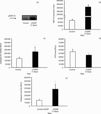
Based on the data shown in Figure 1, we selected left cochleae from different group B animals, in order to focus specifically on the seventh day after cisplatin administration and to establish whether or not heat shock protein 70 expression correlated to cisplatin-induced apoptosis in the rats' cochleae. To this end, we analysed and compared heat shock protein 70 expression, using Western blotting, and the activity of pro-apoptotic markers such as caspase-3 and/or Caspase-7 and caspase-9, using luminescence assay.
A representative Western blot for heat shock protein 70 is shown in Figure 2(a) for an animal sacrificed seven days after cisplatin treatment. Densitometry analysis demonstrated significantly increased heat shock protein 70 expression in the cochleae of cisplatin-exposed rats (420 810 ± 55 730 relative light units), compared with controls (26 520 ± 13 400 relative light units) (p ≤ 0.05), seven days after exposure (Figure 2b). Analysis of the same cochlear extracts also demonstrated significantly increased caspase-3 and/or Caspase-7 (Figure 2c) and caspase-9 (Figure 2e) activities, seven days after cisplatin administration (1.8 ± 0.4 and 3.34 ± 0.14 fold induction, respectively; p ≤ 0.05). In addition, these same cochlear extracts had significantly decreased ATP levels (an indicator of metabolic status; 1.30 ± 0.28 fold reduction; p ≤ 0.05; Figure 2d).
(a) Representative Western blot indicating heat shock protein 70 (HSP-70) expression levels in whole cochlear extracts of rats sacrificed 7 days after cisplatin (CDDP) injection. (b) Quantification of densitometry analysis of HSP-70 expression by Western blotting, in rat cochleae 7 days after CDDP injection (n = 3). (c) Caspase 3 and/or Caspase-7 activity, (d) adenosine triphosphate (ATP) levels and (e) caspase-9 activity, measured in whole cochlea protein extracts from rats sacrificed 7 days after CDDP injection. *p ≤ 0.05 vs controls. AU = arbitrary units; RLU = relative light units

Heat shock protein 70 immunofluorescence staining
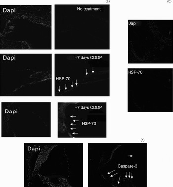
Immunostaining of right cochlea sections from group B animals revealed anti-heat shock protein 70 binding in the spiral limbus, supporting cells, basilar membrane and lateral wall. Interestingly, hair cells showed no significant staining. Heat shock protein 70 immunostaining was especially marked in the interdental cells and spiral prominence (Figure 3a). Immunofluorescence staining for heat shock protein 70 was compared with that for caspase-3 (Figure 3b), using the same material. The main difference between caspase-3 and heat shock protein 70 immunostaining involved the outer hair cells immunostaining was observed for caspase-3 but not for HSP-70. Fluorescence-based secondary antibody immunostaining indicated heat shock protein 70 antibody specificity (Figure 3c).
Photomicrographs showing immunostaining of (a) heat shock protein 70 (HSP-70) and (b) active caspase-3 (immunofluorescence specific antibody control) in the nuclei of rat inner ear cells, in controls and animals sacrificed 7 days after cisplatin (CDDP) injection. (c) Photomicrograph showing fluorescence-based secondary antibody immunostaining, indicating HSP-70 antibody specificity, in animals sacrificed 7 days after CDDP injection. See Methods section for preparation of cochlear sections. Following CDDP injection, increased nuclear HSP-70 and caspase-3 immunostaining is seen in the spiral limbus, supporting cells, basilar membrane and lateral wall. DAPI = 6-diamidino-2-phenylindole (×200)

Serum heat shock protein 70 levels
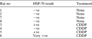
Western blot immunoassay was used to detect and identify heat shock protein 70 in the serum of rats sacrificed seven days after cisplatin injection. Western blotting was positive for 75 per cent of animals injected with cisplatin, compared with none of the control animals (Table I).
Results of Western blot immunoassay for detection of heat shock protein 70* in rat serum

* i.e. Detection of antibodies to the 68 kDa heat shock protein 70 antigen. No = number; HSP-70 = heat shock protein 70; –ve = negative; +ve = positive; CDDP = cisplatin
Auditory steady state and auditory brainstem responses
Tables II and III show mean auditory steady state response and auditory brainstem response thresholds, respectively, in animals from the cisplatin and control groups.
Auditory steady state response thresholds in rats treated with cisplatin and sacrificed after 7 days

Data represent mean threshold ± standard deviation (dB sound pressure level). *n=4. †p ≤ 0.05 vs pre-treatment. CDDP = cisplatin
Auditory brainstem response thresholds in rats treated with saline or cisplatin and sacrificed after 7 days

Data represent mean threshold ± standard deviation. *p≤0.05 vs controls at 7 days, and vs CDDP rats pre-treatment. CDDP = rats treated with cisplatin; SPL = sound pressure level
Auditory steady state response testing for frequencies from 8 to 16 kHz showed an increase in auditory thresholds seven days after cisplatin injection.
Auditory brainstem response testing at 8 kHz also showed a statistically significant increase in response thresholds seven days after treatment, comparing cisplatin-treated animals and controls (24.00 ± 7.7 dB SPL vs 14.70 ± 8.0 dB SPL, respectively; p ≤ 0.005).
Correlation between caspase-3, heat shock protein 70 and superoxide dismutase activities and auditory brainstem response
The study findings indicated that reduced caspase-3 activity was associated with reduced heat shock protein 70 activity in rat cochleae, and also correlated with reduced total superoxide dismutase activity and a raised auditory brainstem response threshold.
Figure 4(a) shows that systemic administration of the caspase-3 inhibitor Z-Asp(OMe)-Glu(OMe)-Val-Asp(OMe)-fluoromethylketone blocked caspase-3 and/or Caspase-7, activity in cisplatin-injected rats sacrificed seven days after injection These results were supported by measuring ATP levels (Figure 4b). In addition, rats injected with this inhibitor plus cisplatin showed clearly reduced heat shock protein 70 expression, as assessed by both indirect immunofluorescence (Figure 4c) and Western blotting (Figure 4d), compared with rats injected with cisplatin alone.
Measurement of (a) caspase 3 and/or Caspase-7 activity and (b) adenosine triphosphate (ATP) levels in whole rat cochlea protein extracts, 7 days after injection of cisplatin (CDDP) alone or CDDP plus Z-Asp(OMe)-Glu(OMe)-Val-Asp(OME)-fluoromethylketone (Casp-3 inh). (c) Heat shock protein 70 (HSP-70) expression in cochlear sections from control rats and those sacrificed 7 days after injection of CDDP alone or CDDP plus Cas-3 inh. (d) Representative Western blot, and quantification of densitometry analysis of HSP-70 expression in whole rat cochlear extracts 7 days after injection of CDDP alone or CDDP plus Casp-3 inh (n = 3). *p ≤ 0.05 vs controls; †p ≤ 0.05 vs CDDP only group. RLU = relative light units; AU = arbitrary units

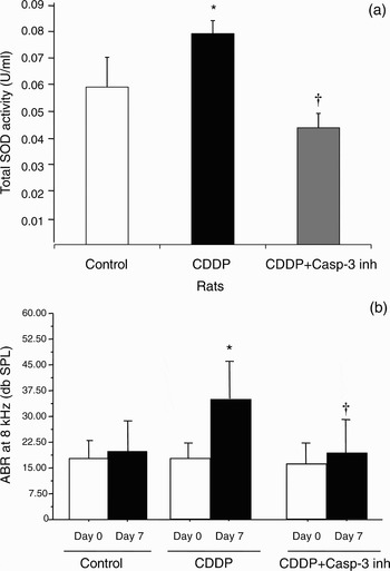
In addition, significantly reduced total superoxide dismutase activity (previously induced by cisplatin injection) was seen in the presence of the caspase-3 inhibitor Z-Asp(OMe)-Glu(OMe)-Val-Asp(OME)-fluoromethylketone (Figure 5a). Likewise, there was a significant reduction in cisplatin-induced auditory brainstem response threshold elevation in the presence of caspase-3 inhibitor (Figure 5b). Caspase-3 inhibitor alone did not show any effect on caspase-3 and/or Caspase-7 or total superoxide dismutase activities (data not shown).
(a) Total superoxide dismutase (SOD) activity in whole rat cochlear extracts 7 days after injection of cisplatin (CDDP) alone or CDDP plus Z-Asp(OMe)-Glu(OMe)-Val-Asp(OME)-fluoromethylketone (Casp-3 inh) (see Methods for definition of units). (b) Auditory brainstem response (ABR) threshold 7 days after injection with CDDP or CDDP plus Casp-3 inh. *p ≤ 0.05 vs controls; †p ≤ 0.05 vs CDDP only group. Day 0 = before treatment; SPL = sound pressure level

Discussion
Cisplatin is a highly effective chemotherapeutic agent; however, it has significant ototoxic side effects. Recent reports have described apoptosis of affected cochlear cells via activation of the intrinsic cell death pathway, following cisplatin administration.Reference García-Berrocal, Nevado, Ramírez-Camacho, Sanz, González-García and Sanchez-Rodríguez9 This finding seems to support reported data suggesting that cis-platin–DNA adducts may kill cells by inducing programmed cell death (i.e. apoptosis).Reference Lee, Nagakawa, Kita, Kim, Iguchi and Endo12
Heat shock proteins are among the diverse molecules potentially related to apoptosis.Reference Sreedhar and Csermely13 They have recently been suggested to be involved in the stress response activated by noxious changes in the cellular environment, such as hyperthermia, ischaemia and environmental toxics (including cisplatin).Reference Oh, Yu, Song, Lim, Koo and Chang14, Reference Cunningham and Brandon15 However, there is little information available on the possible relationship between reversibility of cochlear functions and heat shock protein levels, mainly due to the short follow-up times of the relevant studies.
Heat shock protein 70 is the most highly conserved and stress-inducible heat shock protein in mammals,Reference Martindale and Holbrook16 and is induced in the inner ear in response to heat shock, cochlear ischaemia, noise trauma and cisplatin exposure.Reference Oh, Yu, Song, Lim, Koo and Chang14, Reference Dechesne, Kim, Nowak and Wenthold17, Reference Lim, Jenkins, Myers, Miller and Altschuler18 We found a association between heat shock protein 70 serum levels, histologically observed cochlear cell death and cochlear function, seven days after the injection of both cisplatin alone and cisplatin plus Z-Asp(OMe)-Glu(OMe)-Val-Asp(OME)-fluoromethylketone (an irreversible caspase-3 inhibitor). Cochlear extracts have been reported to exhibit progressively greater expression of caspase-9,Reference García-Berrocal, Nevado, Ramírez-Camacho, Sanz, González-García and Sanchez-Rodríguez9 caspase-3 and heat shock protein 70 following cisplatin exposure, peaking at day seven. These findings correlate with the platinum accumulation found inside rat cochleae in another study employing speciation analysis.Reference Esteban-Fernandez, Gómez-Gómez, Cañas, Verdaguer, Ramirez and Palacios19 In this particular study, platinum accumulation in cochlear tissues was markedly greater seven days after cisplatin administration than 30 days after. Levels of caspases-3 and -9, ATP and heat shock protein 70 were found to have declined on day 30 after cisplatin treatment. In this study animals, we found higher levels of both heat shock protein 70 expression (in cochlear extracts) and serum heat shock protein 70 on day seven after cisplatin injection. This finding supports our previous study in humans concerning the capability of heat shock protein 70 to act as a possible serological marker for cisplatin ototoxicity.Reference Ramírez-Camacho, Citores, Trinidad, Verdaguer, García-Berrocal and Marero4 However, this and other previous studies have shown that the role of heat shock protein 70 as a serological marker is based on its protective effect against cisplatin-induced damage (see Ryback et al.).Reference Rybak, Whitworth, Mukherjea and Ramkumar5
Protection against cisplatin-induced ototoxicity may be provided by endogenous molecules, exogenous agents or a combination of exogenous agents triggering endogenous protective mechanisms. The cochlea contains endogenous protective mechanisms against oxidative stress. It is well accepted that cisplatin generates reactive oxygen species, including superoxide anions (shown in explants of cochlear tissues, and reviewed by Rybak et al.).Reference Rybak, Whitworth, Mukherjea and Ramkumar5 A plausible explanation for the expression of heat shock protein 70 in human and experimental animal sera is that this protein triggers repair and anti-apoptotic mechanisms in response to toxic events; consequently, apoptosis should decrease. Heat shock protein 70 would therefore act as an oto-protective agent in the cochlea. In fact, heat shock protein 70 has been demonstrated to inhibit apoptosis in the presence of aminoglycoside ototoxicity.Reference Taleb, Brandon, Lee, Lomax, Dillmann and Cunningham20 (However, these authors used a transgenic mouse that constitutively overexpressed heat shock protein 70.) Heat shock protein 72 has been proven to have a potential role in protecting outer hair cells from damage,Reference Boettcher, Spongr and Salvi21 noise overstimulationReference Lim, Jenkins, Myers, Miller and Altschuler18 and cisplatin administration.Reference Oh, Yu, Song, Lim, Koo and Chang14 In addition, heat shock induction appeared to provide significant protection in vitro against hair cell death in an utricle preparation.Reference Cunningham and Brandon15 It has even been suggested that blocking critical steps in apoptosis may be a useful strategy to prevent the harmful side effects of cisplatin in patients undergoing chemotherapy.Reference Wang, Ladrech, Pujol, Brabet, Van de Water and Puel22
Our study indicated that administration of inhibitors of caspase-3 and -9 (data not shown) dramatically reduced the extent of hearing loss, apoptosis, and heat shock protein 70 immunostaining and expression in the rat cochleae tested, and also correlated strongly with the observed decrease in cochlear total superoxide dismutase activity. If heat shock protein 70 is considered as an endogenous protective mechanism in cases of cisplatin ototoxicity, reduced expression of this protein in caspase inhibitor treated rats could be due to a decrease in apoptotic events (resulting in a reduced requirement for heat shock protein 70 expression to facilitate cell protection).
• Exposure to cisplatin leads to cochlear cell death by apoptosis, with maximum changes seen seven days after exposure
• Cisplatin-induced ototoxicity may be protected against by endogenous molecules, exogenous agents or a combination of exogenous agents triggering endogenous protective mechanisms
• The cochlea has endogenous protective mechanisms to deal with oxidative stress
• Heat shock proteins are induced in inner ear cells in response to a variety of stimuli; heat shock protein 70 has been proposed as an endogenous protective mechanism against cisplatin ototoxicity
• This study examined the role of heat shock protein 70 in cisplatin-induced cochlear cell death
In the present study, heat shock protein 70 immunostaining was observed in the cochlear spiral limbus, basilar membrane and lateral wall supporting cells. Heat shock protein 70 was especially prominent in the interdental cells and spiral prominence. Hair cells showed some diffuse staining on the second day after cisplatin injection, but no clear staining on days seven or 30. The pattern of heat shock protein 70 staining was similar to that observed after heat stress, ischaemia and noise overstimulation.Reference Gower and Thompson23 However, Oh et al. found that cisplatin preferentially induced the synthesis of heat shock protein 72 in outer hair cells, although they did not rule out any other staining site.Reference Oh, Yu, Song, Lim, Koo and Chang14 Comparing Oh and colleagues' results with our own, discrepancies in heat shock protein staining for outer hair cells may be explained by the theory that cisplatin administration modulates heat shock protein at different times in each cell line. In fact, Oh et al. observed different immunostaining of outer hair cells at different time points, and did not find heat shock protein 72 staining in supporting cells.Reference Oh, Yu, Song, Lim, Koo and Chang14
The oto-protective effect of heat shock protein 70 within the inner ear should be maximised, without compromising the antitumoural effectiveness of cisplatin. Unfortunately, heat shock protein 70 seems to increase the tumourigenicity of cancer cells in rodent models.Reference Aghdassi, Pillips, Dudeja, Dhaulakhandi, Sharif and Dawra24, Reference Phillips, Dudeja, McCarroll, Borja-Cacho, Dawra and Grizzle25 Therefore, strategies for local delivery of heat shock protein 70 to the inner ear should be developed.
Conclusions
Cisplatin seems to induce disturbances in the cellular machinery of the inner ear, most pronounced seven days after administration. Our study suggests that heat shock protein 70 expression could provide a protective mechanism guarding cellular integrity within the inner ear. This protection (observed seven days after cisplatin exposure and later) could be stronger in cochlear structures with greater expression of heat shock protein 70, and is not necessarily focused on the outer hair cells. Indeed, this and other works reported cisplatin-induced morphological changes in the inner ear, among others, at the supporting cells.Reference García-Berrocal, Nevado, Ramírez-Camacho, Sanz, González-García and Sanchez-Rodríguez9, Reference Estrem, Babin, Ryu and Moore26–Reference Ramírez-Camacho, García-Berrocal, Buján, Martin-Marero and Trinidad29 In addition, a previous study from our laboratory alteration of the supporting cell ultra-structure preceded detectable changes in outer hair cells.Reference Ramírez-Camacho, García-Berrocal, Trinidad, González-García, Verdaguer and Ibáñez30
Acknowledgement
This study was supported by Spanish Fondo de Investigaciones Sanitarias (PI05/0673).