Case report
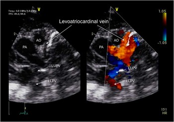
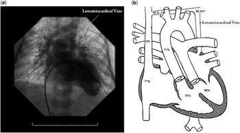
A newborn male infant was diagnosed prenatally with D-transposition of the great arteries and ventricular septal defect. Following delivery at term, transthoracic echocardiography was performed, confirming the diagnosis and revealing a very redundant atrial septum, as well as a new finding of an anomalous venous structure seen ascending from the left atrium to the innominate vein (Fig 1). The baby was taken to the cardiac catheterisation laboratory on day of life 1 to investigate the abnormal venous vessel and perform balloon atrial septostomy via femoral access, after umbilical access revealed a closed ductus venosus. A saturation run revealed a saturation of 90% in the innominate vein, suggesting a pulmonary venous-to-systemic venous connection. This was confirmed on angiography of the left ventricle, which – when followed through to levophase – showed a levoatriocardinal vein (Fig 2). Balloon atrial septostomy was attempted; however, given the very redundant atrial septum, this was not successful. The patient’s systemic saturations remained reasonable despite the lack of unrestrictive atrial shunting, due to shunting at the level of the innominate vein.
Transthoracic echocardiogram: suprasternal notch view, two dimensional (2D) and 2D with color flow Doppler, demonstrating the levoatriocardinal vein. AO=aorta; LLPV=left lower pulmonary vein; LUPV=left upper pulmonary vein; PA=pulmonary artery.

( a ) Levophase of left ventricle angiogram, showing flow from the left atrium to the innominate vein through the levoatriocardinal vein. ( b ) Mullins catheterisation diagram with saturation data.

On day of life 6, the patient was taken to the operating room for surgical repair. Direct visualisation confirmed the presence of the levoatriocardinal vein with connection from the left atrium to the innominate vein, providing a left-to-right shunt. In total, four pulmonary veins were noted to be appropriately entering the left atrium by intra-operative transoesophageal echocardiography and by direct visualisation, excluding total anomalous pulmonary venous return from the diagnosis. Interestingly, left-sided juxtapositioning of the atrial appendages was also discovered. An arterial switch operation with ventricular septal defect closure was successfully performed with the levoatriocardinal vein being ligated. Transoesophageal echocardiography was then used to confirm the results and normal pulmonary venous drainage was documented. The patient had an uneventful postoperative course and was discharged on the 6th postoperative day. At follow-up 2 years later, he continues to do well.
Levoatriocardinal vein, first described by McIntoshReference McIntosh 1 in 1926, is an abnormal persistent connection between the pulmonary and systemic circulations. During early embryogenesis, the lung venous system, which develops from the capillary plexus surrounding the embryonic foregut, is not connected to the heart but is connected to the cardinal and vitalline veins, which give rise to the innominate vein, jugular veins, superior caval vein, and coronary sinus. A connection between the pulmonary venous bed and the left atrium forms when the pulmonary veins join with an outgrowth of the sinoatrial region of the heart. As the pulmonary veins are integrated into the left atrium, their connections with the cardinal and vitalline veins regress and disappear. Persistence of these connections may lead to the presence of a levoatriocardinal vein.Reference Murillo, Cutalo, Jones, Lane, Fleischmann and Restrepo 2 , Reference Bernstein, Moore, Stanger and Silverman 3
The levoatriocardinal vein is a rare entity with ~50 cases being reported in the literature.Reference Cullen, Breen, Rihal, Simari and Ammash 4 , Reference Amoretti, Cerillo and Chiappino 5 Although there are reports of the levoatriocardinal vein occurring in the otherwise normal heart, the majority of cases occur in the presence of left-sided obstructive lesions. These lesions most commonly include cor triatriatum, mitral atresia, aortic atresia, and coarctation of the aorta, with or without atrial communications. In these cases of obstruction to pulmonary venous flow, the persistent connection provides left atrial decompression through the cardinal venous system, resulting in a left-to-right shunt.
The levoatriocardinal vein has not been described in a patient with transposition of the great arteries and ventricular septal defect. Our patient had a redundant, intact atrial septum but no evidence of left-sided obstruction requiring decompression of the left atrium. The pathophysiology resulting in this embryologic abnormality remains unclear in our patient; however, given the difficulty with balloon atrial septostomy, the levoatriocardinal vein served as a beneficial left-to-right shunt, allowing for adequate preductal systemic saturation before surgical repair.
Acknowledgement
None.
Financial Support
This research received no specific grant from any funding agency, commercial or not-for-profit sectors.
Conflicts of Interest
None.