Introduction
One drawback of using wound closure devices is the high tension the materials produced on the wound during healing. This high tension has been observed to generate necrosis, destroy the tissue, and promote eversion or inversion of wound edges [Reference Vogels, Lambertz, Schuster, Jockenhoevel, Bouvy, Disselhorst-Klug, Neumann, Klinge and Klink1, Reference Israelsson and Millbourn2]. The friction performance of wound closure devices plays a vital role in their functionality. In general, the friction generated between wound closure devices and tissue can cause inflammation, pain, and increased recovery time [Reference Zhang, Ren, Zhang, Zeng and van der Heide3]. One possible solution proposed by Vogels et al. is to use materials with the ability to regulate the tension through elasticity, improving the wound healing process. Therefore, in this work, PUs are examined as a candidate material for the use in wound closure devices because of their various desirable properties [Reference Vogels, Lambertz, Schuster, Jockenhoevel, Bouvy, Disselhorst-Klug, Neumann, Klinge and Klink1]. Therefore, the selection of monomers and additives for the synthesis of polyurethanes (PUs) is essential to determine the final properties and therefore the desired application [Reference Arévalo, Uscátegui, Díaz, Cobo and Valero4].
At present, synthetic polymers signify a great development in the design of biomaterials for different applications because of their use in supports for tissue engineering, controlled release systems, implants, artificial skin, devices for osteosynthesis, and also other applications [Reference Vannozzi, Ricotti, Santaniello, Terencio, Oropesa-Nunez, Canale, Borghi, Menciassi and Lenardi5, Reference Meskinfam, Bertoldi, Albanese, Cerri, Tanzi, Imani, Baheiraei, Farokhi and Farè6, Reference Park, Gong, Park, Moon, Wall, Kim, Lee and Knowles7, Reference Mi, Jing, Hagerty, Chen, Huang and Turng8]. This group of biomaterials includes PUs, which are characterized by their wide versatility, resistance to fatigue, elasticity, ease of processing, and biocompatibility [Reference Solanki, Das and Thakore9, Reference Da, Gong, Chen, Zhang, Huang, Guo, Li, Li-Ling, Zhang and Xie10, Reference Zia, Zia, Zuber, Tabasum and Rehman11]. PUs, which contain both hard and flexible segments, are very important polymers because of their desirable engineering characteristics, which permit modification of their mechanical properties and include biocompatibility and biodegradability [Reference Shahrousvand, Sadeghi, Shahrousvand, Ghollasi and Salimi12].
The synthesis of PUs comprises reacting compounds with terminal hydroxyl groups with difunctional or polyfunctional isocyanates [Reference Laube, Weisser, Berger, Börner, Bischoff, Schubert, Gajda, Bräuer and Schnabelrauch13, Reference Gamerith, Herrero Acero, Pellis, Ortner, Vielnascher, Luschnig, Zartl, Haernvall, Zitzenbacher, Strohmeier, Hoff, Steinkellner, Gruber, Ribitsch and Guebitz14]. To obtain a more sustainable synthesis for PUs, current research has been focused on replacing raw materials with petrochemicals [Reference Ng, Lee, Chuah and Cheng15, Reference Jutrzenka Trzebiatowska, Santamaria Echart, Calvo Correas, Eceiza and Datta16, Reference Gil-Castell, Badia, Ontoria-Oviedo, Castellano, Marco, Rabal, Bou, Serra, Monreal, Blanes, Sepúlveda and Ribes-Greus17]. There is currently an increase in the use of raw materials from renewable sources mainly because of economic and environmental reasons. As the price of oil fluctuates, so do polymeric raw materials. On the other hand, because of the growing environmental concern, the use of vegetable oils as raw material for the synthesis of polymers has increased [Reference Mekewi, Ramadan, ElDarse, Abdel Rehim, Mosa and Ibrahim18]. Triglycerides are abundant, renewable, and biodegradable, and they have become an alternative source in the synthesis of polymeric materials [Reference Zhang, Garrison, Madbouly and Kessler19, Reference Madra, Tantekin-Ersolmaz and Guner20]. Vegetable oils contain fatty acids that vary from one plant to another and are characterized by the presence of one, two, or more degrees of unsaturation, which makes them highly reactive and suitable for a wide variety of chemical modifications [Reference Mekewi, Ramadan, ElDarse, Abdel Rehim, Mosa and Ibrahim18]. The composition of the fatty acids depends on the type of vegetable oil [Reference Zhang, Garrison, Madbouly and Kessler19]. The vegetable oils can be modified to increase the functionality of the polyol by means of the addition of hydroxyl groups making use of the unsaturation present in the structure [Reference Pfister, Xia and Larock21]. This is done through various techniques such as alcoholysis, epoxidation, and transesterification, among others [Reference Pfister, Xia and Larock21, Reference Petrović, Milic, Zhang and Ilavsky22, Reference Lligadas, Ronda, Galià and Cádiz23, Reference Jayavani, Sunanda, Varghese and Nayak24].
One major synthetic technique uses the triglycerides of fatty acid groups from the vegetable oils of different seeds, including corn, soy, sunflower, palm, and castor seeds, among others [Reference Ng, Lee, Chuah and Cheng15, Reference Ismail, Motawie and Sadek25, Reference Calvo-Correas, Santamaria-Echart, Saralegi, Martin, Valea, Corcuera and Eceiza26]. Castor oil (Ricinus communis) has several advantages over other oils for use as a polyol, whose ricinoleic acid content is approximately 90% (w/w). Castor oil contains hydroxyl groups in its chain that react with isocyanates polyfunctional to form PUs with different morphological and mechanical properties (good flexibility and elasticity), among others, thanks to the long chain of fatty acids present [Reference Uscátegui, Díaz and Valero27]. Castor oil is not used in the food industry because it contains toxic compounds such as ricin [Reference Meneguelli de Souza, de Carvalho, Araújo, de Melo and Machado28]; thus, its use in PU synthesis would not compete with its use in the food industry [Reference Hejna, Kirpluks, Kosmela, Cabulis, Haponiuk and Piszczyk29]. Castor oil can be incorporated in the synthesis of PUs directly without the need for previous modifications because of the high triglyceride content of the ricinoleic acid present in the structure [Reference Arévalo, Uscátegui, Díaz, Cobo and Valero4]. Furthermore, castor oil contains degrees of unsaturation, which provide the oil with low viscosity, allowing it to be used directly as a polyol [Reference Omonov, Kharraz and Curtis30]. In addition, the presence of hydroxyl groups in the chemical structure of castor oil facilitates chemical modifications of the polyol through which different mechanical properties of the PU are obtained [Reference Uscátegui, Arévalo, Díaz, Cobo and Valero31]. Synthesis of PUs with shape memory using soybean oil and castor oil were characterized as biodegradable and biocompatible materials [Reference Petrović, Milic, Zhang and Ilavsky22]. PUs synthesized with L-Lysine ethyl ester diisocyanate and castor oil are used as memory materials in a way; they were evaluated on the mouse fibroblast cell line L929 for 24, 48, and 72 h and showed cell viability values higher than 70% [Reference Calvo-Correas, Santamaria-Echart, Saralegi, Martin, Valea, Corcuera and Eceiza26].
As mentioned, the use of polymeric materials for the design of implants and medical devices keep rising because of the properties of the polymers. One of the drawbacks of polymeric materials for biomaterials is the formation of biofilms on the surface [Reference dos Santos, Alcaraz-Espinoza, da Costa and de Oliveira32]. The antibacterial activity present in polymeric materials can improve the wound healing process over time [Reference Serrano, García-Fernández, Fernández-Blázquez, Barbeck, Ghanaati, Unger, Kirkpatrick, Arzt, Funk, Turón and del Campo33]. Several techniques that enhance the antibacterial effects of biomaterials have already been developed, including coating materials with antibacterial agents [Reference Serrano, García-Fernández, Fernández-Blázquez, Barbeck, Ghanaati, Unger, Kirkpatrick, Arzt, Funk, Turón and del Campo33], incorporations of antimicrobial agents by means of mixing, grafting, or coating [Reference dos Santos, Alcaraz-Espinoza, da Costa and de Oliveira32], among others. The addition of antimicrobial agents should not modify the properties of the polymers, and the residues of the incorporated material should not generate side effects, that is, the balance between the mechanical and biological properties should be conserved.
Assessing the behavior of mechanical, physicochemical, and thermal properties and biological responses in vitro is essential to ensure functionality [Reference Gil-Castell, Badia, Ontoria-Oviedo, Castellano, Marco, Rabal, Bou, Serra, Monreal, Blanes, Sepúlveda and Ribes-Greus17]. In the same way, biomaterials derived from polymers must be biocompatible because they will be in contact with cells of the human body. They must not generate inflammatory reactions, and the degradation products must be nontoxic to the host [Reference Fabbri, Guidotti, Soccio, Lotti, Govoni, Giordano, Gazzano, Gamberini, Rimini and Munari34].
The interactions that occur between cell lines and materials designed for biomedical applications are currently of interest. A field of study responsible for evaluating the in vitro response of biomaterials based on PUs already exists [Reference Vannozzi, Ricotti, Santaniello, Terencio, Oropesa-Nunez, Canale, Borghi, Menciassi and Lenardi5, Reference Meskinfam, Bertoldi, Albanese, Cerri, Tanzi, Imani, Baheiraei, Farokhi and Farè6, Reference Mi, Jing, Hagerty, Chen, Huang and Turng8, Reference Angeloni, Contessi, De Marco, Bertoldi, Tanzi, Daidone and Farè35, Reference Gossart, Battiston, Gand, Pauthe and Santerre36]. One such study in this field is that of Gossart et al., who evaluated the intrinsic influence of PU chemistry on the modulation and activation of human monocytes in vitro, thus demonstrating the possible application of the tested materials in medical therapies [Reference Gossart, Battiston, Gand, Pauthe and Santerre36]. However, as reported in the aforementioned literature, the observed in vitro responses demonstrated that the PU-based materials show promise for use as biomaterials, but data concerning the biological response of the implanted material in living tissues are insufficient. Few scientific studies have focused on the evaluation of the in vitro and in vivo responses of PU-derived biomaterials. One such study was conducted by Zhang et al., who evaluated the release of peptides in vitro and in vivo from thermoplastic PUs [Reference Zhang, Woodruff, Clark, Martin and Minchin37].
Thus, the objective of this research was to synthesize in vitro and in vivo biocompatible materials, with adequate mechanical properties for biomedical devices. To achieve this objective, PUs were synthesized with modified polyols derived from castor oil and isophorone diisocyanate (IPDI). The effects of the addition of hydroxyl groups to the polyol were evaluated in terms of the mechanical and biological properties in vitro using fibroblasts and in vivo using Wistar rats. The ability to correlate the in vitro and in vivo behavior of the mechanical and biological properties of the synthesized materials provided information on whether PUs could be candidates for use in biomedical devices.
Results and discussion
Characterization of PUs
Mechanical properties
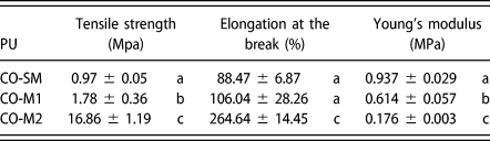
For each of the polyols derived from castor oil, the hydroxyl index was determined after the chemical modification process. For the unmodified polyol, the index was 160 mg KOH g−1 castor oil. In the case of the modified polyols, the values were 191 and 236 mg KOH g−1. High densities of polymeric materials can be related to a higher hydroxyl (OH) content because of increased crosslinking reactions [Reference Carriço, Fraga and Pasa38]. In the research conducted by Carriço et al., it was observed that increasing the castor oil content in foam formulation led to an increase in bulk density, suggesting that the polymer chains were more packed with lower free volume and smaller cells, which in turn increased the rigidity of these materials [Reference Carriço, Fraga and Pasa38]. When evaluating the mechanical properties of the PUs, the strain–strain curves were determined and used to calculate the maximum stress, elongation percentage, and Young’s modulus (Table I).
Mechanical properties of PUs.

Note: The results are presented as the mean ± SD (n = 3). Different letters indicate significant differences between polyols (P < 0.05).
According to the results, the maximum stress and the Young’s modulus were significantly different between polyols (P < 0.05). Table I shows that increasing the hydroxyl groups in the triglyceride chain of the fatty acid of castor oil modified the mechanical properties of the PUs synthesized with IPDI. Thus, the values depended strongly on the degree of crosslinking that occurred in each polyol [Reference Vannozzi, Ricotti, Santaniello, Terencio, Oropesa-Nunez, Canale, Borghi, Menciassi and Lenardi5], and a remarkable difference was observed from the CO-SM PU synthesized with unmodified polyol.
Because the modulus of elasticity is indicative of the rigidity of the material, the stiffness of a material should increase as the modulus of elasticity increases. Table I shows that the material with the highest percentage of elongation (CO-M2) is the one with the lowest modulus. This finding agrees with the results reported by Park et al., who synthesized PUs with polycaprolactone, hexamethylene diisocyanate, and isosorbide with silk additions and determined that the highest percentage of silk used increased stiffness while decreasing the maximum effort. The authors stated that the ability to design flexible and soft polymers allows the fabrication of a wide range of biomaterials for use in the regeneration of soft tissues such as muscles and ligaments [Reference Park, Gong, Park, Moon, Wall, Kim, Lee and Knowles7]. Similarly, Vannozzi et al. reported that in general, soft and deformable substrates represent key features of skeletal muscle tissue engineering [Reference Vannozzi, Ricotti, Santaniello, Terencio, Oropesa-Nunez, Canale, Borghi, Menciassi and Lenardi5].
To design structures that mimic living tissues at the microscale, the modulus of elasticity must exceed 10 kPa [Reference Kim, Kang, Kim, Jiao, Kim and Suh39]. Based on the results obtained in this study, the PUs synthesized could be used as candidates for wound closure devices because the range of values for the modulus of elasticity exceeded 10 kPa. This range of values also accounts for the fact the modulus of elasticity for human skin tissues ranges between 50 kPa and 100 MPa [Reference Nemir and West40, Reference Vatankhah, Semnani, Prabhakaran, Tadayon, Razavi and Ramakrishna41]. The design of a biomaterial must combine flexibility with strength to achieve mechanical properties similar to those of the native skin [Reference Alves, Da Silva, Weber, Luchese, Wilhelm and Fajardo42]. Vannozzi et al. designed porous 3D PU scaffolds and determined that the Young’s modulus of the synthesized materials were greater than 250 kPa. Thus, this group proposed that these materials demonstrated potential in the field of tissue engineering [Reference Vannozzi, Ricotti, Santaniello, Terencio, Oropesa-Nunez, Canale, Borghi, Menciassi and Lenardi5].
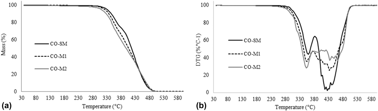
In a previous study [Reference Uscátegui, Arévalo-Alquichire, Gómez-Tejedor, Vallés-Lluch, Díaz and Valero43], PUs were synthesized with hexamethylene diisocyanate. Modification of the polyol via transesterification increased the stress–strain mechanical properties by increasing the number of the hydroxyl groups, thus generating a greater degree of crosslinking. Both the previous study and the results presented in Fig. 1 show that the properties of the PUs strongly depend on the degree of crosslinking of the polymer chain. Therefore, such properties can be manipulated by modifying the crosslink density. Hydrogen bonds produce strong interactions; therefore, the polar nature of a hard segment causes a strong attraction, generating a domain formation [Reference Jutrzenka Trzebiatowska, Santamaria Echart, Calvo Correas, Eceiza and Datta16].
TGs of the synthesized PUs. (a) TG curves of PUs. (b) DTG curves of PUs.

Thermogravimetric analysis
As part of the thermal analysis (TG) performed on the PUs, weight loss was evaluated as a function of temperature. The TG [Fig. 1(a)] and the derivative of the thermograms (DTG) [Fig. 1(b)] of the synthesized PUs showed that the synthesized PUs exhibited three stages of degradation with approximate temperature ranges of 340–360 °C, 420–440 °C, and 450–490 °C. The results indicated that the use of modified polyols did not shift the degradation temperatures of the PUs, and the same tendency as the material produced from unmodified polyol was retained. The synthesized PUs exhibited a high temperature of degradation, >300 °C. These results agreed with the research performed by Jutrzenka et al., who synthesized PUs based on a derivative of glycerin, poly(ethylene-butylene)adipate diol, and 4,4′-diphenylmethane diisocyanate and determined that the materials were stable at temperatures 300 °C and lower. The first weight loss observed occurred at approximately 320 °C and corresponded to urethane bonds in the hard segments. According to the authors, the thermal decomposition of crosslinked PUs was due to the degradation of the side chains [Reference Jutrzenka Trzebiatowska, Santamaria Echart, Calvo Correas, Eceiza and Datta16]. According to the results presented in Fig. 1, the decomposition temperature of the synthesized PUs was higher than 350 °C and most likely occurred because of the increase in the number of the urethane groups generated by the type of polyol used in the synthesis of the material [Reference Zhou, Sheng, Liu, Lin, Ji, Dong, Xu and Yang44]. The second weight loss observed was most likely due to a division and depolymerization at 400 °C, resulting in the degradation of mechanical stability in the soft segments. The second step was slow and dependent on the PU structure [Reference Jutrzenka Trzebiatowska, Santamaria Echart, Calvo Correas, Eceiza and Datta16].
DSC
To determine the effect of polyol modification on the T g of the PUs, DSC tests were performed with each synthesized material. The T g values for CO-SM, CO-M1, and CO-M2 were −14.79 °C, −12.81 °C, and 14.77 °C, respectively. The CO-M2 material synthesized with the higher modification percentage affected the thermal properties of the original PU material (CO-SM). The results indicated the T g was modified by the degree of crosslinking present in the synthesized materials, as the crosslinking affects the mobility of the soft segments because more thermal energy is needed to break additional links in the total network [Reference Vannozzi, Ricotti, Santaniello, Terencio, Oropesa-Nunez, Canale, Borghi, Menciassi and Lenardi5]. Furthermore, the T g of the PUs is dependent on the hydroxyl number of the polyols and, therefore, on the crosslinking density [Reference Jutrzenka Trzebiatowska, Santamaria Echart, Calvo Correas, Eceiza and Datta16]. Saénz-Pérez et al. synthesized PUs with poly(oxotetramethylene) glycol and 4,4′-methylene diphenyl diisocyanate, toluene 2,4-diisocyanate, and butanediol. In determining the T g, they found that the T g increased as the amount of the chain extender increased. Therefore, the authors claimed that the increase in the T g was generated by a reduction in the mobility of the chain segments because of the increased amount of the hard segments [Reference Sáenz-Pérez, Lizundia, Laza, García-Barrasa, Vilas and León45].
Contact angle
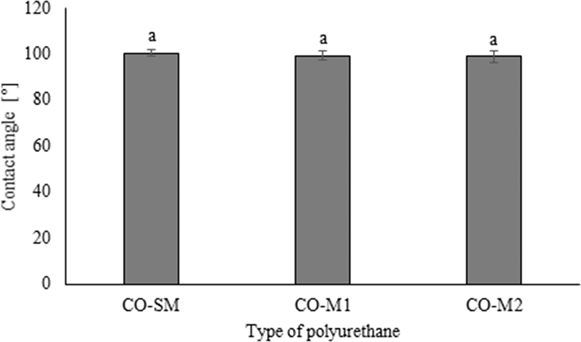
The hydrophilic nature of the synthesized materials was determined by the contact angle, as shown in Fig. 2. The polyols used in the synthesis of PUs did not show significant differences in terms of the contact angle (P < 0.05). Contact angles close to 100° were observed; thus, the materials tended to be hydrophobic. Mi et al. determined the contact angle of thermoplastic PUs based on polycaprolactone and different chain extenders. The contact angles were near 90°. They attributed these contact angle results to the hydrophilic functional groups present in the chain extenders used. The authors claimed that although the PUs maintained the same chemical structure, the variation of the monomers in the synthesis affected the wettability of the material and therefore changed its degradation behavior [Reference Mi, Jing, Hagerty, Chen, Huang and Turng8].
PU contact angles. Notes: The contact angle is presented as the mean ± SD (n = 10). Bars with equal letters (a) do not differ between polyols (P > 0.05).

Gossart et al. evaluated the contact angles of PUs synthesized with L-Lysine diisocyanate, hydroxyethyl methacrylate, and poly(hexamethylene-carbamate) and reported contact angles above 80°. The authors claimed that the common contact angles for PU matrices are in a range of 80–90° and that the contact angle is dependent on the structure of the PU and its interactions with surfaces [Reference Gossart, Battiston, Gand, Pauthe and Santerre36]. The contact angles shown in Fig. 2 were higher than those reported because the crosslinked network generated by the type of polyol used in the synthesis to make the material tends to be more hydrophobic.
Biological evaluation of PUs
In vitro cell viability assay
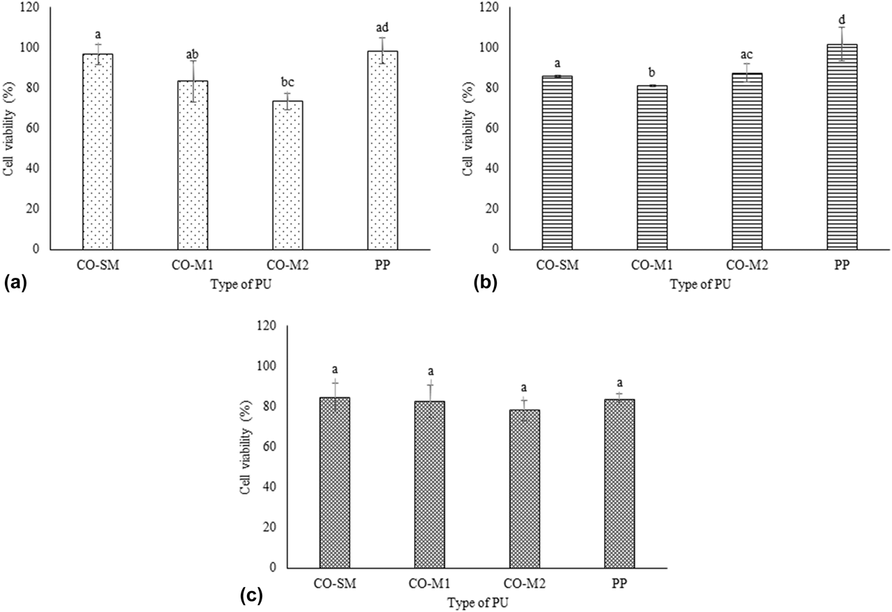
As part of the in vitro biological evaluation of the PUs, the cell viability was determined using three fibroblast cell lines. The results are shown in Fig. 3.
Cell viability at 24 h. (a) L929 mouse fibroblasts, (b) MRC-5 human fibroblasts, and (c) HDFa cells. Notes: Data are presented as the mean ± SD (n = 3). Bars with different letters (a, b, c, and d) indicate significant differences between polyols (P < 0.05).

All polymers tested with the three cell types evaluated showed cell viability percentages greater than 70%. According to the ISO/CD 10993-5 standard, materials with a cell viability result greater than 70% can be considered for studying biomaterials. PP was used as a control material because it is biocompatible and nonabsorbable. Figure 3(a) shows the cell viability results for the PUs for L929 fibroblast cells. The cell viability percentages of the PUs CO-SM and CO-M1 were not different from that of PP (P < 0.05). Significant differences were observed when the CO-M2 polymer was compared with the CO-SM polymer and the control, but the cell viability values were sufficient in demonstrating the synthesized materials could be used as possible biomaterials. The trend observed for fibroblast L929 cells indicated that as the degree of crosslinking of PU increased, the percentage of cell viability decreased. The high degree of crosslinking likely generated a low availability of functional groups, such as –OH groups, in the crosslinked polymer, which could have decreased cell adhesion [Reference Vannozzi, Ricotti, Santaniello, Terencio, Oropesa-Nunez, Canale, Borghi, Menciassi and Lenardi5].
Figure 3(b) shows the cell viability results for human MRC-5 fibroblasts on exposure to the polymers. For this cell line, no trend related to the type of polyol used in the synthesis of PU was observed. Although the cell viability was decreased compared with that of the control material (PP), the materials remained suitable for use in the design of biomaterials because their cell viability remained higher than 80%.
Figure 3(c) shows the results of cell viability of the PUs on the HDFa cells. The materials showed a cell viability greater than 80%. In this case, no significant differences (P < 0.05) were observed between the types of polyols used in the synthesis of PUs. Similarly, no differences were observed between the synthesized materials and the PP control material. Therefore, using the synthesized PUs in the design of biomaterials can be considered.
Coakley et al. evaluated a porcine urinary bladder wall-derived construct cultured with adult human dermal fibroblast HDFa in vitro on as a potential scaffold for tissue engineering. The study results reported an HDFa cell viability greater than 90% [Reference Coakley, Shaikh, O’Sullivan, Kavanagh, Grace and McGloughlin46]. The cytotoxicity of this construct might be related to the relative cell viability of the controls, in which values lower than 30% indicated the material shows severe toxicity, values between 30 and 60% indicated the material shows moderate toxicity, values between 60 and 90% indicated the material shows slight toxicity, and values above 90% indicated the material is not toxic [Reference Shahrousvand, Sadeghi, Shahrousvand, Ghollasi and Salimi12]. Therefore, after reviewing the results related to the evaluation of the cell viability of the fibroblast lines (Fig. 3), the PUs tested herein were determined to be nontoxic to all cell lines evaluated.
In vitro antibacterial activity assay
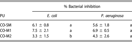
As part of the biological characterization of the PUs, the antibacterial activity was evaluated. The possible reduction in the number of bacteria with respect to the control was determined. The results are presented in Table II.
Bacterial inhibition of PUs.

Note: The percentage inhibition results are presented as the mean ± SD (n = 3). Different letters indicate significant differences between polyols (P < 0.05).
The antibacterial activity of the synthesized materials was determined by calculating the percentage of mortality of the microorganisms. The statistical analysis showed no significant differences between the two evaluated bacteria (E. coli and P. aeruginosa) (P < 0.05). Therefore, the type of polyol did not influence the bacterial response. But the result of CO-M2 against E. coli presents statistical difference with respect to the polyols CO-SM and CO-M1, probably because of the high degree of crosslinking compared with the other polyols, which generates a barrier for the diffusion of materials or fluids within the polymer matrix [Reference Uscátegui, Arévalo-Alquichire, Gómez-Tejedor, Vallés-Lluch, Díaz and Valero43]. The degree of crosslinking of the PUs synthesized can influence the hydrophilic character, as observed in the evaluation of the contact angle in this research. This could affect the results of antibacterial activity presenting values lower than 10% of bacterial inhibition. In a previous study of the research group, the hydrophilic character of PU matrices synthesized with castor oil and IPDI was evaluated, and it was determined that the percentage of water absorption was less than 5% in a period of 15 days [Reference Uscátegui, Arévalo-Alquichire, Gómez-Tejedor, Vallés-Lluch, Díaz and Valero43]. The percentage of degradation of similar materials in an acidic and basic medium was also determined and values lower than 4% of degradation were found after 90 days of evaluation (unpublished results). The degradation of poly(ester-urethane) is mainly produced by a hydrolytic attack of the ester and the urethane bond, respectively [Reference Laube, Weisser, Berger, Börner, Bischoff, Schubert, Gajda, Bräuer and Schnabelrauch13], so it can be inferred that in hydrophobic matrices the diffusion of fluids within the polymer matrix is difficult and therefore the bacterial inhibition has low values.
Totaro et al. evaluated the antibacterial activity of PUs synthesized with polyesters derived from castor oil. The antibacterial activity of the materials was monitored for Gram-positive bacteria. Their antibacterial activity was attributed to the long aliphatic side segments present in the macromolecular chains that can penetrate the cell wall, causing membrane damage and cell death. No antibacterial activity was observed for Gram-negative bacteria, such as E. coli, most likely because of their structure type. Gram-negative bacteria have an additional outer lipopolysaccharide membrane in the cell wall that acts as a permeability barrier and could make them less susceptible to antimicrobial agents, thus resulting in reduced absorption in the cell [Reference Totaro, Cruciani, Vannini, Mazzola, Di Gioia, Celli and Sisti47].
A research goal in recent years has been improving the desirable properties of PUs. For example, increasing the antibacterial activity has been achieved via techniques such as nanocoating PUs with organic and inorganic hybrids and the use of fillers with antimicrobial activity in the polymer matrix, among other techniques [Reference Fu, Wang, Li and Chen48]. However, the functionalization of the materials has resulted in several disadvantages such as incompatibility between the organic and inorganic phases and agglomeration of particles used as fillers, among others [Reference Fu, Wang, Li and Chen48]. The wound healing process is dynamic and complex because of the processes of generating new tissue and cell regeneration [Reference Muxika, Etxabide, Uranga, Guerrero and de la Caba49]. The results obtained for the PUs evaluated herein showed that these materials offer a competitive advantage over other materials. After their synthesis, no additional processes are necessary to successfully design materials with antibacterial activity and infection prevention, as the starting materials already possess these desirable properties.
In vivo PU assays
According to the in vitro cell viability assay results, the synthesized materials were not expected to produce any toxic effects in an in vivo assay because they are nonabsorbable materials that will not release compounds when implanted in a living being. One of the objectives of the in vivo assay was to compare the tissue compatibility response of synthesized PUs with a PP biomaterial. For this study, animal weight was monitored every seven days. At the end of the study, all the animals had gained between 120 and 180 g. No differences between the animals implanted with PUs were observed with respect to those that had been implanted with the PP control material as well as with the animals that had no implanted material (sham). Likewise, no deficiencies were observed related to the physical appearance or motility of the animals, indicating that the implanted materials did not generate adverse effects compared with the control material.
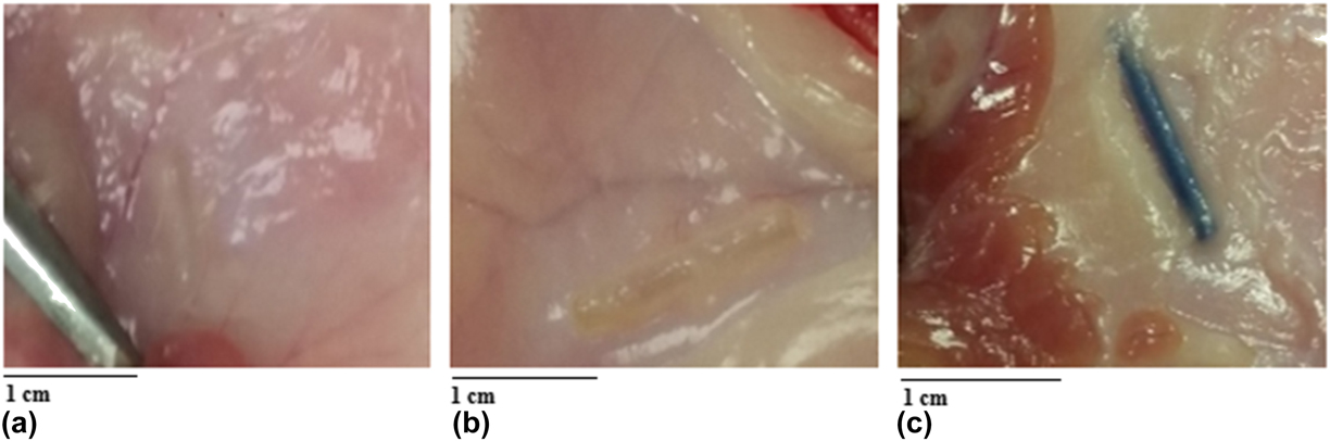
Figure 4 shows images of the tissues in contact with the material 30 days after implantation of three of the evaluated materials, including the control material (PP).
Tissue images of three of the groups of mice at day 30 of the experiment. (a) Skin with the CO-SM material, (b) skin with the CO-M2 material, and (c) skin with the PP material.

Figure 4 shows the tissues of the surrounding area exposed to PUs exhibit no signs of macroscopic lesions. The results of the in vivo assay showed that the three types of PUs were integrated into the tissue microenvironment with mild foreign body reactions. Similar results were observed with the control material (PP). Fibrosis and the formation of granules in the tissue normally occur during the physiological processes of wound closure caused by the formation of eschar in the tissue [Reference Laube, Weisser, Berger, Börner, Bischoff, Schubert, Gajda, Bräuer and Schnabelrauch13].
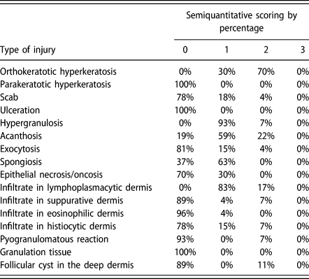
A histopathological diagnosis was made from skin samples. The diagnosis consisted of a construction and validation of the histological scoring system that combined lesion parameters to define each category, in accordance with the stipulations of Gibson-Corley et al. [Reference Gibson-Corley, Olivier and Meyerholz50]. Injury scoring was performed by two independent pathologists for each individual of the experimental group. A semiquantitative scoring (rank evaluation) of the lesions was used. Four ranges were determined that corresponded to the following values: absent = 0, mild = 1, moderate = 2, and severe = 3. Table III shows the statistical analysis of the 27 animals evaluated by the two pathologists.
Lesion score percentages from histopathological diagnoses.

Notes: The results of the semiquantitative scoring of the lesions are presented as the mean ± SD (n = 5). The scoring consists of: 0 = absence, 1 = mild, 2 = moderate and 3 = severe.
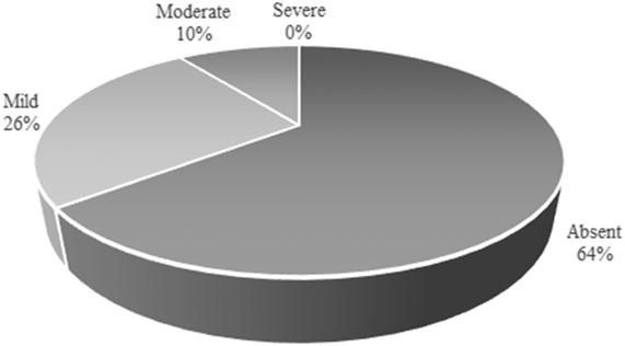
In histopathological analyses, higher scores indicate a greater injury [Reference Inzana, Schwarz, Kates and Awad51]. Table III shows that the highest value corresponded to score 3 (severe). Furthermore, according to the statistical analysis of the histopathological results of the 27 animals, no severe values were observed for the lesions evaluated (Fig. 5). Figure 5 shows that 64% of the lesions were absent in the animals evaluated. Therefore, the lesions generated must have corresponded to reactions triggered by the presence of a foreign material in the living organism.
Classification of the lesions presented in the histopathological analysis of the animals. Note: The results are presented as the mean ± SD (n = 5) and correspond to the scores by two different pathologists.

According to the diagnostic impression, 80% of the Group I animals (CO-SM) presented with mild lymphoplasmacytic dermatitis and the remaining 20% presented with moderate lymphoplasmacytic dermatitis. Furthermore, 20% of the Group II animals (CO-M1) presented with mild mixed dermatitis: 60% of the lesions corresponded to mild lymphoplasmacytic dermatitis and 20% of the lesions corresponded to moderate pyogranulomatous dermatitis. In addition, 80% of the Group III animals (CO-M2) presented with mild lymphoplasmacytic dermatitis, and 20% presented with moderate pyogranulomatous dermatitis. Group IV animals (PP) also showed varied results: 20% exhibited healthy skin, 60% exhibited mild lymphoplasmacytic dermatitis, and the remaining 20% exhibited moderate lymphoplasmacytic dermatitis. Finally, 100% of the Group V animals (sham) presented with mild lymphoplasmacytic dermatitis.
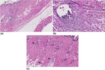
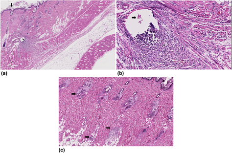
The previous analyses and macroscopic description indicated the lesions that presented during the study corresponded to a foreign body reaction and were not due to adverse reactions to the implanted material. In addition, when comparing the synthesized materials with the commercial material (PP), the obtained responses were similar. The microscopic photographs shown in Fig. 6 present a comparison of the materials causing a high degree of injury with the other materials evaluated.
Macroscopic description of lesions presented in animals. (a) Slight thickening of the epidermis (arrow) and moderate pyogranulomatous dermatitis in response to the foreign body (asterisk) composed of CO-M2 material (H&E 20× stain). (b) Moderate pyogranulomatous dermatitis in response to the foreign body composed of CO-M2 material (arrow), with the presence of epithelioid cells, macrophages, lymphocytes, plasma cells, PMN-eosinophils, and neutrophils (asterisk) (H&E 200× stain). (c) Mild multifocal lymphohistiocytic dermatitis associated with the adnexa and adipose tissue (arrows) with PP commercial material (H&E 80× stain).

Figure 6 shows photographs of two of the evaluated materials, CO-M2 and PP, corresponding to Groups III and IV, respectively. The information obtained via the histological images indicated that the lesions in the animals corresponded to a normal foreign body reaction when compared with the commercial material used as a control (PP).
The results of the in vivo assays indicated the PUs synthesized in this project and the PP control material showed a positive response when implanted in Wistar rats. Similarly, the lesions generated after 30 days of testing did not produce severe lesions or inflammatory responses. Similar values were determined for the in vivo evaluations of PUs synthesized with hydroxyapatite nanoparticles, a polyol derived from the açai berry and hexamethylene diisocyanate. Furthermore, a positive integration between the material and the body of the animal without large-scale inflammatory responses for the control and the PUs was observed [Reference Gabriel, Santos, Jardini, Bastos, Dias, Webster and Maciel Filho52].
Biodegradable PUs and their degradation products are nontoxic and cause a minimal in vivo response, which can be improved by modifying the material composition or preparation methods [Reference Ng, Lee, Chuah and Cheng15]. Thus, the results of this study indicate the synthesized PUs can be used as a matrix for the design of biomedical devices.
Conclusions
The PUs synthesized herein presented properties that may have potential in applications related to systems for wound closure. The mechanical properties were strongly influenced by the type of polyol used: modification of the hydroxyl groups produced polymer chains with higher degrees of crosslinking. Young’s modulus values were within the optimum range needed for the design of biomaterials for use in tissues such as the skin. The percentage of elongation, with a maximum of 260%, indicated that flexible strong materials were fabricated and could facilitate the design of biomaterials that do not cause injuries to soft tissues. The cell viability results showed the materials were not toxic to fibroblast cell lines; thus, such materials could be used for direct contact with the skin without causing damage to the surrounding cells. The in vivo studies related to the grafting of the material in the subcutaneous tissue of Wistar rats showed that no chronic lesions were present, indicating that the materials could safely be in contact with skin tissue. According to the results, PUs synthesized with castor oil polyols could be used as candidates in the design of biomedical devices.
Materials and methods
Reagents
Castor oil (R. communis) was purchased from Químicos Campota y Cía, Ltda. (Bogotá, Colombia). IPDI was purchased from Sigma-Aldrich Chemical Co. (St. Louis, Missouri). Pentaerythritol was obtained from Merck KGaA (Darmstadt, Germany). Phosphate-buffered saline (PBS: 0.01 M KH2PO4–NaHPO4 with 0.8% NaCl, pH 7.4), 3-(4,5-dimethylthiazol-2-yl)-2,5-diphenyl-2H-tetrazolium bromide (MTT), 2.5% trypsin (10×), penicillin–streptomycin (10,000 U penicillin and 10,000 μg streptomycin per milliliter), and Dulbecco’s Modified Eagle Medium (D-MEM, 1×) were obtained from Gibco/Invitrogen (Paisley, U.K.). Fetal bovine serum (FBS) was obtained from Eurobio (Les Ulis, France). Trypticase soy broth (TSB) and trypticase soy agar were obtained from Scharlau Co. (Barcelona, Spain).
Biological materials
Mouse fibroblasts from subcutaneous connective tissue L929 cells (ATCC® CCL-1), human lung fibroblast MRC-5 cells (ATCC® CCL-171™), adult human dermal fibroblast HDFa cells (ATCC® PCS-201-012™), Escherichia coli (ATCC® 2469), and Pseudomonas aeruginosa (ATCC® 27853) were acquired from the strain library of the Universidad de La Sabana (Chía, Colombia).
Animals
Eight-week-old, specific-pathogen-free male Wistar rats with average weights between 200 and 250 g were used in this study. The animals were not subjected to any type of procedure before introduction of the subcutaneous implant. The animals were kept in the ABSL-2 laboratory at the Bioterium of the National Institute of Health of Colombia.
Polyols
The polyols were derived from three modifications of castor oil (CO-SM, CO-M1, and CO-M2). CO-SM corresponded to unmodified commercial castor oil. The polyols CO-M1 and CO-M2 were obtained via transesterifications with pentaerythritol [Reference Valero and Ortegón53]. In the reaction, the castor oil temperature was increased to 120 °C for 10 min. Subsequently, the temperature was further increased to 210 °C, and pentaerythritol (1.32% or 2.64% mol pentaerythritol/mol castor oil for CO-M1 or CO-M2, respectively) and 0.05% lead oxide were added. The temperature was maintained at 210 °C for 2 h. The hydroxyl number of the polyols was determined in accordance with ASTM D1957-86.
Synthesis of PUs
PUs were synthesized by the prepolymer method in which IPDI was maintained in a constant (1:1) NCO/OH ratio. The temperature of the polyol was brought to 60 °C. IPDI was then added, and the reaction was stirred at 60 °C and 300 rpm for 5 min. Subsequently, bubbles were then removed, and the polymer was poured into a steel mold. Curing was performed at 110 °C for 12 h [Reference Valero and Ortegón53].
Characterization of PUs
Mechanical properties
Using a universal traction machine EZ-LX (Shimadzu Corporation, Tokyo, Japan), the maximum stress, the elongation percentage, and the Young’s modulus of the PUs were determined (ASTM D638-10). A load cell of 5 kN was used with a jaw displacement speed of 25 mm/min. Three samples of 40 × 6 × 3 mm3 (length × width × thickness) were tested.
Thermogravimetric analysis
The thermal behavior was evaluated in a thermogravimetric analyzer coupled to a differential scanning calorimeter (DSC), model TGA/DCS1 (Mettler-Toledo, Columbus, Ohio). The method used was in accordance with ASTM D6370 and included a heating rate of 25 °C/min with a temperature range of 25–600 °C in a nitrogen atmosphere and a sample mass of 15 ± 2 mg.
DSC
Glass transition temperatures (T g) were determined using a DSC analyzer, model DSC 3+ (Mettler-Toledo, Columbus, Ohio). The method used a temperature range of −70 to 150 °C in a nitrogen atmosphere with a 20 mL/min flow and a sample mass of 10 ± 2 mg.
Contact angles
Contact angles were determined with a drop shape analysis system (GH11, Krüss, Hamburg, Germany). In accordance with ASTM-D7334-08 (2013), the sessile drop method was used in which 10 μL of distilled water at 20 °C was placed on the PU materials. Ten measurements for each material were obtained.
Biological evaluation of PUs
In vitro cell viability assay
Mouse fibroblast L929 cells, human fibroblast MRC-5, and HDFa cell lines were cultured at 37 °C and 5% CO2 in D-MEM supplemented with FBS (10%) and penicillin–streptomycin (1%) in T75 cell culture flasks. Cell media were changed every 48 h [Reference Rezvanain, Ahmad, Mohd Amin and Ng54]. The cells were trypsinized (trypsin-ethylenediaminetetraacetic acid (EDTA)) at 100% confluence for viability analysis.
The effect of PUs on cell viability was evaluated with the MTT method, defined by ISO/CD 10993-5. Cells were seeded in 96-well plates at a concentration of 4.0 × 10⁴ cells per well in supplemented medium and cultured at 37 °C and 5% CO2 for 24 h. Then, 3 × 2 mm2 (diameter × length) PU cylinders previously sterilized with ultraviolet (UV) light (at 260 nm) were placed in 100 μL of supplemented medium for 30 min on each side [Reference Vannozzi, Ricotti, Santaniello, Terencio, Oropesa-Nunez, Canale, Borghi, Menciassi and Lenardi5]. The materials were left in contact with the polymers for 24 h at 37 °C and 5% CO2. The supernatant and the polymers were then removed, and MTT solution (12 mM in PBS) was added to achieve a total volume of 100 μL. The resulting suspension was incubated for 4 h at 37 °C. Subsequently, the supernatant was removed, 100 μL of dimethylsulfoxide was added, and the resulting suspension was incubated for 15 min at 37 °C. The optical density at 570 nm was then determined using a plate reader (BioTek ELx800 Absorbance Microplate Reader, Highland Park, Winooski, Vermont). A polypropylene (PP) biomaterial was used as a positive control, and doxorubicin was used as a negative control. All tests were performed in triplicate, and cell viability was determined according to Eq. (1):
 $${\rm{Cell}}\;{\rm{viability}}\;\left( \% \right) = {{\left( {{\rm{Ab}}{{\rm{s}}_{{\rm{sample}}}}} \right)} \over {{\rm{Ab}}{{\rm{s}}_{{\rm{control}}}}}} \times 100\quad ,$$
where Abssample corresponds to the absorbance value of the cells after treatment with PU and Abscontrol corresponds to untreated cells.
In vitro assay of antimicrobial activity
The possible antibacterial activity of PUs was determined against E. coli and P. aeruginosa, the main pathogens associated with implant infections [Reference Wu, Cui, Zhu, Qin and Xie55]. Bacterial suspensions were prepared in TSB at 37 °C for 16 h. Aliquots of the bacterial suspension were inoculated in fresh TSB at 37 °C for 6 h. The bacterial cells were removed by centrifugation at 3000 rpm for 10 min at 4 °C. The cells were washed twice with PBS after removing the supernatants. The bacterial suspension was adjusted to the standard absorbance of 0.5 McFarland in PBS [Reference Wu, Cui, Zhu, Qin and Xie55].
After being placed in 96-well plates, 3 × 2 mm2 (diameter × thickness) PU cylinders previously sterilized with UV light (260 nm) were incubated for 30 min on each side. A 100 μL aliquot of bacterial suspension was added to the wells containing the cylinders, and the plates were incubated at 37 °C for 24 h. The optical density at 600 nm was then determined with a plate reader (BioTek ELx800 Absorbance Microplate Reader), and any difference between the material evaluated and the control material was calculated. A bacterial suspension with no PU exposure was used as a control. The tests were performed in triplicate.
In vivo assays of PUs
Twenty-seven eight-week-old adult male Wistar rats (200–250 g) were maintained on a 12:12 h light:dark cycle and were fed with concentrated feed for open formula rodents according to the protocols described by the Bioterium of the National Institute of Health of Colombia.
The experimental protocol was approved by the ethics committee and/or Committee for the Care and Use of Laboratory Animals (CICUAL) and was performed following the guidelines given by the Bioterium of the National Institute of Health of Colombia. The animals were acclimated to the laboratory environment for five days. Subsequently, the animals were randomly divided into the following groups:
Group I: animals undergoing surgery for implantation of synthesized polymer CO-SM.
Group II: animals undergoing surgery for implantation of synthesized polymer CO-M1.
Group III: animals undergoing surgery for implantation of synthesized polymer CO-M2.
Group IV: animals undergoing surgery for implantation of commercial polymer PP.
Group V: animals undergoing surgery with no implantation of materials—sham.
Group VI: animals not undergoing surgery—zero.
Groups I to V used a sample size of n = 5, and Group VI used a sample size of n = 2. Animals from Groups I to V were anesthetized with ketamine (80 mg/kg) and xylazine (10 mg/kg) intraperitoneally. The dorsal region was shaved, and the area was disinfected with Baxedin antiseptic solution. An incision of approximately 1–2 cm was made along the dorsal cervical midline, parallel to the spinal column and between 2 scapulae. By blunt dissection, a subcutaneous pocket was generated to implant the material to be evaluated. The incision was closed with a PP stitch. The animals were then monitored for 30 days.
Group VI animals were sacrificed by inhalation of carbon dioxide, and the tissues of the dorsal mid-cervical area were collected for histopathological evaluation. Additionally, multiple organs (liver, lung, spleen, pancreas, heart, and kidney) were sectioned for histological evaluation. The harvested tissues and organs were stored in 10% buffered formaldehyde.
After 30 days, all Group I–V animals were sacrificed in the same manner as Group VI. Tissues and organs were removed along with the implanted material for histopathological evaluations.
Histopathological analysis
The tissues obtained from the 27 animals were embedded in paraffin, sectioned at 5 μm, and stained using hematoxylin and eosin (H&E). Each section was independently analyzed by two pathologists (double blind study) and rated using four levels (absence, mild, moderate, and severe) [Reference Garg, Sandhu, Sood, Sood and Malhotra56].
Statistical analysis
The results were expressed as the mean ± standard deviation (SD). The data were analyzed using analysis of variance, and differences were considered significant at P < 0.05. For comparison between samples, Student’s t-test was performed using SPSS Statistics 23 software (IBM, Armonk, New York).
Acknowledgments
We would like to thank the Universidad de La Sabana for financing the ING-176-2016 research project and Colciencias for the doctoral grant-loan with the call number 617-2 in 2014. We would also like to thank the Bioterium of the National Institute of Health of Colombia and the Corporación Patología Veterinaria (Corpavet®) for collaboration in conducting the in vivo assays.