Case report
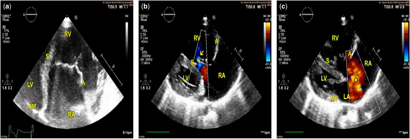
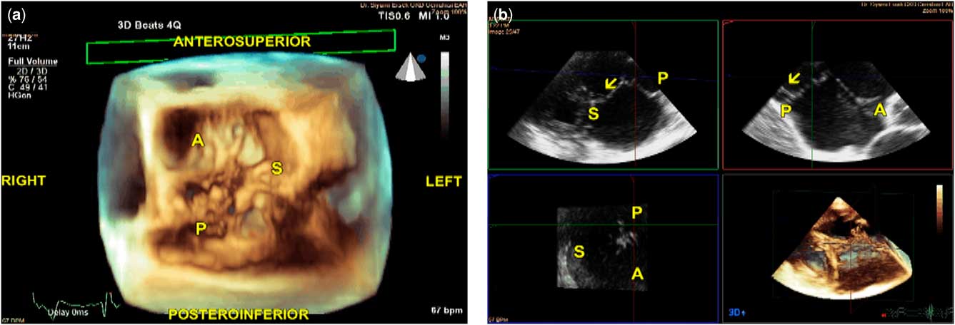
A 17-year-old patient was admitted to the institutional paediatric cardiology outpatient clinic with mild chest pain and shortness of breath on exertion. On physical examination, his blood pressure was 110/70 mmHg and heart rate was 80 bpm. The apical beat was slightly dislocated lateral to the midclavicular line. On auscultation, the second heart sound was widely split, and there was a 1/6 systolic ejection murmur at the left lower sternal border. Lung auscultation was normal, and there was no distention in the jugular veins or any pretibial oedema. An ECG showed normal frontal axis and an incomplete right bundle branch block, and a posteroanterior chest X-ray revealed mild cardiomegaly with a cardiothoracic ratio of 0.55. His oxygen saturation was 98% at room air, as measured with a pulse oximetry. As these findings were suggestive of an atrial septal defect, a transthoracic echocardiogram was performed. On two-dimensional transthoracic echocardiography, the septal leaflet was displaced caudally, and the difference between the origins of the septal tricuspid leaflet and anterior mitral leaflet was measured as 26.0 mm/m2 (Fig 1a, Supplementary video 1). However, there was only mild tricuspid regurgitation on color Doppler examination and the right ventricle was functioning normally, with a tricuspid annular systolic excursion of 20 mm and a lateral tricuspid annular velocity of 12.5 cm/s (Fig 1b, Supplementary video 1). In addition, there was a secundum atrial septal defect on fossa ovalis with a predominant left-to-right shunting, which was measured 32 mm at its widest diameter (Fig 1c). Both the right atrium and the right ventricle were dilated (Fig 1a). Three-dimensional transthoracic echocardiography revealed an abnormally elongated and wide septal leaflet, with a normal-sized anterior leaflet and a slightly small-sized posterior (inferior) leaflet (Fig 2a, Supplementary video 2). Similar to an Ebstein’s anomaly, basal segments of the septal and posterior tricuspid leaflets were tethered against the interventricular septum and the right ventricular free wall, but delamination was not clearly observed on two- or three-dimensional echocardiography (Fig 2b). Despite all these abnormalities, leaflet coaptation was near-normal on three-dimensional echocardiography, thus explaining the lack of severe tricuspid regurgitation. As the patient was not cyanotic and the direction of the atrial septal shunt was from left to right atrium, the atrial septal defect was closed percutaneously with a 36-mm Amplatzer Septal Occluder (St. Jude Medical Inc., Saint Paul, Minnesota, United States of America). No haemodynamic sequelae were observed postoperatively, and the patient was asymptomatic 6 months after the defect closure.
Two-dimensional transthoracic echocardiography findings of the patient. ( a ) The origin of the septal leaflet (S) was displaced caudally compared with the origin of the anterior mitral leaflet (AM). The origin of the anterior tricuspid leaflet (A) was not displaced. ( b ) A mild tricuspid regurgitation was shown (arrow). ( c ) An accompanying atrial septal defect (asterisk). LV=left ventricle; MV=mitral valve; RA=right atrium (including atrialised portion of the right ventricle); RV=right ventricle.

Live/real-time three-dimensional transthoracic echocardiography findings of the patient. ( a ) Shows en face view of the tricuspid valve, as seen from the right ventricular aspect. Note that the septal leaflet (S) was abnormally wide. ( b ) Multiplanar reconstruction of the three-dimensional data set was given. Both the septal (green panel, top left) and posterior (inferior) (red panel, top right) tricuspid leaflets displaced caudally and tethered against right ventricular septum and posterior walls, respectively. Arrows show chordae tendinea tethering the leaflets. Despite the tethering of the basal parts of the leaflets, the overgrowth of the septal leaflet had preserved a near-normal leaflet coaptation. A=anterior tricuspid leaflet. P=posterior tricuspid leaflet.

Ebstein’s anomaly is a rare congenital disorder that is observed in 1:20,000 of all live births.Reference Lupo, Langlois and Mitchell 1 , Reference Pradat, Francannet, Harris and Robert 2 The essential components of an Ebstein’s anomaly are caudal displacement of functional tricuspid annulus owing to the distal dislocation of the attachments of the septal and posterior (anatomically inferior) leaflets, tethering of septal and posterior leaflets, and an elongated and redundant anterior tricuspid leaflet. These abnormalities lead to atrialisation of the basal parts of the right ventricle, abnormal leaflet coaptation, and tricuspid regurgitation.Reference Booker and Nanda 3 Ebstein’s anomaly could be diagnosed incidentally in adults, but at least moderate tricuspid regurgitation is present in the vast majority of cases even if the patient is asymptomatic.Reference Morray 4 , Reference Krieger and Valente 5 Tricuspid regurgitation is a consequence of shortened septal and posterior leaflets secondary to tethering, which prevents normal leaflet coaptation during ventricular systole. In adults, severe tricuspid regurgitation and symptoms of right heart failure are common presentations, along with arrhythmias.Reference Booker and Nanda 3
In the present case, however, coaptation of tricuspid leaflets appeared normal or near-normal despite an apparent caudal dislocation of the functional tricuspid annulus on two-dimensional echocardiography. On three-dimensional echocardiography, septal leaflet was unusually wide and “sail-like”, with a normal-sized anterior leaflet and a slightly small-sized posterior leaflet. We consider that this wide and elongated septal leaflet preserved normal leaflet coaptation and prevented severe tricuspid regurgitation. As the contractility of the right ventricle was also preserved, this balanced abnormality did not cause symptoms compatible with Ebstein’s anomaly. The mild exertional dyspnoea at presentation was attributed to the secundum atrial septal defect, which was subsequently closed.
Several variants of the Ebstein’s anomaly have been reported in the literature. The most common of these variants is the delamination and tethering of the anterior leaflet that leads to displacement of the anterior part of the tricuspid annulus.Reference Tsujii, Kurosaki and Yasuda 6 Similar to the classical Ebstein’s anomaly, this variant also causes abnormal leaflet coaptation leading to severe tricuspid regurgitation.Reference Tsujii, Kurosaki and Yasuda 6 To the best of our knowledge, the present case reported in this manuscript is a novel variant and characterised with an abnormally elongated and “sail-like” septal leaflet that acts to “correct” coaptation defect caused by leaflet tethering and displacement of functional tricuspid annulus. In contrast to the other variants, leaflet coaptation is preserved in this variant that prevents severe tricuspid regurgitation.
Some authors have argued that all essential components of an Ebstein’s anomaly must be present in a patient for the diagnosis, and the rest of cases should be classified as “non-Ebstein’s tricuspid regurgitation”.Reference Ammash, Warnes, Connolly, Danielson and Seward 7 In our opinion, patients with annular displacement and leaflet tethering/delamination should be classified as Ebstein’s variants even if all “classical” findings are not present, as the congenital pathology causing an Ebstein’s anomaly is the leaflet delamination that leads to annular displacement and leaflet tethering. Thus, we consider that the present case represents a variation of the Ebstein’s anomaly rather than representing a novel CHD or “non-Ebstein’s tricuspid regurgitation”.
Acknowledgements
None.
Financial Support
This report received no specific grant from any funding agency or commercial or not-for-profit sectors.
Conflicts of Interest
None.
Ethical Standards
No human or animal experimentations have been performed for this present report.
Supplementary material
To view supplementary material for this article, please visit https://doi.org/10.1017/S1047951117002712