The Holmes’ heart holds a special place in the history of congenital cardiac disease. We report a fascinating variant that raises a number of interesting questions regarding our classification and management of this condition.
Case report
A 59-year-old woman presented with a 1-week history of exertional breathlessness and palpitations. She was a smoker of 10 cigarettes per day and had a history of gallstones and two normal pregnancies. Examination revealed saturations of 97%, an irregular pulse, and blood pressure of 220/130. Her venous pressure was elevated at 7 centimetres H2O above the sternum and she had mild pitting ankle oedema. There was a soft pansystolic murmur at the left sternal edge. Electrocardiography showed right axis deviation, late transition across the chest leads, and delayed atrioventricular conduction (Fig 1). Holter monitoring revealed prolonged periods of atrial flutter with variable block. Chest radiograph showed cardiomegaly. Haemoglobin was 13 grams per decilitre.
Twelve-lead electrocardiogram.

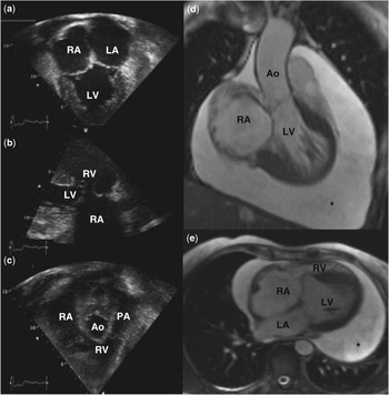
Remarkably, echocardiography showed a double inlet left ventricle with a right atrioventricular valve that straddled and was adhered to the malaligned septum. The greater portion of the right atrioventricular valve was committed to the left ventricle and was almost completely fused with no significant forward flow across it and only trivial regurgitation. The portion that opened into the anterior rudimentary right ventricle functioned normally. Continuous wave Doppler of the regurgitation from the left ventricular portion of the valve showed a peak velocity of 5.8 metres per second, while that from the right ventricular portion (2.8 metres per second) confirmed normal pulmonary artery systolic pressures. Ventriculoarterial connections were concordant. These findings were confirmed at magnetic resonance (Fig 2). There was a large pericardial effusion. Aspiration revealed a transudate with no further indication as to the aetiology. Despite complete drainage, the pericardial effusion re-accumulated. The patient was treated with anti-hypertensives and commenced on aspirin, after declining formal anti-coagulation for the atrial flutter. She remains mildly symptomatic and is under clinical follow-up.
(a) Apical “four-chamber” view of the double inlet left ventricle. (b) Parasternal long-axis right ventricular inlet view showing adherence to the straddling right atrioventricular valve to the malaligned ventricular septum. (c) Modified apical inlet–outlet view of the hypoplastic right ventricle showing the origin of the pulmonary artery. (d) Balanced Steady State Free Precession (SSFP) coronal cine magnetic resonance image showing the origin of the aorta from the left ventricle. (e) Balanced SSFP axial cine magnetic resonance image showing the four chambers (RA = right atrium; RV = right ventricle; LA = left atrium; LV = left ventricle; Ao = Aorta; asterisk indicates pericardial effusion).

Discussion
In 1824, Andrew HolmesReference Holmes1 described the index case of a double inlet left ventricle with concordant ventriculoarterial connections. The case achieved much notoriety when it was re-published some 80 years later by Maude AbbottReference Abbott2 at the behest of William Osler. For many, it heralded the arrival of congenital cardiac disease as a speciality in its own right. In the original description, Holmes described the condition as an intermediate between a true single ventricle and the normal heart. Nowhere is this description better realised than in the present case in which the second chamber is quite clearly a morphological right ventricle.
Our patient unequivocally has the anatomy of the “Holmes’ heart”; however, her heart is distinguished by the straddling right atrioventricular valve that is firmly attached to the crest of the malaligned ventricular septum. The ventricular septal defect, which had been present, is thus closed by a connecting tongue of valvar tissue, analogous to the situation seen in the “ostium primum” defect with a common atrioventricular junction. In addition, the straddling of the right atrioventricular valve results in a valve with two orifices. In this respect, the heart functionally represents a variant of a “double outlet right atrium”, though admittedly it is the ventricular septum rather than the atrial septum that is deviated. Achieving a diagnosis by echocardiography was possible only by using the sequential segmental approach to interpret these connections.Reference Anderson, Becker and Freedom3
In 1951, Lambert first described the straddling of the tricuspid valve in a patient with double inlet left ventricle, a large ventricular septal defect, and ventriculoarterial discordance.Reference Lambert4 Milo et alReference Milo, Ho and Macartney5 later published the largest series of these cases in which they described several sub-types and commented on the disposition of the conducting system. Only once has a straddling tricuspid valve been reported without a ventricular septal defect: in a 4-year-old with pulmonary atresia and intact ventricular septum.Reference Isomatsu, Kurosawa and Imai6 Prolongation of the PR interval in our case supports the supposition, as is usual in the double inlet left ventricle, that there is likely to be an anomalous atrioventricular node located anterolaterally around the right atrioventricular orifice.
Most variants of the double inlet left ventricle present prenatally or in early infancy. Prognosis is poor and not improved by neonatal surgery.Reference Tham, Wald and McElhinney7 The anatomical arrangement described here is of interest because of the patient’s relative lack of symptoms, two normal pregnancies, and late survival. In the absence of a ventricular septal defect, she was not exposed to the damaging effects of cyanosis or the consequences of elevated pulmonary pressures. Despite less than half of the right atrioventricular junction being connected to the hypoplastic right ventricle, it clearly receives almost all right atrial output. This is relevant to the debate regarding indications for one and a-half ventricular repair as it highlights the fact that size alone does not predict a ventricle’s ability to successfully support the pulmonary circulation.Reference Hanley8
In our patient, as is frequent in adults with congenital cardiac disease, clinical deterioration accompanied the onset of atrial arrhythmia almost certainly resulting from chronically elevated right atrial pressure. The presence of the pericardial effusion may be attributable to the haemodynamic situation; however, other aetiologies such as malignancy should also be considered. By virtue of selection, this patient’s greatest future risk remains acquired and not congenital cardiac disease. Occasionally, complex congenital cardiac disease presents at unexpected moments. Such cases can provide fascinating morphological insights and may cause us to re-evaluate conventional understanding of certain conditions and their treatment.
Acknowledgements
The authors thank Professor RH Anderson for his invaluable advice and Dr L Mitchell for the magnetic resonance image. There were no grants or other means of financial support for this study.