Introduction
Physocephalus sexalatus, the type species of the genus Physocephalus was first described as Spiroptera sexalata by Molin (Reference Molin1860). Previously, this species had been misidentified as Spiroptera strongylina by Diesing (Reference Diesing1851) in the helminthological collection of the Vienna Natural History Museum. The material originated from a pig (Sus scrofa) from Vienna (Bremser collection: Johann G. Bremser (1767–1827) was an Austrian medical doctor who collected parasitic worms and donated them to the Imperial Natural-Science Cabinet in Vienna) and from a white-lipped peccary (Tayassu albirostris) from Brazil (Natterer collection: Johann Natterer (1787–1843) was Austrian naturalist and explorer who spent 18 years in Brazil and, after his return in 1835, he enriched the Imperial Natural-Science Cabinet in Vienna with specimens he collected there). Spiroptera strongylina (today: Ascarops strongylina) is known since Rudolphi's (1819) description in which he referred to the Vienna collection, and it is quite possible that the material contained two different species. The genus Physocephalus was introduced by Diesing (Reference Diesing1861) for cylindrical nematodes with an inflated anterior end and a mouth opening with two trilobed lips and three longitudinal wings in the anterior part of the body on each lateral side, and Ph. sexalatus, a stomach parasite of pigs in Europe and peccaries in Brazil, was the first species assigned to this genus.
Two other species, Ph. gracilis (Rudolphi, Reference Rudolphi1819) and Ph. mediospiralis (Molin, Reference Molin1860), which were known at that time but were listed under a different genus name, as well as new species that had been recorded later on were added to the genus Physocephalus. According to the latest synopsis on nematodes (Hallan, Reference Hallan2007) the genus Physocephalus consists of nine species.
Based on a description of Ph. sexalatus in dromedaries from Turkmenistan (Badanin, Reference Badanin1939) and differences in the fauna of intermediate hosts and larval size, Muschkambarowa (Reference Mushkambarowa1967) created the subspecies Ph. sexalatus dromedarii. The aim of this paper is to give a detailed description of a Physocephalus species from dromedaries in Dubai and to upgrade the above-mentioned subspecies to the rank of a species.
Materials and methods
Spirocercid nematodes (n= 35 and n= 425, respectively) were isolated from the abomasum of two adult female dromedaries submitted by Dubai Camel Dairy Farm for necropsy to the Central Veterinary Research Laboratory in July 2011 and October 2012, respectively. Both animals were long-term residents on the farm. To remove mucous and abomasal contents, freshly isolated nematodes were treated for 30 min in warm (40°C) artificial gastric juice and fixed in 10% formalin at 70°C. As males twist their posterior end during hot fixation, making examination of diagnostic structures difficult, some specimens were stretched between two glass slides during the fixation process. Worms were cleared in 30% lactic acid for 12–24 h and examined by light microscopy. Haematoxylin and eosin stained histological sections were made from paraffin-embedded female worms.
For scanning electron microscopy, male and female nematodes were critical point dried using CO2, mounted on a specimen stub and sputter coated with a 10-nm layer of gold–palladium alloy (Polaron Sputter coater SC 7600; Emitech, Montigny-le-Bretonneux, France) before examination with a Supra 40VP scanning electron microscope (Zeiss, Oberkochen, Germany).
Results
Morphology
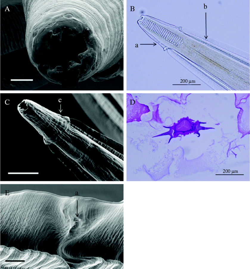
Living worms, which measure between 14.4 and 29.0 mm in length, possess a red-coloured fasciated cuticle which loses colour following fixation. The mouth opening is surrounded by two trilobed lateral lips followed by a pair of flat papillae on each side (fig. 1A). Due to a distinct pocket-like inflation of the cuticle, the anterior end appears like an arrowhead (fig. 1B). An unpaired lateral cervical papilla is situated just prior to this inflation. The shallow toothless oral cavity is followed by a cylindrical pharynx, the walls of which are supported by 19–24 coils arranged like a spiral. The oesophagus consists of a shorter muscular part and a longer muscular part. Three fasciated lateral wings on each side (fig. 1C and D), broader median wings with fine striations, and slender ventral and dorsal wings with coarse striations, on each side, commence right behind the cuticular inflation extending posterior to the oesophagus. They reach their maximum width at the posterior third of the oesophagus. The excretory pore (fig. 1E) is located on the median lateral wing just posterior to the nerve ring on the opposite side to the cervical papilla.
(colour online). The morphology of the anterior region of Physocephalus dromedarii. (A) Mouth opening surrounded by two trilobed lateral lips followed by a pair of flat papillae on each side (scale bar: 20 μm). (B) Arrow-shaped anterior end with cylindrical pharynx supported by spiral-like arranged coils; unpaired cervical papilla (a) on the opposite side to the excretory pore (b). (C) Lateral wings commence posterior to the pocket-like inflation at the anterior end (c); broader median wings with a fine striation, slender dorsal and ventral wings with a more coarse structure (scale bar: 100 μm). (D) Lateral wings in the histological cut through the oesophagus region appear trident-like. (E) The excretory pore (a) occurs as a half-pipe on the dorsal surface of median lateral wing (scale bar: 10 μm).

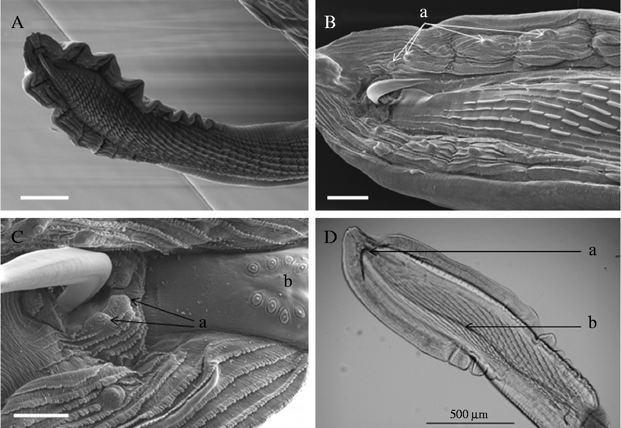
Males (n= 10): body posterior to lateral wings, which stretch over one-third of the total length, cylindrical, 356 (320–400) μm in diameter. Posterior end twisted clockwise in 2–2.5 (3) turns. Caudal (bursal) wings of unequal size: longer wing 1600 (1500–1700) μm in length and 125 (100–150) μm in width, shorter wing 1400 (1200–1600) μm in length and 148 (120–180) μm in width. There are 20–22 interrupted oblique ridges on the ventral body continuing as uninterrupted striations on the wings (fig. 2A). Four pairs of stalked precloacal papillae support the wings: two pairs at a distance and two pairs close to cloaca (fig. 2B). Postcloacal papillae consist of two pairs immediately posterior to cloaca and four very small pairs of papillae situated on the postcloacal plate (fig. 2C). Spicules are of unequal size: 510 (450–540) μm × 15 μm and 2600 (2500–2700) μm × 10 μm, respectively (fig. 2D).
The morphology of the posterior end of male Physocephalus dromedarii. (A) Oblique ridges commence prior to cloaca and proceed beyond the area of caudal wings (scale bar: 200 μm). (B) Precloacal region with four pairs of stalked papillae (a) situated in a row in the centre of caudal wings (scale bar: 50 μm). (C) Postcloacal region with two pairs of papillae (a) right posterior to the cloaca and a further four pairs on the postcloacal shield (b) (scale bar: 20 μm). (D) The short spicule (a) is five times shorter but one-third broader than the long spicule (b).
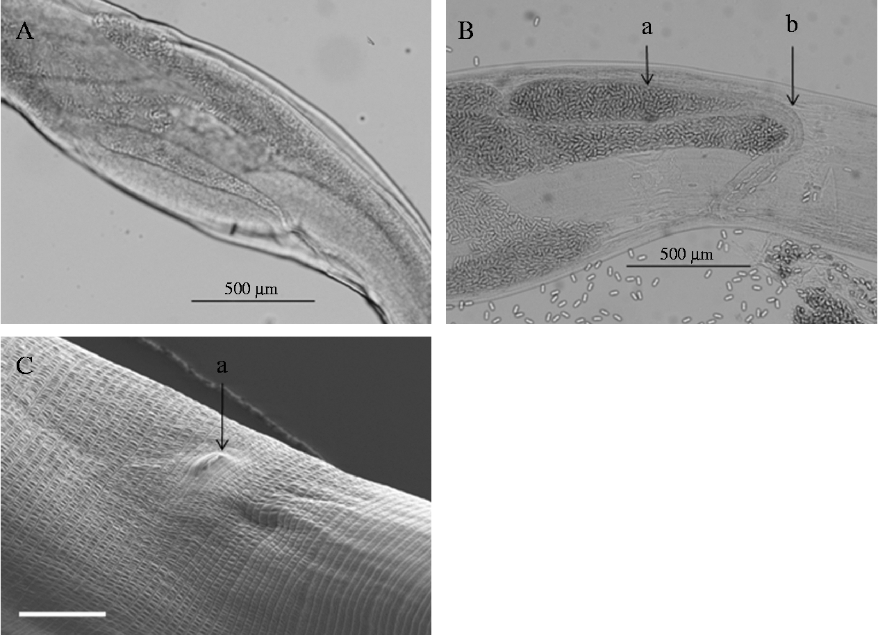
Females (n= 20): lateral wings account for one-quarter of total lengths. Body cylindrical posterior to lateral wings, prior to and after a conspicuous swelling (fig. 3A) situated in the middle of the second third of the body. Posterior end after anus rounded with ventrally bent small tail. Female reproductive system with didelphic uterus; anterior ovary commences near the mid-body, stretches anterior and turns at some distance to terminal part of the oesophagus to be connected to the anterior uterine horn. The latter goes backwards and in the posterior third of the body changes to the forward direction and terminates at the level of the swelling. The posterior part of the female reproductive system takes the opposite way: prior to the anal opening, the posterior ovary fuses with the subsequent uterine horn. The latter is directed anterior and turns backwards at the level of the half-body to terminate at the level of the swelling. The two terminal parts of uterine horns are united into a twisted, narrow tube that is connected to the terminal part of the female reproductive system, the vagina (fig. 3B) – a swelling that merges with a narrow, thick-walled tube containing eggs arranged in a single layer. The female genital opening is inconspicuous (fig. 3C) and may be located immediately prior or posterior to the swelling of the body. Thick-shelled eggs with a dent on one pole are 35–38 μm × 15 μm in size and contain a first-stage larva. The larva has rasp-like mouthparts, similar to those in Habronema larvae, to penetrate the gut of the intermediate host.
The morphology of the terminal part of the female reproduction system of Physocephalus dromedarii. (A) Conspicuous widening of the female's body due to a concentration of uterus loops. (B) The vagina consists of an elongate-piriform widened part (a) and a narrow, thick-walled tube (b). (C) The vulva (a) with transverse cuticular striations in this region (scale bar: 50 μm). The vulva itself appears as an inconspicuous transverse slit.
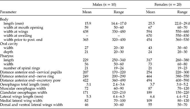
Additional measurements of male and female worms are given in table 1.
Morphometrics (mean values in µm unless otherwise stated) of male and female Ph. dromedarii; n= number of specimens examined.
Discussion
Several authors have studied the morphology of the type species, Ph. sexalatus and Foster (Reference Foster1912) gave the most detailed description of this nematode obtained from stomachs of pigs in the USA. According to this description, male and female specimens measured 6–9 and 13–19 mm, respectively, with a maximum width of 0.24 and 0.33–0.45 mm, respectively. These measurements match those given in the original description by Molin (Reference Molin1860) and are considerably smaller than those of Ph. dromedarii. According to Foster (Reference Foster1912) the length of the two spicules of Ph. sexalatus is 2.1–2.25 and 0.3–0.35 mm, respectively, compared to 2.5–2.7 and 0.45–0.54 mm, respectively in Ph. dromedarii. Physocephalus sexalatus also differs in a smaller width of the lateral wings at the anterior end. In a study of Ph. sexalatus from pigs in Romania, male and female nematodes were slightly longer (10–15 and 16–22 mm, respectively) but spicules were considerably shorter, measuring only 1.2 and 0.2 mm, respectively (Ciurea, Reference Ciurea1912). Foster (Reference Foster1912) mentioned longitudinal striations on the ventral surface between the bursal wings without giving quantification. According to Ciurea (Reference Ciurea1912) there are 7–8 parallel directed, wave-like longitudinal crests prior to the cloaca, interrupted by prongs comparable to the blade of a saw. In our own material, these crests are oblique and number between 20 and 22. While there is an agreement on the four pairs of preanal papillae in Ph. sexalatus, opinions differ regarding the number of postcloacal papillae. Drasche (Reference Drasche von1884), who re-examined the original material of Molin (Reference Molin1860), found and pictured one pair of papillae very close to the cloaca and three pairs of very small papillae on the postcloacal plate. Ciurea (Reference Ciurea1912) described a similar distribution but saw four pairs of small papillae at the tip of the tail, while Foster (Reference Foster1912) failed to see papillae at the edge of the cloaca but depicted four pairs of postcolacal papillae on the postcloacal plate. Like Ph. sexalatus, Ph. dromedarii has four long and stalked preanal papillae but differs in the number of postcloacal papillae. In addition, in Ciurea's description there is a thickening of the cuticle next to the female genital opening. Such a structure is not present in Ph. dromedarii.
Life-cycle studies with Ph. sexalatus showed that coprophagic beetles (Canthon laevis, Geotrupes douei, G. stercorarius, Gymnopleurus sturmi, G. sinnatus, Onthophagus bedeli, O. hecate, O. nebulosus, Phanaeus carnifex, P. vindex, Scarabaeus sacer, S. variolosus) act as intermediate host, and small mammals, birds, reptiles and amphibians as paratenic hosts for Ph. sexalatus (Alicata, Reference Alicata1935). The final host becomes infected by ingesting infected intermediate or paratenic hosts. Infective larvae measured 1.35–1.6 mm in length and 60–68 μm in width.
It is noteworthy that Ph. sexalatus was listed as a parasite of dromedaries in Iran (Mirzayans & Halim, Reference Mirzayans and Halim1980) and Kuwait (Abdul-Salam & Farah, Reference Abdul-Salam and Farah1988).
Seurat (Reference Seurat1912) published a short note on a Physocephalus species (variant cristata) similar to Ph. sexalatus in the abomasum of a dromedary at the Hauts Plateau in Algeria, with males and females slightly longer and with lateral wings broader than in Foster's description. The two unequal spicules measured 2.5 and 0.3 mm. According to Seurat (Reference Seurat1912) the new variation of Ph. sexalatus from dromedary differs mainly in the structure of the head inflation which has four crests formed by curricular folds and the presence of four cuticular grates in the mouth cavity. Other peculiarities were the unscrewed posterior end of the male (bent by just 90°) and the variable position of the female genital opening. Unfortunately, other features were described only superficially and no figures were included. Nevertheless, Railliet (Reference Railliet1915) upgraded this variety to species level. Seurat (Reference Seurat1915) found the larval stage of this nematode in dung beetles (S. sacer, S. variolosus, Geotrupes douei, O. nebulosus, O. bedeli) in the Hauts Plateau of Algeria. Physocephalus cristatus has never been recorded again, but was listed as parasite of camels in Iraq (Altaif, Reference Altaif1974). In a more recent publication on dromedary parasites in Algeria the Physocephalus species found was not specified (Chauve et al., Reference Chauve, Hamza-Cherif and Marfoua1990)
Badanine (Reference Badanine and Neiva1938) gave the first report of Physocephalus sp. in dromedaries in Central Asia and described the nematode as Ph. sexalatus in detail (Badanin, Reference Badanin1939) (regarding the author's name, ‘Badanine’ was created by a typing mistake; the correct family name is Badanin). Based on the difference in the size of the larval stages obtained from beetles collected at camel farms in Turkmenistan, Mushkambarova (Reference Mushkambarowa1967) created the subspecies Ph. sexalatus dromedarii. For the characterization of the adult nematode, she used Badanin's (Reference Badanin1939) description. This description was later completed by Mushkambarova & Dobrynin (Reference Mushkambarova and Dobrynin1972) and matches the measurements established for nematodes found in the dromedaries in Dubai. Males were 12–16 mm in length and the spicules measured 2.5–3 mm and 0.47–0.53 mm. Females had a length of 20–28 mm. The latter authors drew attention to a typical swelling at the beginning of the second half of the body of the female. Such a swelling at the beginning of the second half of the body has also been mentioned in females found in the stomach of donkeys by Ivashkin & Dvojnos (Reference Ivashkin and Dvojnos1984). Other measurements also corresponded to those given by Badanin (Reference Badanin1939) and Mushkambarova & Dobrynin (Reference Mushkambarova and Dobrynin1972). In both sources, five pairs of postcloacal papillae were reported, with one pair immediately posterior to the cloaca and four pairs of smaller papillae on the postcloacal plate. The number of wave-like crests seen at the caudal end of the male prior to the cloaca was indicated to be 14–16, while in our own material the entire number was 20–22.
Mushkambarova & Dobrynin (Reference Mushkambarova and Dobrynin1972) necropsied 58 dromedaries throughout Turkmenistan and found 44 (76%) to be infected. The number of Ph. sexalatus dromedarii isolated ranged between 1 and 883. The same nematodes were also detected in dromedaries in neighbouring Uzbekistan (Sultanov et al., Reference Sultanov, Kabilov, Atachanova and Dadaev1973).
A variety of scarab beetles (Scarabaeus sacer, Gymnopleurus aciculatus, G. mopsus, G. corianius, Chironitis hungaricus, Onitis humerosus, Orthophagus marginalis) serve as intermediate hosts in Turkmenistan (Mushkambarova, Reference Mushkambarova1964, Reference Mushkambarowa1967). Infective larvae found in the beetles collected from camel farms were larger than those of Ph. sexalatus, measuring 1.5–1.85 mm in length and up to 60–90 μm in width. In addition to S. sacer, Sultanov et al. (Reference Sultanov, Kabilov, Atachanova and Dadaev1973) showed Copris lunaris and Adesmia biseriata to be intermediate hosts in Uzbekistan. Contrary to Ph. sexalatus, where pigs become infected when ingesting dung beetles, camels are thought to become infected when drinking water contaminated with larvae that escaped from beetles when they drown in water (Mushkambarova & Dobrynin, Reference Mushkambarova and Dobrynin1972).
The justification of Ph. dromedarii, as a new Spirocercidae species that occurs naturally in the one-humped camel and accidently in equids, is based on a strikingly larger total length of male and female specimens, on longer spicules and the presence of six postcloacal papillae in the males, as well as on the presence of a conspicuous swelling at the beginning of the second half of the female body. There are also differences in the spectrum of intermediate hosts as well as in the size of the infective larvae.
Apart from Ph. sexalatus and Ph. cristatus, Ph. dromedarii has to be distinguished from the remaining species in the genus. The description of P. ellobii is based only on an incomplete female specimen found in a mole rat and must be regarded as dubious. Physocephalus quadrialatus, a species that was found in hamsters in Armenia, Hungary and Mongolia, is characterized by much smaller spicules (0.22 and 0.17 mm) and eggs measuring 54 × 23 μm (Ryzikov et al., Reference Ryzikov, Gvozdev, Tokobaev, Shaldybin, Macaberidze, Merkursheva, Nadtocij, Chochlova and Sharpilo1979). It is worth mentioning also Ph. skrjabini, a stomach nematode of hamsters in Moldowa (Andrejko, Reference Andrejko1964). This species is smaller than Ph. quadrialatus and has spicules measuring 0.3 and 0.2 mm. Physocephalus gracilis is a parasite of sloths in Brazil. While the length is comparable to that of Ph. sexalatus these are very slim nematodes. The male specimen has spicules of nearly equal size and harbours only six pairs of caudal papillae. Another sloth nematode, Ph. leptocephalus, has been transferred to the genus Leiuris due to the presence of teeth in the mouth cavity. Physocephalus nitidulans is a stomach parasite of the Brazilian tapir. This species is larger than Ph. sexalatus but does not have a cuticular inflation, and cervical alae commence at short distance behind the mouth opening (Vicente et al., Reference Vicente, Rodrigues, Gomes and Pinto1997). Males and females of Ph. mediospiralis parasitizing agoutis and pacas in Brazil are also longer than Ph. dromedarii, measuring up to 33 and 54 mm, respectively (Hall, Reference Hall1916; Goncalves et al., Reference Goncalves, Boia, Coura and Pinto2006). Physocephalus lassancei and Ph. raphiceri are parasites of brocket deer in Brazil and steenbuck in South Africa, respectively. Since in both species a cuticular inflation on their anterior ends is missing, they had been described under the genus name of Pereiraia, which later became a synonym of Physocephalus. Both species have spicules of unequal length that are considerably shorter than those in Ph. dromedarii. Skrjabin et al. (Reference Skrjabin, Sobolev and Ivashkin1967) listed also Ph. theodoridesi, a larval stage that was found in the scarabeid beetles Trox perlatus in France and Trox marticini in Mongolia. The final host of this species is unknown. There are also several poorly described Physocephalus spp. mentioned in the literature (Skrjabin et al., Reference Skrjabin, Sobolev and Ivashkin1967).
Acknowledgements
The authors are grateful to Mrs D. Viertel, Leibniz Institute for Zoo and Wildlife Research, Berlin, for her excellent technical assistance in the preparation of scanning electron microscopy samples. Our thanks also to Mr Ch. Vishwanathan for preparing histological preparations. We also thank Dr S. Rehbein, Merial GmbH, Katharinenhof; Dr B. Neuhaus, Museum für Naturkunde, Berlin; Dr H. Sattmann, Naturhistorisches Museum Wien; and Dr P. Kment, National Museum, Prague, for supplying rare literature sources and for fruitful discussions on systematic matters.