Introduction
Gonad-infecting species of Philometra Costa, 1845, are widely distributed in marine fishes of the Atlantic, Indian and Pacific Oceans, sometimes also occurring in brackish-water environments (Rasheed, Reference Rasheed1963; Ivashkin et al., Reference Ivashkin, Sobolev and Khromova1971; Moravec, Reference Moravec2006). These parasites may be severely pathogenic in fish ovaries and can affect reproduction (Sakaguchi et al., Reference Sakaguchi, Yamagata and Sako1987; Clarke et al., Reference Clarke, Dove and Conover2006; Moravec, Reference Moravec2006; Quiazon et al., Reference Quiazon, Yoshinaga and Ogawa2008b).
During recent investigations into the status of Gulf of Mexico populations of the red drum Sciaenops ocellatus (Linnaeus), fish were found with philometrid nematodes in the gonads. These parasites represent a previously undescribed species of the genus Philometra, which is described herein.
Sciaenops ocellatus is an ecologically and economically important sport fish (up to 155 cm long and 45 kg weight) and, according to FishBase (Froese & Pauly, Reference Froese and Pauly2009), ranges in the western Atlantic from Massachussets in the USA to northern Mexico, including Florida, USA. Adults school in nearshore and offshore waters, larvae migrate from nearshore spawning grounds into estuarine nursery habitats, and juveniles mature in estuaries for several years before joining adult schools (Overstreet, Reference Overstreet1983; Peters & McMichael, Reference Peters and McMichael1987). Historically, S. ocellatus has supported commercial fisheries, but concerns about overfishing have prompted significant restrictions on its commercial harvest throughout the USA and, since 1989, a ban on commercial harvest in the US Gulf of Mexico except for a small fishery off Mississippi (Porch, Reference Porch2000). The species is used in aquaculture for food production and stock enhancement in the USA. Martinique, Ecuador, Israel and China are also listed by the Food and Agriculture Organization of the United Nations (Faulk, Reference Faulk2009) as the main producer countries of cultured red drum.
Materials and methods
Fish were collected by a commercial fishing purse-seining vessel contracted by the Florida Fish and Wildlife Conservation Commission to assist with an ongoing study of the population status of red drum in the Gulf of Mexico. All specimens (n = 149) were adults and members of a single school, which was located by a spotter plane. Fish were placed immediately on ice and returned to the laboratory for evaluation. All specimens were measured (standard and total length). Gonads of each specimen were macroscopically evaluated for sex and the presence or absence of nematodes.
One lobe of a gonad from a subsample of 31 infected hosts was saved so that the nematodes could be evaluated. Forceps, needles and brushes were used to extract and unravel female nematode specimens. Male nematodes were rinsed from ovaries with anticoagulant saline (5 g of NaCl and 2 g of sodium citrate dihydrate per litre of ultrapurified water) and isolated in a dish under a Nikon SMZ 1500 dissecting microscope. We counted only the female worms in determining intensity of infection because the males were very small and we could have easily overlooked or missed them during the rinsing process. Female worms occurred in tightly interwoven bundles of one to four individuals, and unravelling specimens was too tedious and time consuming to be practical for every specimen, so the methods of Hesp et al. (Reference Hesp, Hobbs and Potter2002) were followed and worms were ennumerated by counting anterior and posterior ends protruding from bundles.
The recovered nematodes were washed in artificial sea water (30‰ Instant Ocean) or anticoagulant saline and then fixed in 70% ethanol or 5% phosphate-buffered formalin. For light microscopy (LM), nematodes were cleared with glycerine. Drawings were made with the aid of a Zeiss drawing attachment. Specimens examined via scanning electron microscopy (SEM) were postfixed in 1% osmium tetroxide (in phosphate buffer), dehydrated through a graded acetone series, critical-point-dried and sputter-coated with platina; they were examined using a JEOL JSM-7401F scanning electron microscope at an accelerating voltage of 4 kV GB low. All measurements are in micrometres unless otherwise indicated.
Results
Philometra floridensis sp. n
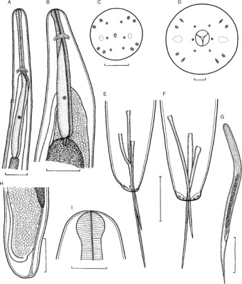
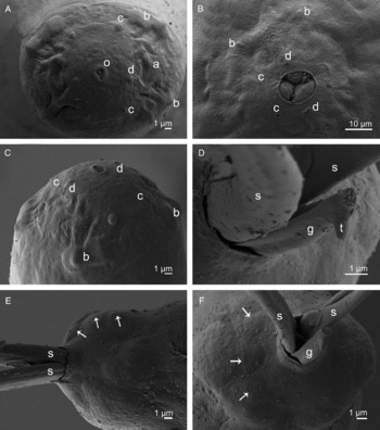
Description. Male (12 specimens; range given; measurements of holotype in parentheses). Body filiform, whitish before and after fixation, 2.79–3.48 (2.82) mm long, maximum width at middle 75–102 (93), somewhat tapering at both ends. Width of cephalic end 36–39 (36), of caudal end 24–36 (36). Cuticle smooth. Cephalic end rounded. Oral aperture triangular, very small, surrounded by 14 minute cephalic papillae arranged in two circles: external circle formed by 4 submedian pairs of papillae, internal circle by 4 submedian and 2 lateral papillae; amphids slightly outlined (figs 1C and 2A, C). Oesophagus 297–393 (378) long, forming 11–13% (13%) of body length, somewhat inflated at anterior end; posterior part of muscular oesophagus overlapped by well-developed oesophageal gland with large cell nucleus in middle (fig. 1A). Oesophageal nucleus and nerve ring 216–306 (285) and 156–180 (162), respectively, from anterior extremity. Excretory pore 201–240 (201) from anterior end. Posterior end of body somewhat narrowed, blunt, with poorly developed flat lateral and subdorsal lobes; one pair of preanal, one pair of adanal and one pair of postanal, very flat and hardly visible caudal papillae present (fig. 2E and F). Spicules slender, needle-like, with somewhat expanded proximal and sharply pointed distal ends (fig. 1E and F); right spicule usually slightly longer than left spicule, rarely both spicules equal in length. Length of right spicule 156–213 (183), of left spicule 144–174 (156); length ratio of spicules 1:1.00–1.39 (1:1.17); length of right spicule representing 5.01–6.73% (6.50%) of body length. Gubernaculum narrow, smooth, 69–84 (69) long, with approximately its proximal half somewhat dorsally bent; length of anterior bent part 33–45 (33), representing 48–54% (48%) of entire gubernaculum length; distal end of gubernaculum with distinct reflected dorsal barb (figs 1E and 2D). Length ratio of gubernaculum and right spicule 1:1.17–2.83 (1:1.17). Spicules and gubernaculum well sclerotized; spicules and posterior part of gubernaculum orange-coloured, anterior part of gubernaculum colourless.
Philometra floridensis sp. n. A, Anterior end of male, lateral view; B, anterior end of largest subgravid female, lateral view; C, D, cephalic end of male and gravid female, respectively, apical views; E, F, posterior end of male, lateral and ventral views; G, larva from uterus; H, posterior end of subgravid female, lateral view; I, cephalic end of subgravid female, lateral view. Scale bars: A, E, F, 50 μm; B, H, 300 μm; C, D, 10 μm; G, I, 100 μm.

Philometra floridensis sp. n., scanning elecron micrographs. A, Cephalic end of male, apical view; B, cephalic end of gravid female, apical view; C, cephalic end of male, sublateral view; D, gubernaculum (distal tip missing); E, F, caudal end of male, subventral and apical views (arrows indicate flat caudal papillae). Abbreviations: a, amphid; b, pair of submedian cephalic papillae of external circle; c, submedian cephalic papilla of internal circle; d, lateral cephalic papilla of internal circle; g, gubernaculum; o, oral aperture; s, spicule; t, dorsal tooth on gubernaculum.

Female (2 largest subgravid specimens; measurements of allotype in parentheses; measurements of additional body fragments of 1 gravid specimen in square brackets): Body filiform, that of fixed specimens brown (red in live specimens), with smooth cuticle; body length 890 (910) mm, maximum width 789 (789) [1360]; width of cephalic end 177 (163) [204], of posterior end 204 (163); maximum width/body length ratio 1:1128 (1:1153); posterior part of body narrower than anterior part. Cephalic end rounded, cephalic papillae very small, indistinct when viewed laterally. Oral aperture large, oval, surrounded by 4 pairs of submedian cephalic papillae of external circle and 6 single papillae (2 lateral and 4 submedian) of internal circle; each external pair consisting of 1 rounded and 1 elongated papilla when viewed apically (figs 1D and 2B). Pair of lateral amphids slightly outlined. Bottom of mouth formed by 3 lobes of oesophageal sectors. Oesophagus including anterior bulbous inflation 1.12 (1.32) [1.13] mm, representing 0.13% (0.14%) of body length; bulb moderately developed, 78 (87) [87] long and 90 (87) [90] wide. Oesophageal gland well developed (fig. 1B); large oesophageal gland nucleus 802 (925) from anterior extremity. Maximum width of oesophagus including oesophageal gland 122 (150) [136]. Nerve ring 245 (299) [218] from anterior end of body. Small ventriculus present. Oesophagus opening into intestine through distinct valve. Intestine dark brown, ending blindly, its posterior end narrow, attached by long ligament ventrally to body wall near caudal end. Vulva and anus absent. Ovaries long, reflected, situated near body ends. Uterus occupying most space of body, filled with numerous eggs 21–30 (27–36) in diameter [with larvae]. Larvae (n = 5) from uterus [423–525] long, maximum width [18–21], length of oesophagus [120–180, representing 28–35% of body length], of sharply pointed tail [78–120, representing 18–24%] (fig. 1G). Posterior end of body rounded, without caudal projections (fig. 1H and I).
Type host. Red drum, Sciaenops ocellatus (Sciaenidae, Perciformes, Osteichthyes) (range standard length: 602–881 mm).
Site of infection. Ovary.
Type locality. Gulf of Mexico, off Treasure Island, Florida, USA; 27°44.464′N, 82°50.263′W (collected 7 October 2008).
Prevalence and intensity. Male (n = 87) and female (n = 62) hosts were 0% and 67.7% infected, respectively. Because female nematodes were counted from only one gonad lobe, the exact infection intensity was not obtained; however, the number of female nematodes counted in a single lobe ranged from 1 to 7 (mean 4 nematodes per fish in 11 host specimens for which worms were counted).
Deposition of specimens. Holotype, allotype and paratypes in the Helminthological Collection of the Institute of Parasitology, Biology Centre of the Academy of Sciences of the Czech Republic, České Budějovice, Czech Republic (Cat. No. N-921); paratypes in the US National Parasite Collection, Beltsville, Maryland, USA (Cat. No. USNPC 101509); additional specimens from these collections are deposited in the Florida Fish and Wildlife Conservation Commission–Florida Wildlife Research Institute (FWC–FWRI), St. Petersburg, Florida, USA (FSBCI067025).
Etymology. The specific name of this nematode refers to Florida, where this parasite was recorded.
Discussion
Moravec et al. (Reference Moravec, de Buron, Baker and González-Solís2008a) reported a total of 23 gonad-infecting species of Philometra, two of which – P. neolateolabracis Rajyalakshmi, Hanumantha Rao & Shyamasundari, 1985 and P. nemipteri Luo, 2001 – were considered species inquirendae. Three additional species, Philometra cyanopodi Moravec & Justine, Reference Moravec and Justine2008; P. fasciati Moravec & Justine, Reference Moravec and Justine2008; and P. lethrini Moravec & Justine, Reference Moravec and Justine2008, have since been described from serranid and lethrinid fishes off New Caledonia (Moravec & Justine, Reference Moravec and Justine2008) and one, P. sawara Quiazon, Yoshinaga & Ogawa, 2008, from a scombrid fish off Japan (Quiazon et al., Reference Quiazon, Yoshinaga and Ogawa2008a). Quiazon et al. (Reference Quiazon, Yoshinaga and Ogawa2008a) also redescribed P. sciaenae Yamaguti, 1941, and P. nemipteri Luo, 2001, revalidating the latter species. Some new data on the female morphology of P. filiformis (Stossich, 1896) have recently been provided by Moravec et al. (Reference Moravec, Gaglio, Panebianco and Giannetto2008b).
Of all these gonad-infecting species, seven are known only by females, whereas conspecific males have not yet been described. These seven species can be distinguished from P. floridensis sp. n. mainly by the body length of gravid females, which do not exceed 48 cm, whereas subgravid females of the new species are up to about 90 cm (gravid females may be about 100 cm long). The new species can be also differentiated from the other seven by some morphological features (e.g. the absence of an anterior oesophageal inflation, presence of caudal projections, or the relative length of the oesophagus to the body length), different host families (Centrolophidae, Platycephalidae, Scombridae, Sebastidae, Serranidae, Sparidae) and different geographical regions (Mediterranean Sea or the Pacific Ocean adjacent to Japan or Peru) (see the key in Moravec et al., Reference Moravec, de Buron, Baker and González-Solís2008a).
From the remaining gonad-infecting Philometra spp. for which males are known, P. floridensis sp. n. can be easily distinguished by the presence of a well-developed reflected dorsal barb on the distal tip of the gubernaculum. A similar dorsal barb has been described only on the gubernaculum of P. carolinensis Moravec, de Buron et Roumillat, 2006, which is parasitic in the ovary of Cynoscion nebulosus (Cuvier) (Sciaenidae) in the estuaries of South Carolina, USA (Moravec et al., Reference Moravec, de Buron and Roumillat2006). The general morphology of both species is similar, their hosts belong to the same fish family, and they both occur in the northwestern Atlantic, so they are likely closely related. However, in contrast to P. floridensis sp. n., P. carolinensis has spicules equally long and measuring only 81–87 μm (vs. 144–213 μm), and the gravid females are only 12–16 cm (vs. about 100 cm) long. Furthermore, the oesophagus length represents 0.8–1.0% of the body length of gravid females in P. carolinensis, as opposed to 0.13–0.14% in subgravid females of P. floridensis sp. n., and larvae from the uterus are mostly shorter in P. carolinensis (399–462 vs. 423–525 μm long).
Philometrid nematodes exhibit a rather high degree of host – and site – specificity (Moravec, Reference Moravec2006). To date, several species of gonad-infecting species of Philometra have been reported from the family Sciaenidae: Philometra carolinensis (see above); P. lateolabracis (Yamaguti, 1935) from species of Johnius, Nibea, Otolithes, Pennahia and Protonibea worldwide; P. neolateolabracis from off India, and P. sciaenae off Japan, both from Pennahia argentata (Houttuyn); Philometra spp. were also reported from Argyrosomus regius (Asso) and Micropogonias undulatus (Linnaeus) from the eastern and western Atlantic, respectively, and from Otolithes ruber (Bloch & Schneider) from the Indian Ocean (Moravec, Reference Moravec2006; Moravec et al., Reference Moravec, de Buron, Baker and González-Solís2008a; Quiazon et al., Reference Quiazon, Yoshinaga and Ogawa2008a, Reference Quiazon, Yoshinaga and Ogawab). Quiazon et al. (Reference Quiazon, Yoshinaga and Ogawa2008b) redescribed P. lateolabracis from its type host, Lateolabrax japonicus (Cuvier) (Percichthyidae), in Japan and indicated that the previous records of P. lateolabracis from other host species were probably misidentifications. Because the host species is identical for both P. sciaenae and the poorly described P. neolateolabracis, the latter may well be a junior synonym of the former. Philometra sciaenae differs distinctly from the new species in the absence of a dorsal barb on the gubernaculum distal tip and by the presence of transverse lamella-like structures on the gubernaculum; its gravid females are much shorter, at most about 10 cm long (Moravec et al., Reference Moravec, Nagasawa and Ogawa1998; Quiazon et al., Reference Quiazon, Yoshinaga and Ogawa2008a). Consequently, P. floridensis sp. n. is the third valid gonad-infecting species of Philometra (in addition to P. carolinensis and P. sciaenae) reported from the perciform family Sciaenidae.
Acknowledgements
Our thanks are due to our colleagues in the Fisheries Independent Monitoring and Fish Biology programmes at the Florida Fish and Wildlife Conservation Commission, Fish and Wildlife Research Institute (FWRI), St. Petersburg, who first detected and provided infected ovaries. We also wish to thank the staff of the Laboratory of Electron Microscopy, Institute of Parasitology, Biology Centre of the Academy of Sciences of the Czech Republic (ASCR): České Budějovice for technical assistance, and Blanka Škoríková and Irena Husáková, Department of Helminthology at the same institute, for help with illustrations. This study was supported partly by grant 524/06/0170 from the Grant Agency of the Czech Republic and by the research projects of the Institute of Parasitology, ASCR (Z60220518 and LC522). Fish collection for this study was funded by grant NA05NMF4331078 to the Florida Fish and Wildlife Conservation Commission from the National Oceanic and Atmospheric Administration (NOAA), USA. Parasite collection was funded jointly by the US Fish and Wildlife Service Sport Fish Restoration Grant Program F-72 and CONACyT Mexico Sabbatical Grant 2007-81 886. We also thank our FWRI colleagues who assisted in specimen processing or who reviewed drafts of this manuscript. We particularly thank Dr Jan Landsberg, whose critical comments were a great help in improving this work. The views expressed herein are those of the authors and do not necessarily reflect the views of NOAA or any of its sub-agencies.