Introduction
Chronic maxillary sinusitis of dental origin is a common disease, and accounts for approximately 10–12 per cent of maxillary sinusitis cases.Reference Mehra and Murad1, Reference Maloney and Doku2
The microbial flora responsible for odontogenic sinusitis differ from those found in acute and chronic maxillary sinusitis, with a clear predominance of anaerobic bacteria in odontogenic sinusitis. This feature is important for the selection of appropriate antibiotic treatment.Reference Brook3
In contrast to rhinogenic sinusitis, the management of chronic maxillary sinusitis of dental origin requires treatment of the odontogenic source in addition to management of the sinus infection.Reference Sandler4
A common cause of chronic maxillary sinusitis of dental origin is a chronic oroantral fistula, which accounts for approximately 60 per cent of odontogenic sinusitis cases.Reference Lopatin, Sysolyatin, Sysolyatin and Melnikov5
Despite the widespread use of functional endoscopic sinus surgery (FESS) to treat chronic rhinosinusitis, external approaches and extensive exploration of the diseased sinus are still widely used in the treatment of chronic maxillary sinusitis of dental origin with oroantral fistula.Reference Al-Belasy6 These methods are traumatic and carry a greater risk of post-operative complication, compared with FESS.Reference Ikeda, Hirano, Oshima, Shimomura, Suzuki and Sunose7
Another important consideration concerns future bony reconstruction of the maxillary sinus, in view of the fact that chronic maxillary sinusitis of dental origin is more often present in the elderly population, who may require prosthetic rehabilitation once the disease is resolved.Reference Guven8, Reference Regev, Smith, Perrott and Pogrel9
In addition, after a Caldwell–Luc procedure the mucosa which grows to re-epithelialise the maxillary sinus lacks normal mucociliary physiology, thus affecting sinus clearance.Reference Costa, Emanuelli, Robiony, Zerman, Polini and Politi10
Particularly in cases of oroantral fistula, surgical intervention is quite often necessary to eradicate the disease.
We present our experience of managing four cases of chronic maxillary sinusitis of dental origin due to oroantral fistula, with a combination of primary oroantral fistula closure and FESS.
Materials and methods
Between January 2004 and August 2009, we encountered four cases of maxillary sinusitis due to oroantral fistula. Patients comprised three men aged 31, 37 and 39 years, variously, and one woman aged 43 years. All complained of facial pain, unilateral nasal discharge and purulent oral discharge, and had a history of upper tooth extraction two, four, five and eight months ago, variously.
Diagnosis was established by means of dental examination, nasal endoscopy, orthopanthomography and computed tomography scan. Mucosal thickening was apparent in the diseased maxillary sinuses. The fistula diameter was 6, 9, 11 and 13 mm, variously.
Patients were treated surgically under general anaesthesia. Initially, a wide middle meatal antrostomy was performed and the diseased sinus cleared using angled antrum forceps and a nasal shaver. Fistulae were debrided, and the buccal lamina of the alveolar bone was infractured to decrease their size. A synthetic, biodegradable, cyanoacrylate-based surgical glue (Glubran 2; GEM, Viareggio, Italy) with adhesive and haemostatic properties was used to seal the fistulae, before final closure using buccal advancement flaps and absorbable sutures.
Results
The post-operative follow-up period ranged from six months to three years. No patient showed any recurrence or other problems over this time.
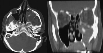
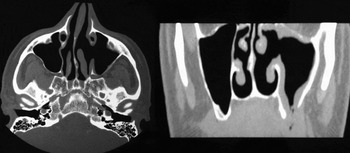
Figures 1 and 2 show pre-operative and two months post-operative imaging for one patient, a 39-year-old man.
Pre-operative, axial and coronal computed tomography head scans of a 39-year-old man with chronic rhinosinusitis and oroantral fistula. The left-sided, opacified lesion comprised foreign material used in a previous attempt to seal the fistula, which has intruded into the maxillary sinus.

Six month post-operative, axial and coronal computed tomography scans of the same patient. The fistula remains closed and the sinus appears free of disease.

Discussion
Oroantral fistulae can be responsible for the development of chronic maxillary sinusitis.
An oroantral fistula is defined as an osteomucosal communication between the oral cavity and either a sinus or the nasal cavity. They are generally iatrogenic and occur after dental procedures (e.g. extractions), intramaxillary cyst removal or external maxillary sinus surgery. They can also be caused by persistent apicodental infection, maxillary tumour necrosis, or surgical correction of a cleft palate or lip.Reference Brook11
Successful management depends largely on primary closure of the defect and concomitant medical therapy. Once a sinus communication has been diagnosed following dental surgery, the size of the defect must be assessed. Defects of 5 mm or less in diameter generally close spontaneously in compliant patients. The use of a resorbable barrier, such as absorbable gelatin sponge, has been suggested in order to cover and protect the defect during the initial stages of healing. In oroantral fistulae of 5 mm diameter or greater, primary closure is indicated. This can generally be accomplished using standard surgical techniques such as buccal advancement flaps, palatal island flaps, full- or split-thickness palatal pedicle flaps, gold foil, or buccal fat pad pedicle flaps.
In refractory cases, other surgical techniques are indicated, such as buccinator flaps, temporalis flaps and tongue flaps. However, it is very important that any surgical closure attempts be performed in a disease-free sinus environment. Performing surgery at an oroantral communication site in the presence of acute sinus infection will probably result in failure.Reference Mehra and Murad1, Reference Laskin12
The microbiology of acute and chronic maxillary sinusitis associated with odontogenic infection is quite unique, in that anaerobes predominate in both types of infection.Reference Brook11 Therefore, selection of appropriate antibiotic coverage is essential for resolution of the infection.
The diseased sinus itself also needs to be addressed, since persistent mucosal disease will probably lead to recurrence of the fistula. Traditionally, a Caldwell–Luc procedure has been performed, with extensive exploration of the diseased sinus and complete removal of the mucosal layer, together with creation of a lower nasal antrostomy for drainage. The disadvantages of this technique have been extensively discussed, and include higher complication rates, greater blood loss, longer operating time and longer hospitalisation for the patient.Reference Ikeda, Hirano, Oshima, Shimomura, Suzuki and Sunose7, Reference Narkio-Makela and Qvarnberg13
More recently, less invasive endoscopic techniques have been advocated for the treatment of chronic maxillary sinusitis of dental origin. These techniques entail endoscopic antrostomy and subsequent removal of irreversibly diseased tissue, polyps and foreign bodies through the antrostomy window.Reference Mehra and Murad1
• Despite widespread use of functional endoscopic surgery (FESS) to treat chronic rhinosinusitis, chronic maxillary sinusitis of dental origin with oroantral fistula is still widely treated using external approaches and extensive exploration of the diseased sinus
• These methods are traumatic and carry a greater risk of post-operative complication, compared with FESS
• This paper recommends the treatment of oroantral fistula accompanying maxillary sinusitis of dental origin using a combination of primary closure and FESS; this is a safe, reliable technique with a lower risk of complications
In the presented patients, we used an endoscopic technique in combination with primary fistula closure for the management of oroantral fistula, with satisfying short and long term results. Several authors have reported similar results using endoscopic techniques for the treatment of odontogenic sinus disease.Reference Kraut and Smith14 In addition to reducing the trauma of disease removal, FESS helps restore the normal patency of the natural ostium, and may facilitate quicker recovery of sinus clearance. This procedure is also safe, with less morbidity compared with the Caldwell–Luc procedure. Endoscopic middle meatal antrostomy rarely has complications, and appears to be effective without requiring significant additional operating time.Reference Ikeda, Hirano, Oshima, Shimomura, Suzuki and Sunose7, Reference Costa, Emanuelli, Robiony, Zerman, Polini and Politi10, Reference Narkio-Makela and Qvarnberg13
The previously published reports of the reliability and safety of this method, together with our own experience, support the treatment of oroantral fistulae with a combination of primary closure and FESS.