Introduction
The inner ear may be particularly sensitive to transient ischaemia because of its high-energy requirements and lack of adequate collateral blood supply. The blood supply to the labyrinth originates from the vertebrobasilar system. The main blood supply to the peripheral auditory system usually arises from the anterior inferior cerebellar artery. Therefore, the most commonly infarcted territory associated with sudden deafness is in the distribution of the anterior inferior cerebellar artery.Reference Lee, Whitman, Lim, Lee and Park1 Few previous reports have described sudden hearing loss associated with cerebellar infarction in the territory of the posterior inferior cerebellar artery.
We observed two cases of posterior inferior cerebellar artery infarction without the prominent cerebellar signs, particularly severe extremity ataxia and abnormal pattern nystagmus. The only presenting complaint was unilateral sudden hearing loss. There was some degree of hearing improvement 3 or 4 days after initial medical treatment.
Case reports
Case one
A 61-year-old man visited our out-patient clinic complaining of rotatory vertigo and sudden hearing impairment on his right side, which occurred just 1 day before his visit. When he visited the clinic, the spinning type vertigo had improved compared to the day before. He had no tinnitus, otalgia or otorrhoea. He had been under treatment for hypertension and atrial fibrillation for five years.
On physical examination, both eardrums were intact. The tuning fork test revealed lateralisation to the left side. The pure tone average of 0.5, 1, 2, and 3 kHz was 61 dB on the right side (Figure 1a). On auditory brainstem response testing, a normal waveform response of 70 dB was evoked on the right side. Neurological testing results for ataxic gait, Romberg's sign, dysmetria, and spontaneous and gaze-evoked nystagmus were all negative.
Pure tone audiograms of case one showing hearing loss on day of admission (a), and improvement 2 days after heparin and steroid treatment (b). ] = Bone conduction (masked) left ear; ×= air conduction (unmasked) left ear; [=bone conduction (masked) right ear; ○ = air conduction (unmasked) right ear; Δ = air conduction (masked) right ear.

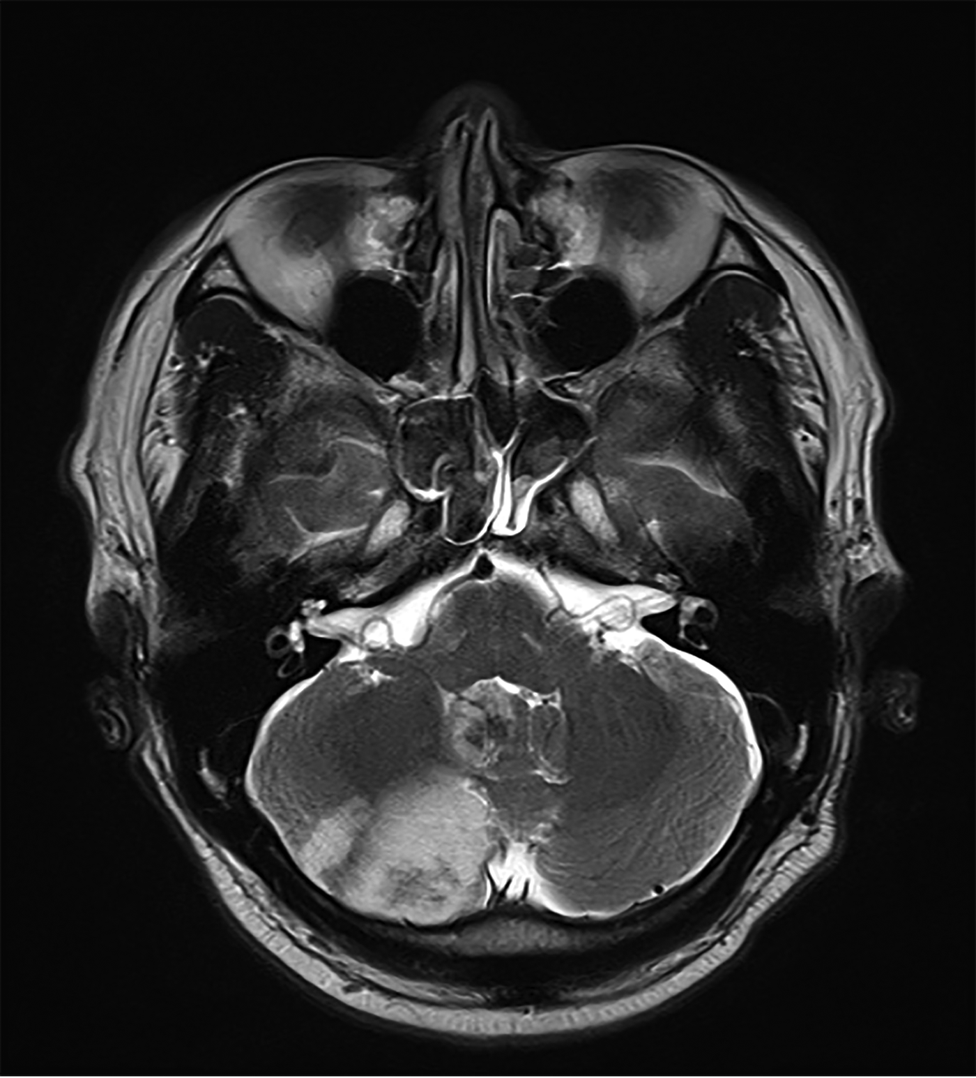
In light of the sudden hearing loss on the right side, the patient was hospitalised immediately and started on steroid treatment. He underwent brain magnetic resolution imaging (MRI) on the first day of admission. T2-weighted MRI demonstrated acute posterior inferior cerebellar artery infarction with haemorrhagic changes on the right side (Figure 2). He was transferred to the neurology department and mannitol and warfarin therapy was started. No other cerebellum-associated neurological signs or symptoms were detected during his period of admission.
Axial, T2-weighted magnetic resonance image of case one showing high-signal intensity in the territory of the right posterior inferior cerebellar artery, suggesting acute infarction.
Follow-up pure tone audiometry conducted on the 3rd day revealed improvement in hearing on the right side, with a response of 42 dB (Figure 1b). His speech discrimination score was 35 per cent.
The patient was discharged on the 10th day, with mild hearing loss on his right side and without any other neurological deficit.
Case two
A 69-year-old man visited our out-patient clinic with a 2-day history of hearing loss on the right side and rotatory vertigo which was improving. He had a 10-year history of right-sided tinnitus, but he had no subjective hearing impairment, otalgia or otorrhoea. He had suffered a transient ischaemic attack about 10 years earlier, and was under treatment for diabetes mellitus, hypertension and atrial fibrillation.
On physical examination, both eardrums were intact. The tuning fork test revealed lateralisation to the left side. A pure tone audiogram showed moderate sensorineural hearing loss of 77 dB on the right side (Figure 3a). The speech discrimination score was 35 per cent on his right side. A normal waveform response of 70 dB was detected on auditory brainstem response testing. No spontaneous or gaze-evoked nystagmus was noted, and no neurological signs or symptoms were detected.
Pure tone audiograms of case two showing hearing loss on day of admission (a), and improvement 8 days after heparin and mannitol treatment (b). ] = Bone conduction (masked) left ear; ×= air conduction (unmasked) left ear; [=bone conduction (masked) right ear; Δ = air conduction (masked) right ear; ○ = air conduction (unmasked) right ear.
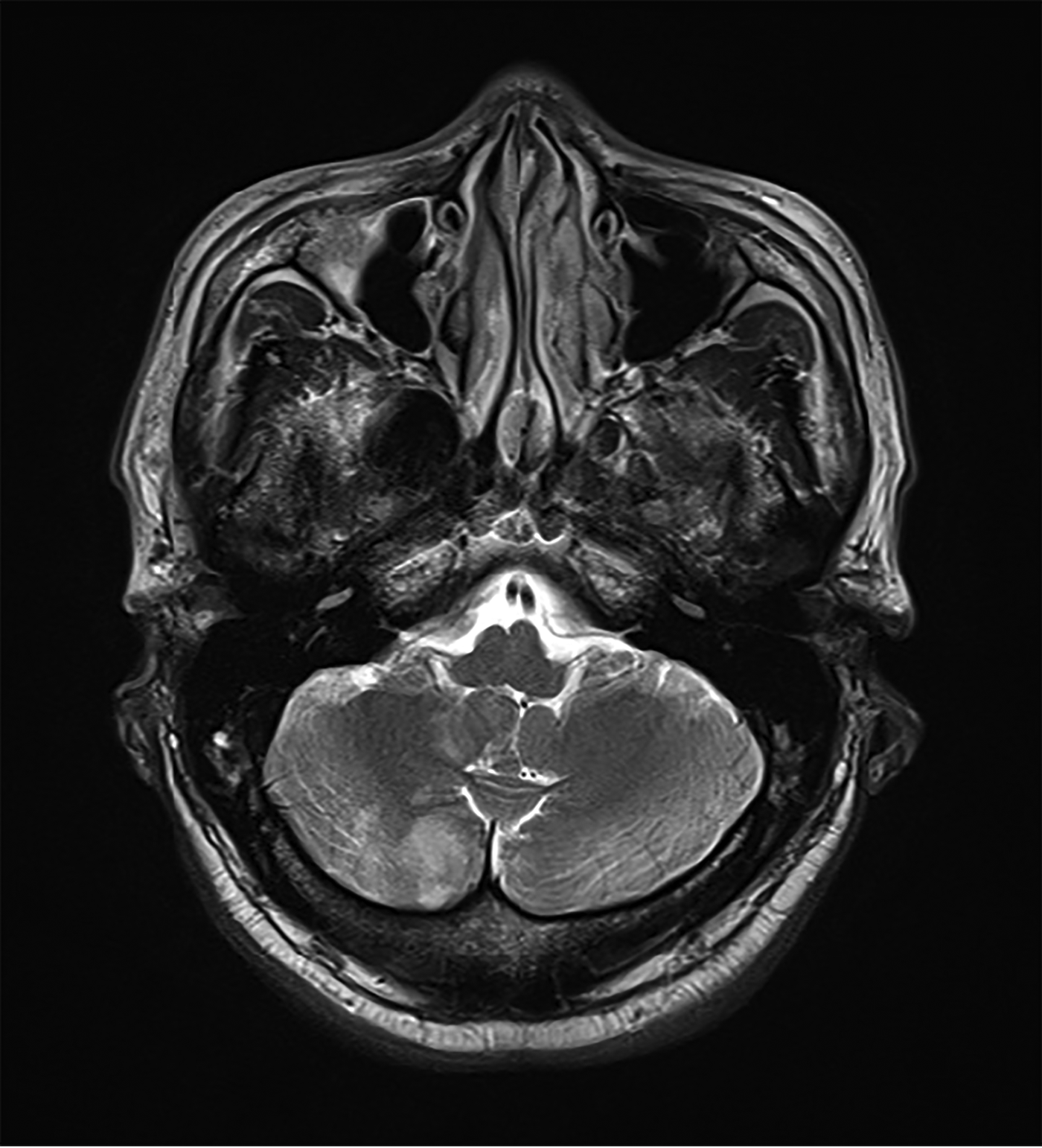
The patient was admitted immediately and medical therapy was commenced for sudden hearing loss. He underwent a brain MRI on the 3rd day after admission. T2-weighted axial imaging showed acute cerebellar infarction in the territory of the right posterior inferior cerebellar artery (Figure 4). Based on a diagnosis of acute posterior inferior cerebellar artery infarction, the patient started treatment with mannitol and heparin.
Axial, T2-weighted magnetic resonance image of case two showing increased signal intensity in the territory of the right posterior inferior cerebellar artery, suggesting acute infarction.
Follow-up pure tone audiometry conducted on the 4th day after admission showed slightly improved air conduction. Subsequent pure tone audiometry performed on the 8th day showed further improvement on the right side, with a response of 53 dB (Figure 3b). His speech discrimination score on day 8 was 40 per cent. No other neurological symptoms or signs were noted during his admission period.
The patient was discharged following 10 days of treatment.
Discussion
Two common aetiologies for sudden deafness are circulatory disturbance, most often vertebrobasilar ischaemia, and inflammation, most often viral.Reference Haberkamp and Tanyeri2 The blood supply to the auditory system including the labyrinth originates from the vertebrobasilar system, so cochlear audiometric features do not preclude a vascular cause in sudden deafness.Reference Lee and Baloh3 The inner ear is particularly vulnerable to low flow within the vertebrobasilar system as it has a complete absence of collateral circulation and very high-energy metabolism.Reference Lee, Whitman, Lim, Lee and Park1
It is important for otolaryngologists to recognise and detect sudden deafness of vascular cause because the management and prognosis generally differ from that of hearing loss associated with viral infection. Furthermore, delayed management may result in a permanent severe neurological deficit and threaten the patient's life. Therefore, early diagnosis should be made based on prompt and exact patient history, and a physical examination followed by an appropriate imaging study.
In 1962, Williams and Wilson reported that only 1 (1.5 per cent) of 65 patients with basilar insufficiency had hearing impairment.Reference Williams and Wilson4 In 1967, Fisher noted that 10 (8.9 per cent) of 112 patients with vertebrobasilar insufficiency complained of acute hearing loss.Reference Fisher5 In 1989, Grad and Baloh reported that 6 of 42 patients (14.3 per cent) had acute hearing impairment as a symptom of vertebrobasilar insufficiency.Reference Grad and Baloh6
The most commonly infarcted territory on brain MRI associated with sudden deafness is in the distribution of the anterior inferior cerebellar artery.Reference Lee, Whitman, Lim, Lee and Park1 In fact, it is reported that the main blood supply to the peripheral auditory system usually arises from the anterior inferior cerebellar artery.Reference Hausler and Levine7–Reference Oas and Baloh9 Cerebellar infarction in the territory of the posterior inferior cerebellar artery can rarely cause acute hearing loss because the internal auditory canal artery sometimes originates from the posterior inferior cerebellar artery or directly from the basilar artery.Reference Sunderland10 Mazzoni described a posterior inferior cerebellar artery origin of the labyrinthine artery in 3 out of 100 temporal bone dissections.Reference Mazzoni11 In 2008, Lee reported that 7 of 685 patients (1.0 per cent) with vertebrobasilar ischaemic stroke had acute unilateral hearing loss associated with non-anterior inferior cerebellar artery territory vertebrobasilar ischaemic stroke, in which the posterior inferior cerebellar artery territory was the most commonly affected site (5 out of 7; 71.4 per cent).Reference Lee12 Based on these reports, the posterior inferior cerebellar artery could be the region related to sudden hearing loss among the non-anterior inferior cerebellar artery infarctions.
Typical cases of posterior inferior cerebellar artery infarction show severe vertigo, vomiting, dysarthria, postural instability, limb ataxia and gait disturbance.Reference Lee13 The major signs of cerebellar infarction are truncal ataxia and nystagmus. In our two patients, MRI showed that the extent of infarction was sufficient to present the typical neurological signs of posterior inferior cerebellar artery infarction. Nevertheless, the patients’ only complaining symptom was unilateral sudden hearing loss. Before their first visit, both patients complained of whirling vertigo. As in the natural course of idiopathic sudden hearing loss, the dizzy symptom disappeared after a few days, and no nystagmus was detected during the patients’ period of admission.
Isolated infarction in the territory of the posterior inferior cerebellar artery usually results from emboli originating from the heart or great vessels.Reference Amarenco, Lévy, Cohen, Touboul, Roullet and Bousser14 Both of the patients described here suffered from atrial fibrillation, and we assume the cause of the emboli was related to irregular heartbeats.
Sudden hearing loss of cerebrovascular origin is treated with an antiplatelet agent such as aspirin, or with anticoagulants such as heparin or warfarin.Reference Rubenstein, Norman, Schindler and Kaseff15 Our patients started antiplatelet treatment just after acute cerebellar infarction was detected by MRI. Gradual reperfusion was reflected in the partial recovery of hearing detected by follow-up audiogram a few days later.
• Magnetic resonance imaging should be considered in any patient with cardiovascular risk factors who presents with sudden sensorineural hearing loss, to rule out vascular insult
• Posterior inferior cerebellar artery infarction could be one differential diagnosis of sudden deafness
Otologists have to be aware of sudden deafness of vascular cause that presents without any neurological symptoms or signs, especially in patients in a high-risk group for cerebrovascular diseases such as diabetes mellitus, hypertension, atrial fibrillation and dyslipidaemia. Prompt and accurate diagnosis should be made via patient history, physical examination and imaging such as brain MRI. Once acute cerebellar infarction is detected as a cause of sudden deafness, proper management should be started as soon as possible to increase the likelihood of prompt recovery without complications.
In conclusion, we observed two patients with a very rare clinical manifestation of posterior inferior cerebellar artery infarction who presented with acute unilateral hearing loss only. Otologists should consider conducting MRI to rule out vascular insult in any patient with cardiovascular risk factors who presents with sudden hearing impairment. Posterior inferior cerebellar artery infarction could be one differential diagnosis of sudden deafness.
Acknowledgement
This paper was supported by research funds awarded by Chonbuk National University in 2013.