Introduction
The capacity of the human brain for functional and structural reorganization throughout life is now well recognized (Kheirbek and Hen, Reference Kheirbek and Hen2013), contrasting with a long-held view that this is not possible beyond critical periods during development. Earlier experimental studies were conducted in animals or had to rely on post-mortem analysis. More recently the developments in the field of neuroimaging enabled non-invasive investigation of the living human brain, while learning of new skills or novel experience were used as paradigms for investigating different aspects of brain plasticity.
We aimed to review the strongest neuroimaging evidence for experience-dependent plasticity in adult humans and therefore focused on longitudinal structural studies only. Longitudinal studies not only provide causal evidence for learning-induced plasticity, but also have the potential of uncovering dynamic properties of experience-dependent changes and allow detection of subtle changes that cannot be identified in cross-sectional studies because of anatomical differences between brains. On the other hand, changes in brain structure in result of learning can be seen as more robust evidence for brain plasticity compared to functional changes and it is likely that they are the substrate of lasting intervention effects.
Structural scans are also better suited for studying small changes. A number of studies have quantified the test-retest reliability of MRI measures of cortical thickness, with absolute thickness differences averaging 0.03 mm (Han et al., Reference Han2006) and ≤0.05 mm (Wang et al., Reference Wang2008). Wonderlick et al. (Reference Wonderlick2009) found that surface-based measures of cortical thickness, white matter volume, and gray matter volume were particularly reproducible, with all computed test-retest ICCs falling above 0.95. In a recent multi-centric study, Jovicich et al. (Reference Jovicich2013) evaluated reproducibility as absolute changes relative to the mean and found that with longitudinal segmentation analysis all structures had absolute volume reproducibility errors in the range 1.8–2.2% (average across sites), with the exception of smaller structures such as the amygdala that yielded higher errors. Similarly, studies evaluating the test-retest variability of measures derived from DTI have generally reported very good reproducibility, with coefficient of variation typically below 6.5% (Ciccarelli et al., Reference Ciccarelli2003; Heiervang et al., Reference Heiervang, Behrens, Mackay, Robson and Johansen-Berg2006) and even below 2% (Pfefferbaum et al., Reference Pfefferbaum, Adalsteinsson and Sullivan2003; Bisdas et al., Reference Bisdas, Bohning, Besenski, Nicholas and Rumboldt2008; Danielian et al., Reference Danielian, Iwata, Thomasson and Floeter2010; Vollmar et al., Reference Vollmar2010; Wang et al., Reference Wang, Abdi, Bakhadirov, Diaz-Arrastia and Devous2012). The interval between scans in different studies ranges from minutes (Wang et al., Reference Wang2008) to one year (Danielian et al., Reference Danielian, Iwata, Thomasson and Floeter2010).
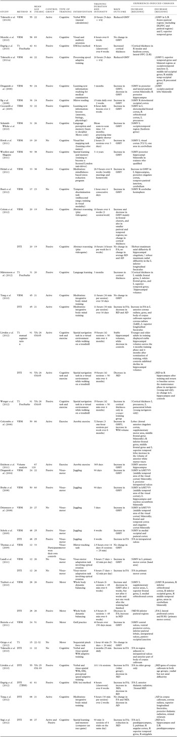
Over the last few years the number of plasticity studies is continuously growing, reflecting not only the greater sophistication of imaging techniques, but also the importance of research in this area. There are a number of recent reviews, each with focus on different aspects of the subject (Fields, Reference Fields2011; May, Reference May2011; Johansen-Berg, Reference Johansen-Berg2012; Zatorre et al., Reference Zatorre, Fields and Johansen-Berg2012; Lövdén et al., Reference Lövdén, Wenger, Martensson, Lindenberger and Backman2013; Park and Bischof, Reference Park and Bischof2013; Thomas and Baker, Reference Thomas and Baker2013). We hope to extend them by including more studies and discussing the evidence for plasticity in gray as well as white matter. We also focus on the various moderators that impact on learning and can affect the results, concluding by directions for future research. We searched Embase, PsychINFO, and PubMed from 1990 to June 2013 for the terms [learning OR training] AND [gray matter OR white matter OR brain] AND [changes OR plasticity]. In addition, we conducted a free-text search for “structural neuroplasticity” AND “MRI.” Two hundred eighty-nine papers were identified, and following screening of titles and abstracts, we reviewed the suitable full text papers. Due to the varied set of studies, the initial search retrieved a small number of relevant publications and we identified additional studies by hand-searching reference lists. In total, we identified 33 studies in healthy volunteers (mean age over 18 years) that employed a longitudinal design (participants were scanned before and after training) and used MRI-based techniques to measure morphological changes. Their baseline characteristics are summarized in Table 1.
Baseline characteristics of studies

Notes: YA = younger adults; OA = older adults; WM = working memory; EM = episodic memory; GMV = gray matter volume; R = right; L = left; DLPFC = dorsolateral prefrontal cortex; OFC = occipito-frontal cortex; FA = fractional anisotropy; MD = mean diffusivity; AD = axial diffusivity; RD = radial diffusivity.
Neuroplasticity induced by learning in younger adults
Gray matter changes
In their seminal study, Maguire et al. (Reference Maguire2000) demonstrated that experience shapes the human brain. They found that London taxi drivers with their detailed knowledge of London's complex street plan had enlarged posterior hippocampi. The size of the hippocampi correlated with the number of years spent driving taxi, suggesting that the observed difference was indeed a result of experience, rather than a pre-existing brain characteristic that predisposed people to become taxi drivers.
The first longitudinal studies in this area investigated visuo-motor learning. Draganski et al. (Reference Draganski, Gaser, Busch, Schuierer, Bogdahn and May2004) reported that learning to juggle over a three-month period is associated with gray matter increases in the visual motion areas bilaterally. The increase persisted three months later, although it was reduced compared to that seen soon after learning. Driemeyer et al. (Reference Driemeyer, Boyke, Gaser, Buchel and May2008) used the same design to investigate the temporal aspects of the changes and found that they can be detected after as little as a week of training. They also showed that neither performance nor exercise alone could explain these changes and concluded that to induce structural changes learning of a new task itself is more important than continuous training of an already-learned task. A different research group attempted to replicate the earlier juggling studies. After six weeks of training in juggling, Scholz et al. (Reference Scholz, Klein, Behrens and Johansen-Berg2009) reported an increase in GMV as well, but in different brain regions, namely medial occipital and parietal cortex; however, the increase was sustained after four weeks without practice. Other motor training regimes shown to increase GMV in specific brain regions include 40 hours of golf practice over 150 days (Bezzola et al., Reference Bezzola, Merillat, Gaser and Jancke2011) and five hours over seven days of practicing a visuo-motor task involving optical rotation (Landi et al., Reference Landi, Baguear and Della-Maggiore2011).
Several studies have extended the findings in procedural learning by investigating structural changes related to cognitive interventions. Gains in neural volume relative to a control group were demonstrated by Woollett and Maguire (Reference Woollett and Maguire2011) who focused on spatial learning and memory. They imaged trainee taxi drivers at the beginning and at the end of their training (after 3–4 years) and compared the results to controls. Increased GMV in posterior hippocampi was observed only in trainees who qualified as taxi drivers, but not in trainees who failed to qualify or controls. Kwok et al. (Reference Kwok2011) reported an increase in GMV in the left visual cortex after two hours of learning color names, while Bueti et al. (Reference Bueti, Lasaponara, Cercignani and Macaluso2012) found an increase in GMV in the right cerebellar hemisphere after one hour of temporal discrimination training. The last two studies did not use any control group, which makes interpretation of the results more difficult.
Further, there is evidence that learning of more abstract information results in structural alterations as well. Two studies investigated this possibility in medical students: after three months of intensive studying for an exam (Draganski et al., Reference Draganski2006) and after two weeks of eight hours a day classes (Ceccarelli et al., Reference Ceccarelli, Rocca, Pagani, Falini, Comi and Filippi2009). After the study phase, both studies reported increases in GMV in several regions, including posterior and inferior parietal cortices bilaterally and the posterior hippocampus (Draganski et al., Reference Draganski2006), the left dorso-medial frontal cortex, right orbitofrontal cortex, as well as the left precuneus (Ceccarelli et al., Reference Ceccarelli, Rocca, Pagani, Falini, Comi and Filippi2009). Draganski et al. (Reference Draganski2006) observed differential temporal effects of learning on brain morphology – a third scan performed three months later revealed that the structural changes in posterior parietal cortices were sustained while the increase in GMV in the posterior hippocampus became even more pronounced over time. Although all these cortical regions are associated with reasoning and declarative memory, it is not clear why different regions were found to increase in volume in the two studies. Methodological differences such as the differences in time points of analysis (15 days vs. 3 months) or the different methods used to assess change in volume (tensor-based morphometry vs. VBM) could account for the discrepancies. Consistent with the above two studies, three months of language learning also resulted in increased cortical thickness in frontal and temporal regions in the left hemisphere, as well as an increase in hippocampal volumes (Mårtensson et al., Reference Mårtensson2012). Studies on learning abstract information then suggest that the morphometric brain changes reported in plasticity studies are not due only to increased automatization of the underlying processes, which is a possibility in skill learning.
Negative studies
In contrast to these positive results, there are negative studies that did not observe any change in gray matter measures after training. For instance no change in GMV was found after 11 hours of integrative body–mind training (Tang et al., Reference Tang, Lu, Geng, Stein, Yang and Posner2010), as well as following one hour 40 minutes of motor training on a sequential pinch force task (Gryga et al., Reference Gryga2012). Two other negative studies however observed alterations in function. After two weeks training on a motor adaptation task, there were fMRI changes in brain activity but not structural changes (Thomas et al., Reference Thomas, Marrett, Saad, Ruff, Martin and Bandettini2009). Further, Mozolic et al. (Reference Mozolic, Hayasaka and Laurienti2010) found that although a training program that reduces cross-modal distraction in older adults resulted in greater increases in CBF than a control program, it did not induce significant change in GM volume. In both studies the functional changes were associated with improved performance, suggesting that they may precede structural changes and are therefore more sensitive marker for experience-dependent plasticity; however, methodological limitations cannot be excluded.
One possible explanation of these negative results is related to the type of interventions. For instance, Fields (Reference Fields2013) argued that the task used in the study by Thomas et al. (Reference Thomas, Marrett, Saad, Ruff, Martin and Bandettini2009), i.e. learning to operate a joystick when the direction of the controls is reversed, was less challenging than many of the tasks used in studies reporting positive findings, and therefore improving on this task might not require structural remodeling of the brain. Also the task was more ecologically valid, making it likely that the participants had encountered similar tasks in their daily life. Second, these negative studies have relatively short duration and/or intensity of the interventions, ranging from one hour 40 minutes over five days (Gryga et al., Reference Gryga2012) to 11 hours (30 minutes per day over 30 days; Tang et al., Reference Tang, Lu, Geng, Stein, Yang and Posner2010). However, a study of mirror reading involved two weeks of 15 minutes a day practice only, and still the authors reported an increase in the gray matter volume (Ilg et al., Reference Ilg2008). It is possible that the duration and intensity of training required to induce structural changes in the brain depends on the task and respectively the brain region involved. Finally, methodological issues such as short follow-up or lack of statistical power to detect changes in GM volume can account for the results.
To complicate interpretation further, two studies conducted by the same research group and using the same duration and intensity of training found reductions in GMV. The training period comprised of five days involving four hours practice each day. The first study used a mental calculation task to train working memory and found reductions in GMV in the bilateral DLPFC, right inferior parietal lobule, left paracentral lobule, and left superior temporal gyrus (Takeuchi et al., Reference Takeuchi2011b), while the second study found reductions in GMV in the left superior temporal gyrus and the occipito-temporal junction after training processing speed (Takeuchi et al., Reference Takeuchi2011a). The authors speculated that usage-dependent selective elimination of synapses that helps to sculpt neural circuitry is one possible mechanism explaining the results. It has also been suggested that increases in white matter density could account for the regional loss in GMV (Golestani et al., Reference Golestani, Paus and Zatorre2002; Draganski et al., Reference Draganski2006), but this potential underlying mechanism was not tested in these studies.
White matter changes induced by learning
Structural reorganization following training is not limited to gray matter. Cross-sectional studies using DTI (diffusion tensor imaging) showed that compared to non-pianists, pianists had higher white matter integrity in the corticospinal tract and corpus callosum (Bengtsson et al., Reference Bengtsson, Nagy, Skare, Forsman, Forssberg and Ullen2005). They were followed by the first longitudinal study in this area, which showed increased fractional anisotropy (FA) in the white matter underlying the intraparietal sulcus as early as six weeks after training in juggling (Scholz et al., Reference Scholz, Klein, Behrens and Johansen-Berg2009). In line with these results, Landi et al. (Reference Landi, Baguear and Della-Maggiore2011) observed increased FA underneath the hand area of the contralateral primary motor cortex following daily training on a complex visuo-motor rotation task for one week. The reported WM changes in both studies (Scholz et al., Reference Scholz, Klein, Behrens and Johansen-Berg2009; Landi et al., Reference Landi, Baguear and Della-Maggiore2011) were in regions adjacent to increases in GM volume. However, in the study of Scholz et al. (Reference Scholz, Klein, Behrens and Johansen-Berg2009) there was no correlation between the magnitude of gray-matter and white-matter changes across participants.
Several studies investigated the effects of cognitive interventions on white matter integrity. In young adults, two months of working memory training was associated with increased white matter integrity in regions adjacent to the intraparietal sulcus and the anterior part of corpus callosum, both of which are important in WM (Takeuchi et al., Reference Takeuchi2010). There is evidence that visual and verbal WM have different neural correlates, with a region along the inferior parietal sulcus showing greater response during visual stimuli. The task in this study was in a visual modality and indeed much greater association was found between FA change adjacent to inferior parietal sulcus and training compared to the association between the FA change in corpus callosum and training, suggesting that the observed effects were training-specific. Meditation practice (11 hours over four weeks) has also been found to result in increased FA in corpus callosum and corona radiata, which communicate with regions involved in attentional control (anterior cingulate), accompanied by decrease in axial and radial diffusivity (Tang et al., Reference Tang, Lu, Geng, Stein, Yang and Posner2010). No change in GMV was found, suggesting independent mechanisms or different time-course of gray and white matter plasticity. More recent study by the same group (Tang et al., Reference Tang, Lu, Fan, Yang and Posner2012) using the same training paradigm except that the intervention lasted for only two weeks (5 hours) found reduced axial diffusivity, but no change in radial diffusivity or FA, demonstrating a dynamic pattern of white matter change over time.
Negative: Contrary to these results, Taubert et al. (Reference Taubert2010) observed a decrease in FA following balance training over six weeks. They proposed that methodological issues such as the relatively short duration of the intervention (one training session lasting 45 minutes each week) or the introduction of a novel step in FA analysis with adjustment for the hypothesized and detected volume changes may account for the discrepancy between previous findings of learning-dependent FA increases and their results. Another study reported no change in FA and increase in both axial diffusivity (right hippocampal cingulum) and radial diffusivity (left inferior longitudinal fasciculus) after four weeks of videogame practice (Colom et al., Reference Colom2012). These results are difficult to interpret since increase in axial diffusivity has been associated with alterations in axonal coherence whereas increase in radial diffusivity has been related to demyelination. Changes in anisotropy indices in the opposite direction may result from axonal reorganization or rewiring of crossing fibers, but their integrity was not investigated in these studies (Jbabdi et al., Reference Jbabdi, Behrens and Smith2010).
Studies in older participants
The capacity of the older brain to change its structure in response to environmental demands is a question with wide implications in terms of devising interventions to slow the age-related decline in cognitive functions. Several studies now suggest that the aged brain exhibits such plasticity. Boyke et al. (Reference Boyke, Driemeyer, Gaser, Buchel and May2008) trained older adults to juggle for 90 days, using the same paradigm as in the other juggling studies (Draganski et al., Reference Draganski, Gaser, Busch, Schuierer, Bogdahn and May2004; Driemeyer et al., Reference Driemeyer, Boyke, Gaser, Buchel and May2008). Only 23% of the trained older adults (compared to 100% of younger group) were able to master the juggling skill, but those who did showed a significant increase in gray matter volume in the middle temporal region, which is associated with visual motion processing; the increase reversed after the training had ceased, suggesting that to maintain gains the training should continue. The training-related changes were described as “slightly smaller” than those seen in young adults (Draganski et al., Reference Draganski, Gaser, Busch, Schuierer, Bogdahn and May2004), but whether they were significant was not reported. Further, in older adults changes in the hippocampus and nucleus accumbens were observed; these two regions had not shown changes in young adults, implying that different regions may be involved in the same task depending on age. In addition to juggling, 40 hours of playing golf as a leisure activity have been found to increase GM in task-relevant regions including sensorimotor regions and areas in the dorsal stream in a group of 40–60-year-olds (Bezzola et al., Reference Bezzola, Merillat, Gaser and Jancke2011).
Further support for preserved structural plasticity of the aged brain has come from studies investigating the effects of aerobic exercise. Colcombe et al. (Reference Colcombe2006) reported increases in gray matter volume in inferior frontal gyrus, anterior cingulate, and superior temporal gyrus in older adults assigned to a six-month cardiovascular training program. The fact that exercise has an effect on the prefrontal cortex, which is very sensitive to age-related changes, might have important clinical implications. In a more recent study, Erickson et al. (Reference Erickson2011) found significant effects of aerobic exercise on anterior, but not posterior hippocampal volume in elderly participants; these effects were evident after six months, and increased linearly over time. Both studies did not find any effect in active control groups performing stretching exercise suggesting that only training that improves cardiovascular fitness has an impact on the brain.
Finally, morphological brain changes in older people have also been demonstrated in response to cognitive interventions. For instance both younger and older participants who played a spatial navigational game while walking on a treadmill exhibited stability of hippocampal volume over the four-month training period, whereas control participants declined. Importantly, the increase in structural hippocampal integrity was maintained for four months after the training had stopped (Lövdén et al., Reference Lövdén2012). This study highlights that as the natural course of aging is one of decline, interventions in the elderly can be considered effective not only if they result in improvement, but also if they prevent deterioration. Wenger et al. (Reference Wenger2012) did further analysis on the data from this study and found an increase in cortical thickness in the left precuneus and left paracentral lobule but in the younger training group only. Although these results suggest that plasticity is region-specific, another study provides evidence that experience shapes cortical structures in older adults as well. Engvig et al. (Reference Engvig2010) demonstrated that eight weeks of intensive memory training resulted in increased cortical thickness in the right fusiform and lateral orbitofrontal cortex, which correlated positively with improvement in source memory performance.
The same sample and training paradigm were later used to investigate learning-induced white matter plasticity (Engvig et al., Reference Engvig2012). Following training no change in FA in frontal areas was observed in the practice group, while a significant decrease in the untrained elderly was found, suggesting that training might protect against age-related changes. Further analyses showed less increase in radial diffusivity in the trained elderly compared with controls, while changes in axial diffusivity did not differ significantly between the two groups. Consistent with this, in the spatial navigational game study, Lövdén et al. (Reference Lövdén2012) also found changes in radial diffusivity but not in axial diffusivity – a pattern that has been linked to myelination (Song et al., Reference Song2005; Alexander et al., Reference Alexander, Lee, Lazar and Field2007). The possibility that experience may induce an increase in myelination is supported by animal studies that found that neuronal activity promotes myelination (Ishibashi et al., Reference Ishibashi2006; Engvig et al., Reference Engvig2012). With regard to mean diffusivity, a significant increase in frontal mean diffusivity, with larger effects in anterior compared with posterior regions, was demonstrated (combined with increased FA) by Engvig et al. (Reference Engvig2012), while Lövdén et al. (Reference Lövdén2012) found decreased mean diffusivity, interpreted as increased “hippocampal barrier density,” in right hippocampus after training.
Finally, Lövdén et al. (Reference Lövdén2010) compared younger with older adults and reported that an intensive cognitive training program (101 one-hour sessions over 180 days) on tests of working memory, perceptual speed, and episodic memory, led to an increase in FA and decrease in mean diffusivity, in the anterior corpus callosum. Importantly, the magnitude of the training-related changes in the WM was as great for older adults as for younger adults. Since the white matter tracts in the anterior part of corpus callosum show more pronounced age-related decline compared with more posterior regions (Burzynska et al., Reference Burzynska2010), and this difference may account for age-related effects on cognitive performance (Madden et al., Reference Madden2009), this apparent preserved plasticity has important implications for aging research.
In summary, there is evidence that training can induce structural plasticity in older adults. However, all studies included healthy young–old (65–75-year-olds), so whether such plasticity is present in very old age is not clear. Whether the aging brain has reduced capacity for such changes relative to a young adult brain remains unknown, because there are not many studies that directly compare younger with older people using the same paradigm. Age-comparative studies are needed because they will increase our understanding of the different compensatory processes in the elderly brain, which may involve recruitment of additional structures to perform the same task. Consistent with this, fMRI studies showed increased frontal activation in older compared with younger participants, suggesting that frontal networks are activated to compensate for deteriorating brain structure. This increase or shift in activation is functionally relevant because it is associated with better cognitive performance across multiple cognitive tasks (Park and Reuter-Lorenz, Reference Park and Reuter-Lorenz2009). The capacity of the brain to compensate for age-related changes is an important determinant of resilience and respectively successful aging; therefore, this is a crucial question for future research.
Relationship between structure and function
Studies that combine structural with functional data are of particular interest, because they provide opportunity to investigate whether GM increase in a specific brain area overlaps with functional changes in the same area, or alternatively different areas in the same network. Even more importantly, changes in task processing with practice may result in different patterns of brain activity (Kelly and Garavan, Reference Kelly and Garavan2005), which in turn may have an impact on the time-course of learning-induced structural brain changes, highlighting the importance of considering structural and functional changes simultaneously.
Ilg et al. (Reference Ilg2008) combined functional MRI and VBM to investigate the effects of a two-week training period of mirror reading. Practice-related GMV increase in the right occipital cortex was found, and importantly, this increase in GMV overlapped with the peak of the activation increase as a function of training, suggesting at least some consistency between the changes in task-specific activation and structural changes. A more recent study on volunteers who learnt to decipher Morse code found increased gray matter density in the left occipito-temporal region together with an increase in task-specific neural activation in brain regions thought to be involved in language perception and memory. The changes in structure and function were broadly in the same region, but not in the same location (Schmidt-Wilcke et al., Reference Schmidt-Wilcke, Rosengarth, Luerding, Bogdahn and Greenlee2010). Schmidt-Wilcke et al. (Reference Schmidt-Wilcke, Rosengarth, Luerding, Bogdahn and Greenlee2010) speculated that the morphological changes were induced by an activity such as writing down words, which was not required in the scanner, but was an essential part in the learning process. Other studies did not find any structural changes although training resulted in functional alterations. They have been discussed above (Thomas et al., Reference Thomas, Marrett, Saad, Ruff, Martin and Bandettini2009; Mozolic et al., Reference Mozolic, Hayasaka and Laurienti2010; Takeuchi et al., Reference Takeuchi2011a). Taken together, these results suggest that structural and functional changes do not always follow the same time course after training, nor do they occur in the same brain region. Although it is possible that changes in brain structure and function reflect different aspects of neuroplasticity, it is of note that interpreting fMRI data has important limitations, including the fact that the bold signal reflects neuronal mass activity and it is unable to differentiate between excitatory and inhibitory activity.
Time course and maintenance of experience-dependent changes
The exact time course of experience-dependent morphological changes remains unknown. We do not know how long an intervention has to last to yield structural changes, nor how long such changes last. These questions have been extensively discussed by Lövdén et al. (Reference Lövdén, Wenger, Martensson, Lindenberger and Backman2013). In summary, the evidence is mixed, with some studies demonstrating experience-dependent plasticity in humans after a few months of intervention (Draganski et al., Reference Draganski, Gaser, Busch, Schuierer, Bogdahn and May2004; Engvig et al., Reference Engvig2010; Takeuchi et al., Reference Takeuchi2010; Mårtensson et al., Reference Mårtensson2012), with others finding changes much faster than this (Driemeyer et al., Reference Driemeyer, Boyke, Gaser, Buchel and May2008; Kwok et al., Reference Kwok2011; Landi et al., Reference Landi, Baguear and Della-Maggiore2011; Bueti et al., Reference Bueti, Lasaponara, Cercignani and Macaluso2012). Most of the studies that reported fast changes lack a control group. Sagi et al. (Reference Sagi, Tavor, Hofstetter, Tzur-Moryosef, Blumenfeld-Katzir and Assaf2012) found significant microstructural changes after only one day of 90 minutes of practice on a spatial learning and memory task. They observed increased FA and reduced mean diffusivity in structures of the limbic system including the hippocampus and parahippocampus. Both passive and active control groups were used. The active group showed some effect in the parahippocampus, but that was significantly smaller than the effect for the experimental group. The results were confirmed in a supporting rat study.
The maintenance of training-induced structural brain changes is another question without a clear cut answer. Following termination of training, the earlier juggling studies found that the gray matter changes reversed after three months (Draganski et al., Reference Draganski, Gaser, Busch, Schuierer, Bogdahn and May2004; Boyke et al., Reference Boyke, Driemeyer, Gaser, Buchel and May2008) or two months (Driemeyer et al., Reference Driemeyer, Boyke, Gaser, Buchel and May2008). Contrary to this, Scholz et al. (Reference Scholz, Klein, Behrens and Johansen-Berg2009) reported that the increase in GMV continued after four weeks without juggling practice, although white matter did not continue to increase after the second scan. The stability in hippocampal volumes observed by Lövdén et al. (Reference Lövdén2012) was evident four months post-training, while controls exhibited further decline in the volume of this structure, demonstrating maintenance of learning-induced changes for this time period. The controversy in interpreting the results is illustrated by a study that found both increase and decrease in GMV in different regions after two weeks of training on a whole body balancing task, but the changes could no longer be detected at four weeks of training, nor one week after the training had finished (Taubert et al., Reference Taubert2010).
Neurobiological mechanisms of structural changes
We currently lack detailed knowledge about the nature of the underlying cellular mechanisms, which can be broadly categorized into neuronal changes in gray matter, neuronal changes in white matter, and extra-neuronal changes (Zatorre et al., Reference Zatorre, Fields and Johansen-Berg2012). A great difficulty poses the fact that MRI findings are not biologically specific, i.e. they can be influenced by any of these cellular changes, and only histological studies can make direct links between imaging measures and neurobiological mechanisms.
With regard to gray matter, the observed changes in volume or cortical thickness may reflect neurogenesis, synaptogenesis, or changes in neuronal morphology. Most likely, different mechanisms play a role depending on the time frame (fast vs. slow learning) or the involved brain regions. For instance, Xu et al. (Reference Xu2009) demonstrated dendritic spines growth and associated synaptogenesis within one hour after onset of motor training in rats. At the same time it has been shown that stem cells might take up to three months to differentiate into neurons (Cummings et al., Reference Cummings2005), suggesting that mechanisms such as neurogenesis and genesis of glial cells are slower and may involve structural changes occurring over months. It is also possible that depending on the brain region different specific mechanisms are responsible for neuroplasticity. For example, neurogenesis has been proven only in the hippocampus, but not in the cerebral cortex (Eriksson et al., Reference Eriksson1998). When it comes to the relevance of different mechanisms in humans, up until now there have been concerns that adult neurogenesis might not impact behavior because of its low frequency. However, a recent study measuring the concentration of nuclear bomb test-derived 14C in genomic DNA found that one-third of human hippocampal neurons (numbers comparable to those seen in middle-aged rodents) are subject to exchange throughout adulthood. The substantial and continuous rate of generation of new neurons suggests that hippocampal neurogenesis may have functional significance (Spalding et al., Reference Spalding2013).
Potential mechanisms underlying modifications of white matter integrity include axonal changes and altered myelination. In DTI studies it is often assumed that different indices are associated with specific morphological changes. Several studies have investigated indices related to FA, including axial diffusivity (AD), which is linked to morphological axonal changes such as changes in the number, density, or diameter of axons, their branching or trajectories, and radial diffusivity (RD), which is associated with myelination. However, a recent review (Madden et al., Reference Madden, Bennett, Burzynska, Potter, Chen and Song2012) concluded that radial diffusivity is sensitive but not specific to myelination and may be considered as a marker of overall tissue integrity. Furthermore, Zatorre et al. (Reference Zatorre, Fields and Johansen-Berg2012) proposed that changes of axon diameter and myelination interact, and therefore axial diffusivity and radial diffusivity are not independent components of FA. He argued that experience-induced changes in axon diameter can induce changes in the myelin sheaths through activation of oligodendrocytes, whereas myelinating glia in turn can regulate axonal diameter, illustrating the close interrelationship between different cell types.
Finally, extra-neuronal changes such as increases in glial cell size and number or angiogenesis may play an important role in inducing morphological changes in both gray and white matter, thus suggesting the possibility for a common underlying mechanism driving experience-related plasticity (Scholz et al., Reference Scholz, Klein, Behrens and Johansen-Berg2009).
Correlation with behavior
Establishing the correlation between structural changes and behavior is important, because the ultimate aim of all interventions is a meaningful improvement in performance that can be transferred to everyday tasks. When considering improvement in performance it is essential to distinguish between task-specific improvements, which often follow practice on a task, and enhancement of an underlying ability, as the latter is more likely to generalize to everyday behavior. Several studies report a significant relationship between structural changes and behavior, including aerobic exercise (Erickson et al., Reference Erickson2011), juggling performance (Draganski et al., Reference Draganski, Gaser, Busch, Schuierer, Bogdahn and May2004), improvement in memory (Engvig et al., Reference Engvig2010; Reference Engvig2012), or temporal discrimination task (Bueti et al., Reference Bueti, Lasaponara, Cercignani and Macaluso2012); they suggest that the observed changes were functionally relevant. However, many studies using a range of training paradigms failed to find any significant correlation between change in behavior and change in structure (Draganski et al., Reference Draganski2006; Boyke et al., Reference Boyke, Driemeyer, Gaser, Buchel and May2008; Driemeyer et al., Reference Driemeyer, Boyke, Gaser, Buchel and May2008; Scholz et al., Reference Scholz, Klein, Behrens and Johansen-Berg2009; Lövdén et al., Reference Lövdén2010; Schmidt-Wilcke et al., Reference Schmidt-Wilcke, Rosengarth, Luerding, Bogdahn and Greenlee2010), whereas others suggest that improvement in some behavioral measures might not require morphological changes (Thomas et al., Reference Thomas, Marrett, Saad, Ruff, Martin and Bandettini2009). To some extent, the inconsistencies in findings may reflect the fact that it is uncertain which meaningful behavioral measures should correlate with the structural changes (e.g. performance peak, amount of improvement, amount of practice, or accuracy).
Factors that moderate the effects of training
Behavioral studies have identified several factors that moderate the effect of cognitive interventions. These factors are most likely moderators of the effect of training on brain structure as well. For instance, strategy training where participants are trained on strategies to increase performance typically result in large effects on the trained tasks, but limited transfer, whereas studies using multimodal approaches consisting of complex interventions have broad benefits. These findings suggest that as different types of training result in differential behavioral responses, they probably also have different morphological substrates. Other characteristics of the task such as task difficulty (Jolles et al., Reference Jolles, Grol, Van Buchem, Rombouts and Crone2010), variability, and novelty of the task stimuli may be important. Fields (Reference Fields2013) argued that the negative results of Thomas et al. (Reference Thomas, Marrett, Saad, Ruff, Martin and Bandettini2009) may be accounted for by such factors (see above). Furthermore, the type of skill or learning task will determine the brain regions involved, and research shows divergent plastic responses depending on the region, e.g. neurogenesis in the adult human brain have been demonstrated only in the hippocampus but not in the cortex (Eriksson et al., Reference Eriksson1998).
Training-related factors should also be considered, including the duration, intensity, and the total amount of training (Hempel et al., Reference Hempel2004). The duration of training ranges from one day (Sagi et al., Reference Sagi, Tavor, Hofstetter, Tzur-Moryosef, Blumenfeld-Katzir and Assaf2012) to three years (Woollett and Maguire, Reference Woollett and Maguire2011), while the intensity ranges from 15-minute daily practice in mirror-reading (Ilg et al., Reference Ilg2008) to eight-hour daily lessons (Ceccarelli et al., Reference Ceccarelli, Rocca, Pagani, Falini, Comi and Filippi2009). In behavioral studies, adaptive training where the difficulty of the task is adjusted according to individual performance is associated with greater gains, while with regard to maintenance of gains the material learned under distributed practice is retained longer than the material learned by massed practice. Most likely different interventions require different amount of training to have an effect on brain morphology.
Finally, baseline individual differences such as age (Karbach and Kray, Reference Karbach and Kray2009), gender, variations in pre-existing ability (Bellander et al., Reference Bellander2011) in training performance (Jaeggi et al., Reference Jaeggi, Buschkuehl, Jonides and Shah2011), or person's previous experience with the particular task, together with motivational processes (Jaeggi et al., Reference Jaeggi, Buschkuehl, Jonides and Shah2011), arousal, and feedback are all likely to interact with the effects of practice.
Methodological issues related to neuroimaging studies of experience-dependent structural plasticity
The methodological issues related to imaging studies of brain plasticity have been discussed elsewhere (Thomas and Baker, Reference Thomas and Baker2012; Reference Thomas and Baker2013; Draganski and Kherif, Reference Draganski and Kherif2013; Erickson, Reference Erickson2013; Fields, Reference Fields2013). Here, we provide a brief overview. First, the lack of appropriate controls in plasticity studies limits the interpretation of results. Some studies do not have any control condition (Driemeyer et al., Reference Driemeyer, Boyke, Gaser, Buchel and May2008; Takeuchi et al., Reference Takeuchi2010; Kwok et al., Reference Kwok2011; Landi et al., Reference Landi, Baguear and Della-Maggiore2011; Bueti et al., Reference Bueti, Lasaponara, Cercignani and Macaluso2012; Gryga et al., Reference Gryga2012), while others use passive control groups that control for effects such as changes in scanner performance over time. However, the strongest evidence that the observed changes are task-specific rather than general effects is provided by studies using an active control group. This controls for non-specific effects of interventions, such as adherence to a training schedule and use of computer and other equipment, as well as for effects of expectance, which may affect cognitive performance (Colcombe et al., Reference Colcombe2006; Mozolic et al., Reference Mozolic, Hayasaka and Laurienti2010; Tang et al., Reference Tang, Lu, Geng, Stein, Yang and Posner2010; Erickson et al., Reference Erickson2011; Takeuchi et al., Reference Takeuchi2011b; Lövdén et al., Reference Lövdén2012).
Second, considerable heterogeneity in study methodology is reflected in the variety of outcomes and poses additional challenges in understanding the findings. Studies differ significantly in the types of training paradigms employed. Motor training in juggling or golf; cognitive interventions targeting working memory, processing speed, or attention; meditation; and aerobic exercise have all been used. To add to the diversity, duration and intensity of training varies much between studies, as well as the number and time-points of MRI measurements, as some studies performed scans only before and after training, while others imaged participants at mid-points or at follow-up. Comparison between studies employing different MR techniques, e.g. voxel-based morphometry versus tensor-based morphometry or different methods of image analysis, is also not particularly straightforward.
Another issue relates to the inconsistencies in replicating findings. Even when separate studies have used a similar training paradigm, there is limited consistency across studies with regard to the location or the time course of learning-dependent morphological changes. For example, the earlier juggling studies, conducted by the same research group, reported changes in different areas, except the hMT/V5 (middle temporal area of the visual cortex) for which all studies have found increase in GMV following training (Draganski et al., Reference Draganski, Gaser, Busch, Schuierer, Bogdahn and May2004; Boyke et al., Reference Boyke, Driemeyer, Gaser, Buchel and May2008; Driemeyer et al., Reference Driemeyer, Boyke, Gaser, Buchel and May2008). However, a juggling study from a different research group using a similar training paradigm found no evidence for structural changes in hMT/V5 (Scholz et al., Reference Scholz, Klein, Behrens and Johansen-Berg2009). Further, Scholz et al. (Reference Scholz, Klein, Behrens and Johansen-Berg2009) observed increases in GMV, even after practice was terminated, while the other three juggling studies (Draganski et al., Reference Draganski, Gaser, Busch, Schuierer, Bogdahn and May2004; Boyke et al., Reference Boyke, Driemeyer, Gaser, Buchel and May2008; Driemeyer et al., Reference Driemeyer, Boyke, Gaser, Buchel and May2008) reported that after the training had stopped, the effects decreased, suggesting different time-course of morphological changes.
Apart from juggling, other paradigms that have been tested in more than one study include training in working memory (Lövdén et al., Reference Lövdén2010; Takeuchi et al., Reference Takeuchi2010), meditation (Tang et al., Reference Tang, Lu, Geng, Stein, Yang and Posner2010; Holzel et al., Reference Holzel2011), learning abstract information (Draganski et al., Reference Draganski2006; Ceccarelli et al., Reference Ceccarelli, Rocca, Pagani, Falini, Comi and Filippi2009), and aerobic exercise (Colcombe et al., Reference Colcombe2006; Erickson et al., Reference Erickson2011). However, the replicability in these studies cannot be assessed because of differences in the training protocols, imaging techniques, and statistical analysis.
In addition, methodological limitations common to all MR imaging studies such as different sources of noise and variability in image acquisition and analysis can distort results. For instance, in a study by Thomas et al. (Reference Thomas, Marrett, Saad, Ruff, Martin and Bandettini2009), when different scans in the sequence were used for alignment (e.g. positioned at a mid-point between time-points instead of using a single time-point), the structural changes seen initially when standard methods were used were not found, emphasizing that minor differences in analysis technique can lead to spurious results.
Conclusions
Experience-dependent structural changes have been demonstrated in different samples and in a variety of training contexts. This review supports the notion that plasticity is an inherent property of the human brain regardless of age (Pascual-Leone et al., Reference Pascual-Leone, Amedi, Fregni and Merabet2005) and highlights the complexity of investigating these processes. Despite the increasing number of studies over the last years, there is currently no clear pattern of results. There are experience-dependent structural changes, but increase in GMV, decrease in GMV, both increase and decrease in GMV, or no change have all been reported. With regard to white matter changes most studies found increased integrity as measured by increased FA following training, but other patterns of results have also been reported. The inconsistent pattern of findings is partly due to the heterogeneity in studies’ methodologies, but it is also likely that different factors, not considered in the studies so far, moderate some of the effects.
Many questions remain to be answered, such as differentiating between use-dependent effect resulting from increased metabolic demand and learning-specific effects, the underlying neural correlates of learning, the temporal dynamic of training-dependent changes, the relation between structure and function, the upper limits of improvement, and how to maintain gains and ultimately to confer benefits to everyday behavior.
Future research will need to include much larger samples to allow consideration of possible moderators and sufficient statistical power to detect small effects. To overcome heterogeneity it will be crucial to validate methods, standardize training paradigms, use robust experimental designs, and compare structural studies done at different research centers. Greater knowledge about what aspects of learning experiences drive the observed changes and how they interact with individual factors will provide the foundation for designing specific interventions with increased efficacy.
A major challenge in plasticity studies is the lack of understanding about the cellular mechanisms underlying the macrostructural observations. Animal studies combining imaging with histological or electrophysiological studies can be very useful in this regard. Greater understanding of the neurobiological mechanisms underlying learning is important when choosing intervention duration and intensity or intervals between scans. Finally, a multi-modal imaging approach where measures derived from various modalities complement each other can help to distinguish among several possible types of cellular changes (e.g. magnetization transfer is thought to be specific to myelination), as well as to establish converging evidence across tissue classes and structural indices. The question whether neuroplasticity will contribute to the treatment of dementia needs to be posed again at that stage.
Conflict of interest
Dr Klaus P. Ebmeier reports consultation fees received from Lily in relation to Amyvid™.
Description of authors’ roles
Vyara Valkanova contributed to concept, literature search, and data extraction; produced first draft of the paper; and contributed to revisions and final version of the paper. Rocio Eguia Rodriguez and Klaus P. Ebmeier contributed to concept, literature search, data extraction, and revisions and final version of the paper.
Acknowledgments
Klaus P. Ebmeier was supported by UK Medical Research Council (G1001354), the Gordon Edward Small's Charitable Trust (SC008962), and the HDH Wills 1965 Charitable Trust.