Endothelial cell heterogeneity and tropism of infectious agents
Much like the skin, the endothelium as a whole can be viewed as an organ. However, the tissue microenvironment contributes significantly to the development of a widely diverse array of endothelial cell phenotypes throughout the body (Aird et al., Reference Aird, Edelberg, Weiler-Guettler, Simmons, Smith and Rosenberg1997; Chi et al., Reference Chi, Chang, Haraldsen, Jahnsen, Troyanskaya, Chang, Wang, Rockson, van de Rijn, Botstein and Brown2003; Aird, Reference Aird2006). Endothelial cells from different vascular beds often display diverse characteristics in both physiological and pathologic settings. Endotheliotropic infectious agents may target the vasculature as a whole, or specific vascular beds, via utilization of receptors or adhesins that are selectively expressed. Furthermore, the ability of vascular beds to respond differentially to systemic inflammation can have a direct impact on the development or progression of clinical disease.
A prominent example of the role of vascular bed heterogeneity in infection is provided by the toxin-mediated effects observed in edema disease in swine. Edema disease begins as an intestinal Shiga toxin-producing Escherichia coli (STEC) infection, but the more serious clinical symptoms result from toxin-mediated vascular damage, particularly in the CNS (Linggood and Thompson, Reference Linggood and Thompson1987; Gannon and Gyles, Reference Gannon and Gyles1990; Ray and Liu, Reference Ray and Liu2001). During intestinal infection, (STEC) release Shiga-like toxins (Stx) into the intestine. The toxins are absorbed across the gut epithelium into the circulation, and transported to small vessel endothelial cells. Edema disease has been proposed as a model for hemolytic-uremic syndrome (HUS) in humans, a disease also characterized by the extra-intestinal effects of Stx (Cornick et al., Reference Cornick, Matise, Samuel, Bosworth and Moon1999; Ray and Liu, Reference Ray and Liu2001). Interestingly, the interaction between Stx and endothelial cells and the development of vascular lesions depend on the level of the globotriaosyl ceramide (Gb3) and globotetraosyl ceramide (Gb4) on the endothelial cell (Louise and Obrig, Reference Louise and Obrig1991, Reference Louise and Obrig1992). Human renal endothelial cell express 50 times more Gb3 than human umbilical vein endothelial cells and are comparably more susceptible to Stx-induced cytotoxicity in vitro (Obrig et al., Reference Obrig, Louise, Lingwood, Boyd, Barley-Maloney and Daniel1993). However, in one study Gb3 and Gb4 were not expressed in normal porcine brain tissue (Boyd et al., Reference Boyd, Tyrrell, Maloney, Gyles, Brunton and Lingwood1993). Matise et al. (Reference Matise, Sirinarumitr, Bosworth and Moon2000) proposed that either cytokine activation of porcine brain endothelial cells triggers expression of Gb3 or Gb4, or the Stx binds an alternate receptor in the porcine brain. Similarly, in ETEC infections leading to HUS in people, the sensitization of brain endothelial cells to Stx by circulating cytokines has been proposed to be a factor that contributes to the development of CNS signs. In support of this hypothesis, treatment of human brain endothelial cells with TNF-α or IL-1β increases the expression of Gb3 and Stx-induced cytotoxicity in vitro (Stricklett et al., Reference Stricklett, Hughes, Ergonul and Kohan2002). During (STEC) infections of humans or swine, the response of endothelial cells to systemic inflammatory mediators produced during gastrointestinal infection appears to play a role in determining the site of Stx-induced vascular damage, and the more severe consequences of the disease. Future mechanistic studies to identify the host-response factors responsible for promoting Stx interactions with endothelial cells in edema disease of swine could lead to novel therapeutic intervention strategies and potentially shed light on HUS in humans.
African horse sickness (AHS), a viral disease of equids, is characterized by clinical signs that result predominantly from selective vascular damage and permeability. There are four forms of AHS. In order of severity they are; horse sickness fever, subacute or cardiac form, mixed or cardiopulmonary form, and the peracute or pulmonary form (Mellor and Hamblin, Reference Mellor and Hamblin2004). Endothelial cells have been identified as the primary target of the AHS virus by in situ-hybridization (Brown et al., Reference Brown, Meyer and Grubman1994), immunohistochemical staining for viral antigen (Wohlsein et al., Reference Wohlsein, Pohlenz, Salt and Hamblin1998), and electron microscopy (Gomez-Villamandos et al., Reference Gomez-Villamandos, Sanchez, Carrasco, Laviada, Bautista, Martinez-Torrecuadrada, Sanchez-Vizcaino and Sierra1999). In horse sickness fever, viral antigen is found predominantly in the endothelial cells of the spleen, whereas in the peracute form of the disease, viral antigen is found throughout the cardio-vascular and lymphatic systems (Wohlsein et al., Reference Wohlsein, Pohlenz, Salt and Hamblin1998). The viral and host factors that determine the degree of viral tropism for various vascular beds, and subsequent viral replication, are currently unknown. However, the degree to which endothelial cells are affected clearly contributes to clinical disease.
Unlike AHS virus, Equine arteritis virus (EAV), an arterivirus that replicates in macrophages and endothelial cells, does not appear to target a particular vascular bed. Consequently, EAV infection results in panvasculitis and vascular necrosis with lymphocytic infiltration of medium and small arteries throughout the infected horse (Henson and Crawford, Reference Henson and Crawford1974; MacLachlan et al., Reference MacLachlan, Balasuriya, Rossitto, Hullinger, Patton and Wilson1996). The interaction of EAV with the endothelial cell is central to disease pathogenesis, as demonstrated by the direct relationship of both endothelial cell replication of EAV and cytopathic effect, with strain virulence (Moore et al., Reference Moore, Balasuriya, Hedges and MacLachlan2002).
Another example of the heterogeneity displayed by endothelial cells from different anatomical sites is provided by Ehrlichia ruminantium (formerly Cowdria ruminatium) infection of endothelial cells, as occurs in heartwater disease in ruminants (Logan et al., Reference Logan, Whyard, Quintero and Mebus1987; Mebus and Logan, Reference Mebus and Logan1988). E. ruminatium infection of endothelial cells alters trafficking of MHC II molecules, resulting in decreased MHC II surface expression and antigen presentation (Vachiery et al., Reference Vachiery, Trap, Totte, Martinez and Bensaid1998). A higher level of infection was required to achieve a similar inhibition of MHC II expression in bovine brain endothelial cells (BBECs), than in bovine umbilical vein endothelial cells. This difference in cellular responsiveness may be clinically relevant, as endothelial cells of the brain are a target in heartwater disease. Perhaps other endothelial cell signaling pathways are differentially affected by intracellular infection in ways that influence the host's immune response.
Endothelial cells also play a key role in the progression of hemorrhagic nephritis enteritis of geese, a syndrome attributed to infection with Goose hemorrhagic polyoma virus (Guerin et al., Reference Guerin, Gelfi, Dubois, Vuillaume, Boucraut-Baralon and Pingret2000). In experimentally infected goslings, arteriolitis and endothelial cell nuclear enlargement are the first lesions observed. Endothelial cells appear to be the most regularly infected cell type based on electron microscopy (Lacroux et al., Reference Lacroux, Andreoletti, Payre, Pingret, Dissais and Guerin2004). The clinical and histological progression of hemorrhagic nephritis enteritis make the endothelial cells the target of initial infection, and a source of viral dissemination. The bacterial adhesins and endothelial cell receptors involved in this process have not been identified, nor has the mechanism(s) of dissemination.
Endothelial cells from different species can also display disparate responses to infection, despite similar burdens of the infectious agent in the host. Bluetongue virus (BTV) is an example of an infectious agent that causes very different clinical symptoms in various species, despite achieving similar viral burdens. BTV actively infects and replicates in cattle and sheep, yet despite prolonged viremia, clinical disease is rare in cattle (MacLachlan, Reference MacLachlan1994). In sheep, BTV replication in endothelial cells leads to degeneration and necrosis of the vascular wall (Erasmus, Reference Erasmus1975; Mahrt and Osburn, Reference Mahrt and Osburn1986). BTV-infected white-tailed deer develop a clinical syndrome similar to sheep, and have coagulation abnormalities suggestive of a consumptive coagulopathy (Howerth and Tyler, Reference Howerth and Tyler1988). In a histomorphological analysis of tissues from experimentally infected deer, Howerth and Tyler (Reference Howerth and Tyler1988) reported evidence of BTV within endothelial cells at 4 days post-infection, followed by endothelial cell degeneration and necrosis leading to denudation of the vascular wall. Thrombosis, hemorrhage and vessel rupture were observed subsequent to endothelial cell damage. The authors proposed that viral-induced endothelial cell damage was the trigger for disseminated intravascular coagulation in infected animals. Although direct damage to the endothelium and exposure of subendothelial collagen could trigger coagulation, activation of endothelial cells and paracrine action of cytokines could also play a key role in progression of BTV-induced DIC. Why similar disease seldom develops in cattle is an area of active investigation. BTV replicates to high titers in pulmonary artery endothelial cells from cattle, sheep, and deer in vitro (McLaughlin et al., Reference McLaughlin, DeMaula, Wilson, Boyce and MacLachlan2003), suggesting that disease expression is likely not due to permissiveness to viral replication, but rather a differential response of the endothelial cells to infection. BTV-infected bovine lung microvascular cells produced higher levels of prostacyclin than BTV-infected ovine lung endothelial cells (DeMaula et al., Reference DeMaula, Jutila, Wilson and MacLachlan2001). Furthermore, the plasma thromboxane to prostacyclin ratio was significantly higher in experimentally infected sheep than cattle (DeMaula et al., Reference DeMaula, Leutenegger, Bonneau and MacLachlan2002), indicating a procoagulative state in infected sheep. The potent vasodilatory and antithrombotic actions of prostacyclin may counteract some of the proinflammatory changes associated with infection in cattle, and explain why they develop less severe vascular disease than sheep. Perhaps BTV infection of endothelial cells from different ruminant species could be used as a model to dissect apart the signaling pathways and endothelial cell responses that contribute to vascular disease. Information from such studies might identify ways to protect the endothelium from damage during BTV, and possibly other intracellular infections.
The endothelial cell and coagulopathy
Coagulation is classically presented as two series of protein interactions and cleavages (the extrinsic and intrinsic pathways) that converge on a common pathway ending in thrombin generation and fibrin clot formation. This traditional view facilitates an understanding of the clinical tests used to assess hemostasis, but likely does not reflect what actually occurs in vivo. The more recently developed model of Hoffman and Monroe (Hoffman et al., Reference Hoffman, Monroe and Roberts1996; Hoffman and Monroe, Reference Hoffman and Monroe2001; Hoffman, Reference Hoffman2003), provides a more accurate depiction of hemostasis in vivo, and facilitates a better understanding of how endothelial cells regulate coagulation. According to this model, coagulation occurs in three overlapping phases: (1) initiation by a tissue factor bearing cell surface, (2) amplification as proteins localize to the initiating surface and spread to platelets, (3) propagation as proteases and cofactors combine on the activated platelet surface to stimulate a thrombin burst sufficient to cause fibrin polymerization. The tissue factor bearing surface could be extravascular tissue that is exposed upon damage to the vessel wall, activated endothelial cells, or even circulating activated monocytes. Coagulation activated by tissue factor bearing monocytes can occur in a completely intravascular fashion (making the term extrinsic pathway somewhat of a misnomer). Importantly, endothelial cell activation can result in increased expression of adhesion molecules responsible for monocyte adherence to the vessel wall (Veltrop et al., Reference Veltrop, Thompson and Beekhuizen2001), which localizes coagulation to the inflamed endothelium.
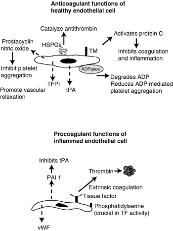
Figure 1 is a simplified diagram of membrane-bound and secreted factors that play a role in endothelial cell regulation of coagulation. Under physiologic conditions, endothelial cells maintain an anti-thrombotic surface characterized by high levels of heparan-sulfate proteoglycan (HSPG) expression. HSPGs act to repel negatively charged molecules and cells, catalyze the action of circulating antithrombin III, and promote the release of prostacyclin from the endothelial cell (Wiedermann Ch and Romisch, Reference Wiedermann Ch and Romisch2002). Another weapon in the endothelial cells anticoagulant arsenal is tissue factor pathway inhibitor, which directly inhibits tissue factor-mediated coagulation. Trials using tissue factor pathway inhibitor to limit sepsis-induced coagulopathy have met with mixed results (Creasey et al., Reference Creasey, Chang, Feigen, Wun, Taylor and Hinshaw1993; Carr et al., Reference Carr, Bild, Chang, Peer, Palmier, Frazier, Gustafson, Wun, Creasey, Hinshaw, Taylor and Galluppi1994; Abraham, Reference Abraham2000; Hoffman and Faist, Reference Hoffman and Faist2000; de Jonge et al., Reference de Jonge, Dekkers, Creasey, Hack, Paulson, Karim, Kesecioglu, Levi, van Deventer and van der Poll2001; Chen et al., Reference Chen, Giannopoulos, Shiels, Webster, McVey, Kemball-Cook, Tuddenham, Moore, Lechler and Dorling2004; Dang et al., Reference Dang, Navarro and David2004). Endothelial cell thrombomodulin also aids in limiting coagulation, by binding to and inactivating thrombin, and promoting the activation of protein C (Boffa and Karmochkine, Reference Boffa and Karmochkine1998). Activated protein C has potent anti-inflammatory and antiapoptotic effects (Esmon, Reference Esmon2003; D'Angelo et al., Reference D'Angelo, Della Valle, Giudici and Vigano D'Angelo2004). Therapy with activated protein C has been shown to improve survival of human sepsis patients (Dhainaut et al., Reference Dhainaut, Yan, Margolis, Lorente, Russell, Freebairn, Spapen, Riess, Basson, Johnson and Kinasewitz2003). Finally, endothelial cells also secrete nitric oxide and prostacyclin, which are both antithrombotic and vasodilatory (Gryglewski et al., Reference Gryglewski, Botting and Vane1988).
Endothelial cell's role in regulating coagulation. The surface of endothelial cells is coated in HSPGs that repel negatively charged surfaces, and act to catalyze the action of antithrombin III. Endothelial cell thrombomodulin (TM) binds and inactivates circulating thrombin, and plays a key role in activating circulating protein C. Activated protein C inhibits coagulation co-factors Va and VIIIa, and also has potent anti-inflammatory and antiapoptotic properties. Endothelial cells also secrete (indicated by dashed arrows) prostacyclin and nitric oxide, which promote vascular relaxation and also inhibit platelet aggregation, as well as tissue factor pathway inhibitor (TFPI) and tissue plasminogen activation (tPA) which inhibit tissue factor-mediated coagulation and promote fibrinolysis, respectively. Cell surface bound ADPases destroy ADP, which normally serves as a potent stimulus for platelet aggregation. When inflamed, these anticoagulant functions are often down-regulated, or lost entirely. Additionally, the endothelial cells begin to express tissue factor, which activates the extrinsic coagulation pathway. Cell-surface phosphatidyl serine promotes the activity of tissue factor. Activation of endothelial cells can also result in the release of von Willebrand factor (vWF) from storage granules and plasminogen activator inhibitor 1 (PAI-1) which inhibits the fibrinolytic activity of tPA. Cumulatively, these changes alter the endothelial cell surface from a non-thrombotic, to a prothrombotic state.

The endothelial cell functions mentioned above are often lost in the face of inflammation. An activated endothelium expresses receptors for coagulation proteins, secretes proinflammtory and prothrombotic cytokines, and loses its ability to inhibit thrombus formation. Inflamed endothelial cells secrete TNF-α, IL-1β, and IL-6, all of which promote coagulation (Edgington et al., Reference Edgington, Mackman, Fan and Ruf1992; Dosquet et al., Reference Dosquet, Weill and Wautier1995; Levi et al., Reference Levi, van der Poll and ten Cate2006) and they often express high levels of tissue factor on their surface (Carson et al., Reference Carson, Johnson and Tracy1993; Levi et al., Reference Levi, van der Poll and ten Cate2006).
Tissue factor, a member of the cytokine receptor superfamily and the central activator of coagulation in vivo, plays a prominent role in the pathogenesis of sepsis (Carraway et al., Reference Carraway, Welty-Wolf, Miller, Ortel, Idell, Ghio, Petersen and Piantadosi2003; Welty-Wolf et al., Reference Welty-Wolf, Carraway, Ortel, Ghio, Idell, Egan, Zhu, Jiao, Wong and Piantadosi2006). Tissue factor expression on an activated endothelium, and the subsequent activation of coagulation, contribute to vascular dysfunction in sepsis (Pawlinski et al., Reference Pawlinski, Pedersen, Schabbauer, Tencati, Holscher, Boisvert, Andrade-Gordon, Frank and Mackman2003; Levi et al., Reference Levi, van der Poll and ten Cate2006; Strukova, Reference Strukova2006). Furthermore, inhibition of tissue factor attenuated organ dysfunction and improved survival in murine and baboon models of sepsis (Creasey et al., Reference Creasey, Chang, Feigen, Wun, Taylor and Hinshaw1993; Dackiw et al., Reference Dackiw, McGilvray, Woodside, Nathens, Marshall and Rotstein1996; Abraham, Reference Abraham2000; Carraway et al., Reference Carraway, Welty-Wolf, Miller, Ortel, Idell, Ghio, Petersen and Piantadosi2003; Pawlinski et al., Reference Pawlinski, Pedersen, Schabbauer, Tencati, Holscher, Boisvert, Andrade-Gordon, Frank and Mackman2003).
Activation of endothelial cells can also result in loss of thrombomodulin from the cell surface (Faust et al., Reference Faust, Heyderman and Levin2001), and decreased production of vasodilators and anticoagulant proteins (Wu and Thiagarajan, Reference Wu and Thiagarajan1996). These events disarm the endothelial cell, making it less capable of preventing localized coagulation along the vessel wall. In the face of infection, endothelial cell tissue factor can be like a match, igniting the coagulation fire. Cytokines act like gasoline by promoting additional tissue factor expression and microparticle release from endothelial cells and circulating monocytes. The platelets are wood for the fire, providing an additional source of coagulation co-factors and structural support. Finally, the systemic inflammatory response alters the function of the vascular wall (i.e. cytokine-mediated loss of HSPGs and thrombomodulin) that essentially is like walking off with the body's only fire extinguisher.
Infection-induced coagulopathy
A number of human pathogens, including Neisseria meningitidis (Heyderman et al., Reference Heyderman, Klein, Daramola, Hammerschmidt, Frosch, Robertson, Levin and Ison1997) and group A streptococci (Bryant et al., Reference Bryant, Hayes-Schroer and Stevens2003) up-regulate tissue factor on endothelial cells and promote coagulation. Rickettsia rickettsii, the causative agent of Rocky Mountain Spotted Fever (RMSF), induces a procoagulant state, although DIC rarely occurs (Elghetany and Walker, Reference Elghetany and Walker1999). R. rickettsii infection of human umbilical endothelial cells in culture triggers the release of large multimers of von Willebrand factor (Sporn et al., Reference Sporn, Shi, Lawrence, Silverman and Marder1991), and increased tissue factor activity (Sporn et al., Reference Sporn, Haidaris, Shi, Nemerson, Silverman and Marder1994). The mechanisms involved in infection-induced tissue factor expression are not well defined. Several reports focused on the role of lipopolysaccharide (LPS), or the truncated form of LPS known as lipooligosaccharide (LOS), in inducing tissue factor expression and activity (Colucci et al., Reference Colucci, Balconi, Lorenzet, Pietra, Locati, Donati and Semeraro1983; Erlich et al., Reference Erlich, Fearns, Mathison, Ulevitch and Mackman1999; Brandtzaeg et al., Reference Brandtzaeg, Bjerre, Ovstebo, Brusletto, Joo and Kierulf2001). LPS signals through its receptor complex, TLR-4, CD14 and MD2, which leads to activation of nuclear factor-kappaβ and the mitogen-activated protein kinases. For a detailed discussion of LPS signaling in endothelial cells, the reader is referred to a current review by Dauphinee and Karsan (Reference Dauphinee and Karsan2005).
Despite the ubiquitous presence of LPS, or LOS, on gram-negative bacteria, all gram-negative pathogens do not have equivalent tendencies to promote coagulopathy. There are at least three potential explanations for this discrepancy. Firstly, some pathogens associated with profound thrombosis, such as N. meningitidis and Haemophilus somnus, release large amounts of LOS in the form of outer membrane vesicles (Stephens et al., Reference Stephens, Little, Wilkie and Barnum1984; Mirlashari et al., Reference Mirlashari, Hoiby, Holst and Lyberg2001). Perhaps the local concentration of LPS or LOS is critical in promoting tissue factor activity on endothelial cells. Second, structural differences or modifications of LPS/LOS, may affect activation of endothelial cells. This could explain in part, why some gram-negative pathogens are more strongly associated with coagulopathy. Third, the ability of the host immune response to control bacterial survival in the bloodstream would determine the amount of LPS available to contact the endothelium.
Gram-positive bacteria are also capable of triggering coagulation disturbances. A good example is the coagulative activity of Staphlococcus aureus superantigens and coagulases (Mattsson et al., Reference Mattsson, Herwald and Egesten2003; Bjerketorp et al., Reference Bjerketorp, Jacobsson and Frykberg2004). The ability of bacterial pathogens to trigger coagulation likely depends on more than one factor, but clearly often involves interaction with the endothelium.
Production of inflammatory cytokines by endothelial cells or circulating leukocytes also contributes to sepsis-induced coagulopathy. For example, TNF-α has been shown to increase endothelial cell tissue factor expression and enhance the ability of thrombin to induce tissue factor activity in vitro (Aderka, Reference Aderka1991; Salgado et al., Reference Salgado, Boveda, Monasterio, Segura, Mourelle, Gomez-Jimenez and Peracaula1994; Wada et al., Reference Wada, Otu, Wu, Abid, Okada, Libermann, Kodama, Shih, Minami and Aird2005). Furthermore, anti-TNF therapy was effective in reducing coagulation disturbances in a baboon model of sepsis (Aderka, Reference Aderka1991; Emerson et al., Reference Emerson, Lindsey, Jesmok, Duerr and Fournel1992; Terry and Callahan, Reference Terry and Callahan1996). Similarly, IL-1 infusion in rabbits resulted in a 10-fold increase in tissue factor activity in 3–5 h (Nawroth et al., Reference Nawroth, Handley, Esmon and Stern1986), and IL-1 containing conditioned media directly increased the procoagulative activity of cultured endothelial cells in vitro (Stern et al., Reference Stern, Bank, Nawroth, Cassimeris, Kisiel, Fenton, Dinarello, Chess and Jaffe1985). Because proinflammatory cytokines are produced by an array of cells, we do not know the relative contribution that endothelial cells make to cytokine-induced coagulopathy. However, cytokines produced by endothelial cells might have potent autocrine or paracrine effects that are disproportionate to the amount of cytokine secreted. Alternatively, the effects of pathogens that interact with both leukocytes and endothelial cells could elicit stronger procoagulative response due to higher cumulative cytokine levels. One in vitro strategy to address this question would be to selectively inhibit the production of specific cytokines from either endothelial cells or leukocytes in co-culture models of infection.
Vasculitis and thrombosis in H. somnus septicemia
Vasculitis is characteristic of H. somnus septicemia in cattle (Andrews et al., Reference Andrews, Anderson, Slife and Stevenson1985). In its most severe presentation, thrombotic meningoencephalitis (TME), vasculitis and thrombosis occur throughout the central nervous system (Roberts et al., Reference Roberts, Wood, Hunter, Munro and Imray1979; Momotani et al., Reference Momotani, Yabuki, Miho, Ishikawa and Yoshino1985; Thompson et al., Reference Thompson, Vickers, Stevenson and Davidson1987). In vitro studies support a role for both endothelial cells and platelets in the progression of TME, and the associated thrombus formation. Previous work done in our laboratory has shown that H. somnus causes caspase-dependent apoptosis of large vessel endothelial cells (Sylte et al., Reference Sylte, Corbeil, Inzana and Czuprynski2001). Apoptotic cells have been shown to be procoagulant as the result of membrane alterations (Bombeli et al., Reference Bombeli, Karsan, Tait and Harlan1997). Although apoptotic endothelial cells have not yet been definitively demonstrated in infected cattle, we hypothesize that H. somnus-induced apoptosis contributes in part to thrombosis that characterizes TME. We have also demonstrated that H. somnus causes platelet aggregation, and H. somnus-activated platelets induce endothelial cell apoptosis (Kuckleburg et al., Reference Kuckleburg, Sylte, Inzana, Corbeil, Darien and Czuprynski2005). Bovine brain-microvascular endothelial cells incubated with H. somnus secrete TNF-α and IL-1β, express increased tissue factor activity and exhibit a reduced ability to activate protein C (manuscript in revision). We hypothesize that the interactions among H. somnus, platelets, and endothelial cells contribute significantly to the vasculitis and thrombosis associated with H. somnus septicemia.
Abortion and thrombosis
Equine herpes virus-1 (EHV-1) abortion is associated with infection of endometrial endothelial cells and thromboischemic damage. Placental vascular lesions in infected animals contain viral antigen (Smith et al., Reference Smith, Whitwell, Mumford, Gower, Hannant and Tearle1993). EHV-1 infection of endothelial cells is thought to promote thrombus formation. However, in a recent study, Chiam et al. (Reference Chiam, Smid, Kydd, Smith, Platt and Davis-Poynter2006) found no difference in the procoagulant activity of EHV-1 infected equine endothelial cells and non-infected controls. This study quantified the ability of equine endothelial cells, grown on microcarrier beads, to coagulate fresh equine plasma as a measure of coagulopathy. Equine endothelial cells treated with LPS for 16 h showed a significant decrease in clot time, compared to untreated control cells, a response that was not observed in EHV-treated cells. Because the purpose of the study was to develop an in vitro assay to compare pathogenicity of different EHV strains, no investigation of tissue factor or adhesion molecule up-regulation, or loss of anticoagulant functions on EHV-treated endothelial cells was performed. Due to the relatively high dose of LPS and incubation time (0.1 μg/ml−1 and 16 h) required to significantly reduce clot times, and the relative insensitivity of measuring clot times in general, it is difficult to conclude whether EHV-infection promoted a prothrombotic state in endothelial cells that was not detected in this study.
Endothelial cells in vasculitic syndromes
Feline infectious peritonitis (FIP) is a progressive and fatal condition in cats resulting from infection with feline coronavirus (FCoV) (Jones et al., Reference Jones, Hunt and King1997). The wet, or effusive, form of the disease is characterized by a fibrinous to granulomatous serositis with protein rich effusions into body cavities, and granulomatous to necrotizing phlebitis (Montali and Strandberg, Reference Montali and Strandberg1972; Weiss and Scott, Reference Weiss and Scott1981). The vascular lesions in affected cats can be found in the liver, pancreas, kidney, lymph nodes, muscular layers of the gastrointestinal tract, meninges and eyes (Jones et al., Reference Jones, Hunt and King1997). The disease progresses as circulating monocytes are infected and disseminate the virus (Weiss and Scott, Reference Weiss and Scott1981; Jacobse-Geels and Horzinek, Reference Jacobse-Geels and Horzinek1983; Horzinek et al., Reference Horzinek, Ederveen, Egberink, Jacobse-Geels, Niewold and Prins1986).
In a study by Weiss et al. (Reference Weiss, Dodds and Scott1980), intraperitoneal inoculation of cats with FeCoV induced DIC. The mechanisms responsible for triggering DIC in cats with FIP are not clearly defined. Boudreaux et al. (Reference Boudreaux, Weiss, Toivio-Kinnucan, Cox and Spano1990) observed that platelets isolated from FeCoV infected cats displayed an elevated responsiveness to ADP and epinephrine. Clearly, activated platelets could contribute to DIC. FeCoV is known to induce hepatic necrosis (Jones et al., Reference Jones, Hunt and King1997), which would lead to depletion of coagulation factors. At this time, it is difficult to tease apart the relative contributions of monocyte-induced vasculitis, endothelial cell alterations, platelet activation, and reductions in coagulation factor production by a diseased liver, in the coagulopathy that has been reported in FIP.
The vascular damage seen in FIP historically has been attributed to immune-mediated mechanisms. In the previously mentioned study by Weiss et al., FeCoV inoculation of seropositive kittens resulted in a more rapid onset of clinical disease than in seronegative cats. This finding supports the contribution of an immune-mediated process to the disease. The granuloma formation and vasculitis associated with FIP were previously classified as a type III hypersensitivity reaction (Jacobse-Geels and Horzinek, Reference Jacobse-Geels and Horzinek1983; Pedersen et al., Reference Pedersen, Evermann, McKeirnan and Ott1984). However, this classification recently has been brought into question in a recent study by Kipar et al. (Reference Kipar, May, Menger, Weber, Leukert and Reinacher2005). These authors characterized the morphological aspects of FIP associated vasculitis by performing a detailed histopathological analysis on five cats found to have both virally infected monocytes and multifocal granulomatous vasculitis by immunohistochemical analysis. The authors reported the presence of monocytes adherent to endothelial cells, or emigrating from the blood vessel at lesion sites. Inflammatory cells were found between layers of the basal lamina of affected vessels. The authors concluded that evidence for a type III hypersensitivity reaction was lacking, as they did not identify any cell-independent immune complex deposition within vascular lesions. They propose that virally-infected and activated monocytes initiate phlebitis in FIP. The authors contend that widespread expression of endothelial cell MHC II in affected cats reflects a disseminated response to monocyte-induced viremia, and endothelial cell activation could actively contribute to the overall pathogenesis.
Interestingly Granger and Kubes (Reference Granger and Kubes1994) suggest, that the restriction of FIP-induced vascular inflammation to veins could reflect the increased tendency for leukocytes to adhere to venous endothelial cells, in comparison to arterial endothelial cells. Whether this is the result of distinct differences in the adhesive properties of endothelial cells in different vascular beds, or due to mechanical factor such as sheer stress, remains to be elucidated. In a recent study, Mairey et al. (Reference Mairey, Genovesio, Donnadieu, Bernard, Jaubert, Pinard, Seylaz, Olivo-Marin, Nassif and Dumenil2006) elegantly demonstrated that adhesion of N. meningitidis to human brain endothelial cells only occurred at sheer stress levels equivalent to those detected in cerebral capillaries, which is the anatomical site of N. menigitidis attachment in vivo. The interaction between FeCoV-infected monocytes and the endothelium could likewise be governed partially by mechanical and rheologic factors.
A vascultic syndrome in animals that is clearly an example of a type III hypersensitivity reaction is equine purpura hemorrhagica. Equine purpura hemorrhagica is an acute disease that is commonly associated with streptococcal infections, characterized by edema and hemorrhage as the result of a leukocytoclastic vasculitis (Bennet and King, Reference Bennet and King1948; Jones et al., Reference Jones, Hunt and King1997). Unlike classical purpura hemorrhagica, which is characterized by thrombocytopenia, studies of equine purpura have not demonstrated thrombocytopenia, or coagulation defects in association with the disease (Jones et al., Reference Jones, Hunt and King1997). Edema formation and hemorrhage in the equine patient is therefore, more likely the result of direct physical compromise of the vascular barrier. Galan and Timoney (Reference Galan and Timoney1985) were the first to demonstrate the presence of immune complexes, containing IgA and the M antigen of Streptococcus equi, in the sera of horses with post-streptococcal infection purpura hemorrhagica. The streptococcal M-protein is an important virulence factor, as it enhances survival in equine blood by limiting the deposition of complement component C3 (Boschwitz and Timoney, Reference Boschwitz and Timoney1994). Antibodies that react against the M-protein also recognize myosin. This cross-reaction has been implicated in the pathogenesis of rheumatic fever in humans (Cunningham et al., Reference Cunningham, McCormack, Fenderson, Ho, Beachey and Dale1989). Likewise, antibodies that recognize the S. equi M-protein may react with antigens on the vascular wall. We could find no published studies investigating potential cross-reactive target antigens between streptococci and endothelial cells. Furthermore, purpura hemorrhagica can follow a variety of insults that cause tissue necrosis. Therefore, the cross reactivity between antibodies recognizing a particular bacterial protein and a cross-reacting antigen on host tissue does not likely explain all cases of the disease. At this time, we do not know whether antibody mediated (i.e. complement driven) destruction of the vascular wall, or autoreactive T-cells contribute to its pathogenesis.
Endothelial cell dysfunction in equine laminitis
Equine laminitis is a devastating disease that leads to separation of the hoof wall from the bone of the digit. A detailed discussion of the pathogenesis of laminitis is beyond the scope of this review. Rather, we would like to emphasize the potential contributions of endothelial cells to the onset and progression of this disease. Recent studies have associated leukocyte emigration into the tissues surrounding the dermal microvasculature of the laminae with the onset of clinical lameness in black walnut extract (BWE)-induced laminitis (Black et al., Reference Black, Lunn, Yin, Hwang, Lenz and Belknap2006; Hurley et al., Reference Hurley, Parks, Reber, Donovan, Okinaga, Vandenplas, Peroni and Moore2006). The degree to which leukocytes emigrate to other organs in the horse following administration of BWE, or during the course of other laminitic-inducing conditions, is unknown at this time. Additional factors that contribute to emigration of leukocytes in the equine digit following systemic administration of BWE are unknown. Considering the key role that endothelial cells play in regulating leukocyte trafficking, studies aimed at elucidating the changes that occur in the endothelial cells of lamellar vasculature in BWE-induced laminitis could elucidate key steps in the progression of the disease. For example, are the endothelial cells of the lamellar vasculature more prone to up-regulation of adhesion molecules following a systemic inflammatory event? Are inter-endothelial cell junctions in the equine digit different (i.e. more susceptible to proteolytic degradation, display lower rates of protein turnover, etc.) than inter-endothelial cell junctions in other vascular beds? Alternatively, is there a chemotactic factor that is being produced locally in the digit to attract circulating leukocytes? Answering these questions would help us to discern whether the pathology that occurs in the equine digit results from selective emigration of leukocytes and tissue damage, or a relative inability to resolve tissue damage due to decreased blood flow once the condition is initiated.
Recent studies have identified some potentially important differences in the vasculature of the equine digit. Peroni et al. (Reference Peroni, Moore, Noschka, Grafton, Aceves-Avila, Lewis and Robertson2006) reported that isolated equine laminar veins displayed higher responsiveness to vasoconstrictors than laminar arteries. The authors proposed that this may be a contributing factor to the development of laminitis. Because intact vessels bathed in test solutions were used to evaluate contractile responses in their study, their observations could be due to differential responses of the endothelium, or the surrounding pericytes and smooth muscle cells. In an earlier study, Mallem et al. (Reference Mallem, Gogny, Gautier, Bucas and Desfontis2003), demonstrated reduced beta3-adrenoreceptor-mediated vascular relaxation in endotoxin-treated equine digital veins. These authors suggested that endotoxin treatment triggered cyclooxygenase-induced prostanoid production, which in turn reduced vasorelaxation. It would be interesting to know if endothelial cells from different vessels in the digit displayed unique responses to endotoxin, such as production of metalloproteases, nitric oxide, or prostacyclin. Insulin resistance has been proposed to be a contributing factor to laminitis. Evaluation of plasma glucose, insulin, triglyceride, nonesterified fatty acids and corsitol levels identified as a ‘prelaminitic metabolic syndrome’ predicted 11 of 13 cases of clinical laminitis in pastured ponies (Treiber et al., Reference Treiber, Kronfeld and Geor2006). In humans, hyperglycemia-induced shedding of the endothelial cell glycocalyx leads to impaired flow-mediated dilation of vessels and an increased tendency to form blood clots (Gouverneur et al., Reference Gouverneur, Berg, Nieuwdorp, Stroes and Vink2006; Nieuwdorp et al., Reference Nieuwdorp, van Haeften, Gouverneur, Mooij, van Lieshout, Levi, Meijers, Holleman, Hoekstra, Vink, Kastelein and Stroes2006). Perhaps hyperglycemia, from carbohydrate overload, insulin resistance, or a combination of the two, results in a similar response in vessels of the equine digit. Although the pathogenesis of laminitis is complex and is an area of active investigation, there is disproportionately little in the literature regarding the potential contributions of endothelial cells to disease progression.
Endothelial cell regulation of vascular permeability
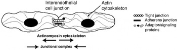
Vascular barrier function is governed by the continuity of the endothelium, which in turn is regulated by the balance between contractile forces in individual endothelial cells, and the tethering forces of intercellular junctions (Garcia et al., Reference Garcia, Davis and Patterson1995; Lum and Malik, Reference Lum and Malik1996; Dudek and Garcia, Reference Dudek and Garcia2001). This balance is illustrated in Fig. 2. Active cellular contraction places tension on inter-endothelial cells junctions that can lead to leakage of fluid and even extravasation of cells between the endothelial cells. Vascular leakage can also occur subsequent to direct damage to the constituents of the inter-endothelial cell junctions. A detailed discussion of the components of these junctions and their regulation is beyond the scope of this review, and the reader is referred to previous reviews (Bazzoni, Reference Bazzoni2006; Mehta and Malik, Reference Mehta and Malik2006).
Regulation of vascular permeability. The continuity of the endothelial cell barrier is determined, in part, by the connectivity of adjacent endothelial cells. Connectivity in turn, is regulated by the balance of forces generated by the inward contraction of the actinomysoin cytoskeleton, and the anchorage sites at inter-endothelial cell junctions. An imbalance in these two counter-acting forces can lead to gap formation, vascular leakage and edema.

Intracellular infection of endothelial cells and vascular permeability
Infections caused by agents that are capable of causing widespread and rapid alterations in vascular permeability often prove quickly fatal. A previously mentioned example is provided by AHS virus. The severe pulmonary edema associated with the peracute form of AHS is thought to result from a combination of direct infection of the pulmonary endothelium, and cardiac dysfunction resulting from infection of the endocardium (Gomez-Villamandos et al., Reference Gomez-Villamandos, Sanchez, Carrasco, Laviada, Bautista, Martinez-Torrecuadrada, Sanchez-Vizcaino and Sierra1999). Detailed transmission electron microscopy studies of infected capillaries found separation of adjacent endothelial cells that varied from mild to severe (Gomez-Villamandos et al., Reference Gomez-Villamandos, Sanchez, Carrasco, Laviada, Bautista, Martinez-Torrecuadrada, Sanchez-Vizcaino and Sierra1999). The mechanisms responsible for the formation of inter-endothelial cell gaps in AHS have not been studied, and warrant future investigations.
Another example of a group of pathogens notorious for infecting endothelial cells and inducing vascular leakage are the Rickettsia spp. Rickettsia are obligate intracellular pathogens that specifically target the endothelium, resulting in diffuse and often life-threatening increases in vascular permeability (Walker et al., Reference Walker, Valbuena and Olano2003). For a detailed review of human Rickettsioses, the reader is referred to the current review by Parola et al. (Reference Parola, Paddock and Raoult2005). In an early study in dogs, R. rickettsii infection increased retinal vascular permeability that persisted well beyond clinical recovery, and was associated with thrombocytopenia, hyperfibrinogenemia, and slightly prolonged partial thromboplastin times (Davidson et al., Reference Davidson, Breitschwerdt, Walker, Levy, Carlson, Hardie, Grindem and Nasisse1990). The mechanistic relationship between intracellular Rickettsia cells and vascular permeability has not been clearly defined. Vasoactive prostaglandin production by Rickettsia-infected endothelial cells may contribute to vascular permeability. Rydkina et al. (Reference Rydkina, Sahni, Baggs, Silverman and Sahni2006) demonstrated COX-2 mRNA induction and elevated secretion of 6-keto-PGF and PGE in R. rickettsii infected endothelial cells. In a recent study, Valbuena and Walker (Reference Valbuena and Walker2005) observed gap formation in adherens junctions of Rickettsia infected endothelial cells. The destruction of the adherens junction could be the result of a direct interaction between junctional proteins and intracellular bacteria, production of inflammatory cytokines by the infected endothelial cell, or cell contraction triggered by either the bacterium itself, or a component of the host response.
Extracellular pathogens and vascular permeability
Work done in our laboratory demonstrated that the extracellular bacterial pathogen H. somnus elicited contraction, and hence paracellular permeability, in BBEC monolayers (manuscript in revision). We have demonstrated that incubation with H. somnus elicits rapid cytoskeletal rearrangements in BBECs in vitro that coincide with increased monolayer permeability. The ability of H. somnus to induce monolayer permeability is dependent on myosin-light chain kinase driven-cell contraction, as demonstrated by the use of pharmacological inhibitors (Behling-Kelly et al., Reference Behling-Kelly, Kim, McClenahan and Czuprynski2007). We hypothesize that because BBEC monolayers do not similarly respond to purified H. somnus LOS, recombinant cytokines, or formalin-fixed or heat-killed H. somnus, a labile surface molecule (i.e. outer membrane protein) serves as the initial stimulus for BBEC contraction. We also hypothesize that similar interactions between other pathogens and endothelial cells might alter vascular permeability in the absence of direct endothelial cell infection. Furthermore, circulating factors released during sepsis may have additive or synergistic effects on vascular function. Perhaps pharmacologic interference with a common functional endpoint, such as myosin-light chain-induced cell contraction, could be exploited as a strategy to prevent vascular leakage, and dissemination of bacterial pathogens. There is a precedent for this strategy in modulating the response to sepsis in animal models. Pharmacological inhibition of myosin light chain kinase using a selective myosin light chain kinase inhibitor (ML-7) protected rats from LPS-induced colonic permeability and bacterial translocation (Moriez et al., Reference Moriez, Salvador-Cartier, Theodorou, Fioramonti, Eutamene and Bueno2005). Perhaps a similar approach could limit the dissemination of pathogens across vascular barriers.
Summary
The unique and irreplaceable role of the endothelium in maintaining adequate blood flow and nutrient exchange to all organs makes it is essential that it receives the same investigational and clinical attention as other organs. At the present time, there are no commonly used clinical tests to detect vascular dysfunction in veterinary medicine. For example, exams like fluorescein angiography could be used to detect widespread alterations in vascular permeability. If we gain a better understanding of what cellular mechanisms underlie vascular leakage in various pathogenic situations, then new therapeutic interventions might be devised. A treatment aimed at modulating endothelial cell contraction would be entirely different from one focused on preventing proteolytic digestion of cellular junctions. In infection-induced coagulopathies, perhaps supplementation with substances like hyaluron could help to repair an injured endothelial glycocalyx, while avoiding some of the undesirable side-effects associated with administration of heparin. Hopefully the future will bring, a better understanding of how infection alters vascular function and novel therapeutic approaches to address some of the more devastating clinical outcomes of infectious disease.