Introduction
Lymphatic filariasis caused by lymphatic-dwelling nematodes is a serious public-health problem in many tropical countries, including India. It affects about 120 million people globally (Michael et al., Reference Michael, Bundy and Grenfell1996) and is listed as the second leading known cause of disability (World Health Organization, 1995). Individuals living in filarial endemic areas can be grouped into: (1) asymptomatic microfilaraemic carriers who are often free of overt clinical manifestations; (2) chronic patients who display chronic manifestations such as elephantiasis and/or hydrocoele with or without patent infection; (3) acute cases, individuals who suffer from periodic episodes of acute symptoms characterized by adenolymphangitis; and (4) endemic normals, subjects who remain free of demonstrable filarial infection and do not present any of the acute and chronic filarial disease manifestations. Members of this latter group of individuals, although continually exposed to infection, remain apparently normal, can be considered as putatively immune subjects, and are the only available group of subjects in endemic areas for whom issues on protective immunity may be addressed. The concept of endemic normals as a basis for the existence of acquired protective immunity (resistance to infection) has been utilized to identify protective antigens in lymphatic filariasis (Freedman et al., Reference Freedman, Nutman and Ottesen1989) and in onchocerciasis (Elson et al., Reference Elson, Guderian, Aranjo, Bradley, Days and Nutman1995). Animal models have provided evidence for studies on protective immunity in filariasis (Pfaff et al., Reference Pfaff, Schulz-key, Soboslay, Geiger and Hoffman2000; Hoffman et al., Reference Hoffman, Pfaff, Schulz-key and Soboslay2001; Babayan et al., Reference Babayan, Ungeheuer, Martin, Attout, Belnoue, Snounou, Renia, Korenaga and Bain2003, Reference Babayan, Attout, Harris, Taylor, Le Goff, Vuong, Renia, Allen and Baino2006) which was induced either by irradiated larvae or immunization with defined antigens. Several filarial antigens with protective efficacy in animal models and/or preferential responses in putatively immune individuals have been described (Freedman et al., Reference Freedman, Nutman and Ottesen1989; Chenthamarakshan et al., Reference Chenthamarakshan, Reddy and Harinath1995; Dabir et al., Reference Dabir, Dabir, Krithika, Goswami and Reddy2006). We have reported earlier that the antibody response to a water-insoluble, detergent-soluble filarial surface glycoprotein (Dssd1) was significantly elevated in endemic normals (microfilaria-negative individuals) in comparison to that of microfilaria-positive individuals (Bal & Das, Reference Bal and Das1999). Furthermore, the antibody response to Dssd1 was shown to be positively associated with the circulating-filarial-antigen-negative (infection-free) individuals, irrespective of their disease status (Bal et al., Reference Bal, Beuria, Mandal and Das2003). The study also reported the importance of carbohydrate epitopes in immunoreactivity of Dssd1 in infection-free individuals. The present paper describes the ability of this glycoprotein (Dssd1) in clearing microfilaria in a Setaria digitata–Mastomys coucha model.
Materials and methods
Preparation of Dssd1 antigen
The Dssd1 antigen was prepared from water-insoluble components of adult Setaria digitata (cattle filarial parasite) as described previously (Bal & Das, Reference Bal and Das1999; Bal et al., Reference Bal, Beuria, Mandal and Das2003). Briefly, the insoluble component was collected as a pellet after centrifuging an aqueous extract of worms in saline. The pellet was solubilized in detergent NP-40 (0.5% NP-40 in 0.01 m Tris–HCl, pH 8.0), centrifuged again and ammonium sulphate (50%) was added to the supernatant to precipitate the proteins. The precipitate was then dissolved in 0.01 m phosphate buffered saline (PBS, pH 7.4), dialysed extensively against PBS and fractionated in a Sephadex G-200 column using a chromatographic system (BioRad, Richmond, California, USA). Dssd1 was revealed as single band of size 210 kDa on SDS-PAGE, and was found to contain 45 μg of carbohydrate per mg of protein.
Implantation of parasites
Mastomys coucha individuals, procured from the Central Drug Research Institute, Lucknow, India and bred in our animal facility, were used for all the experiments. Implantation of parasites into the peritoneal cavity of mastomys was performed as described earlier (Mukhopadhaya et al., Reference Mukhopadhaya, Dash and Ravindran1996). Briefly, fresh, gravid female S. digitata worms collected from cattle in Dulbecco's Modified Eagle's Medium (DMEM; Sigma Chemical Company, St Louis, Missouri, USA) were used for implantation. Animals weighing about 60 g were anaesthetized with ketamine hydrochloride and three gravid female worms were aseptically implanted into the peritoneal cavity of each animal through a small incision.The incisions were stitched with surgical catgut immediately after implantation. Induction of microfilaria was monitored by taking 20 μl blood samples from the tail vein.
Immunization schedule
Experiments were performed in three groups of eight M. coucha. In the first experiment, group I, mastomys of either sex (n = 8) were immunized by the intraperitoneal route with two doses of 75 μg Dssd1, with an interval of 15 days. Worms were implanted in the peritoneal cavity of mastomys 10 days after the last immunization. In the second experiment, group II, mastomys (n = 8) were implanted with the parasites and then immunized twice, intraperitoneally, with Dssd1 antigen (75 μg/mastomys without adjuvant) at 7 and 22 days after implantation. In the third experiment, group III (control), animals (n = 8) were implanted with parasites and received saline alone.
Enzyme-linked immunosorbent assay
Antibody levels to Dssd1 were determined as described earlier (Bal & Das, Reference Bal and Das1999). Briefly, enzyme-linked immunosorbent assay (ELISA) plates were coated with 2 μg ml− 1 of Dssd1 antigen in alkaline buffer, pH 9.2, and incubated for 5 h at 37°C and then overnight at 4°C. Plates were blocked with 0.4% bovine serum albumin in the buffer for 1 h at room temperature, and then 100 μl of serum (1:100 dilutions) was added to the wells and incubated for 3 h at 37°C. After washing with PBS–Tween (0.1%), 100 μl of horseradish peroxidase (HRP)-conjugated goat anti-mouse IgS (1:2000, Sigma) was added to the wells and kept at 37°C for 3 h. After washing with PBS–Tween, the presence of antibodies was detected with OPD substrate (o-phenylenediamine containing H2O2). The enzymatic reaction was stopped by adding a drop of 4N sulphuric acid and the absorbance was read at 492 nm using an ELISA reader (BioRad). The antibody level in the test sample was calculated by subtracting the optical density of normal mouse sample (day zero).
Statistical analysis
Comparison of groups was done with the non-parametrical Mann–Whitney U-test. P values < 0.01 were considered significant.
Results and discussion
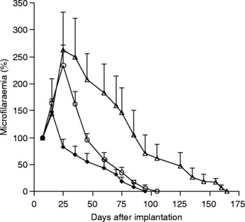
Intraperitoneal implantation of S. digitata gravid female worms was found to induce microfilaraemia in mastomys. The microfilariae (Mf) could be detected in peripheral circulation as early as 4 days after implantation. Microfilaraemia in animals lasted about 165 days, with the peak Mf counts of about 40/20 μl of blood by 25 days (fig. 1). We have observed that implanted adult worms survive in the recipients for less than 10 days. The persistence of microfilaraemia with high Mf density for longer periods may be influenced by the implanted adult female worms. It has been shown in a Litomosides sigmodontis–mouse model that the presence of an adult immature female worm for a few hours was enough to make a host susceptible for microfilaraemia, irrespective of the genetic background (Hoffman et al., Reference Hoffman, Pfaff, Schulz-key and Soboslay2001). Active immunization with Dssd1 antigen either before or after implantation caused the rapid clearance of Mf in implanted mastomys. Immunization of animals with Dssd1 antigen before implantation (group I) resulted in significant suppression of Mf density and a total clearance by about 95 days post implantation. Induction of anti-Dssd1 antibodies (mean OD ± SD) was observed to be 0.65 ± 0.14 at the time of implantation. Reduction of Mf density (about 18%) was observed in group I versus 160% increase in group III (non-immunized) animals on day 25 as compared to day 7. The microfilaria (mean ± SD) count on day 25 was observed to be 14 ± 3.66 and 38.9 ± 5.51 in the immunized and control groups of mastomys, respectively. The circulating Mf in group I were found to be eliminated faster than in group III animals, indicating that microfilarial clearance in vivo is very significantly potentiated by antibodies to Dssd1. Mf density was observed to decline by about 45% on the 45th day, 55% on the 60th day and 80% on the 75th day after implantation in pre-immunized animals. The microfilarial density was more than 100% until the 85th day in non-immunized control animals.
Effect of immunization with Dssd1 antigen on microfilaraemia in Setaria digitata-implanted Mastomys coucha. Group I, animals immunized with two doses of Dssd1 25 and 10 days before implantation of worms (♦, n = 8); group II, animals immunized with two doses of Dssd1 antigen 7 and 22 days after implantation of worms (○, n = 8); group III, normal microfilaraemic controls (△, n = 8). The results are expressed as mean percentage of microfilaraemia (mean ± SD) for each group calculated with reference to microfilarial (Mf) density for each of the animals. The Mf density on day 7 was taken as 100% for each of the animals. Mann–Whitney U test: group I versus group III, P < 0.001 on days 25, 35, 45, 60 and 75; group II versus group III, P < 0.01 on day 35 and P < 0.001 on days 45, 60 and 75.

The effect of immunization with Dssd1 antigen was studied in animals with ongoing microfilaraemia (group II). Intraperitoneal immunization with Dssd1 antigen resulted in suppression of Mf density and a total clearance by 105 days. Anti-Dssd1 antibody level (mean OD ± SD) was observed to be 0.57 ± 0.10 and 0.09 ± 0.02 on day 35 in the immunized and control group of mastomys, respectively. The antibody level was found to be maintained in immunized animals until day 105 (0.45 ± 0.08). The reduction in Mf density was marked after day 30, resulting in a sharp decline. A significant reduction in Mf density was observed: about 40% and 65% of the original microfilaraemia by 60 and 75 days, respectively, indicating that active immunization with Dssd1 antigen is effective against a pre-existing filarial infection.
Extensive studies have been conducted in experimental models of filariasis to address potential protective immunity, since various animals are permissive to several species of filarial parasite that infect humans or animals (Rajan et al., Reference Rajan, Ganley, Paciorkowski, Spencer, Klei and Shultz2002; Hartmann et al., Reference Hartmann, Sereda, Sollwedel, Kalinna and Lucius2006; Anand et al., Reference Anand, Murugan, Prabhu, Anandaraman, Reddy and Kaliraj2008; Dabir et al., Reference Dabir, Dabir, Goswamy and Reddy2008; Lamb et al., Reference Lamb, Harris, Legeoff, Read and Allen2008). Mastomys coucha, a multimammate rat, has been used extensively to study the protective immune response for both Brugia malayi and Brugia pahangi infection. It has been shown that induction of long-lasting microfilaraemia can be obtained in M. coucha by intraperitoneal implantation of live gravid female S. digitata worms (Mukhopadhaya et al., Reference Mukhopadhaya, Dash and Ravindran1996). It is reported that active immunization of microfilaraemic animals with methyl biperazine carboxylic acid, an acid hydrolysis product of diethyl carbamazine citrate (DEC), coupled to bovine serum albumin, followed by administration of subcurative doses of DEC resulted in rapid clearance of microfilaraemia in S. digitata infected M. coucha (Mukhopadhaya & Ravindran, Reference Mukhopadhaya and Ravindran1997).
The present report demonstrates the immunoprophylactic evaluation of a water-insoluble, detergent-soluble glycoprotein (Dssd1) in S. digitata-infected mastomys. Immunization with Dssd1 antigen not only resulted in a significant reduction in Mf density but also cleared microfilaria completely from the circulation. Immunization of animals with Dssd1 antigen before or after implantation yielded more or less similar results. Complete clearance of Mf was achieved within 95 days and 105 days in pre-immunized and post-immunized mastomys, respectively, compared to 165 days in non-immunized animals. A significant reduction in Mf density was noticed in the immunized group when compared with the control group of animals. Elimination of microfilaria from the circulation might be due to the induction of anti-microfilarial immunity in the immunized group of animals. Dssd1 has been shown earlier to be surface associated, as the antisera to Dssd1 showed binding to the sheath of Wuchereria bancrofti and S. digitata microfilaria (Bal & Das, Reference Bal and Das1999). The involvement of surface proteins in immune elimination has been suggested (Zahner, Reference Zahner1995). Presence of circulating antibodies to the Mf sheath has been shown to correlate well with the absence of circulating Mf in infected animals (Tanner & Weiss, Reference Tanner and Weiss1978). A similar association has also observed in Brugian and Bancroftian filariasis, suggesting that antibodies to Mf sheath could be instrumental in eliminating circulating Mf (Ravindran et al., Reference Ravindran, Satapathy, Sahoo and Babbu Geddam2000). Several other filarial antigens with protective potential in animal models have been studied (Kazura et al., Reference Kazura, Maroney, Pearlman and Nilsen1990; Dixit et al., Reference Dixit, Gaur, Khan, Saxena, Murthy and Murthy2004; Thirugnanam et al., Reference Thirugnanam, Pandiaraja, Ramaswamy, Murugan, Gnansekar, Nandakumar, Reddy and Kaliraj2007).
The Dssd1 antigen recognized preferentially by sera from an endemic normal population was shown to induce microfilaria clearance in infected mastomys in the present study, suggesting its protective potential both in human and experimental filariasis.
Acknowledgements
We thank Dr S.K. Kar, Director, Regional Medical Research Centre, Bhubaneswar, for constant support and encouragement. The constant assistance of Mr H.S. Naik and Mr K.C. Parichha is gratefully acknowledged.