Pulmonary atresia with intact ventricular septum is a rare congenital heart defect with a spectrum of severity ranging from simple membranous pulmonary atresia to hypoplasia of the right ventricle with abnormal right ventricle to coronary artery connections. Reference Chikkabyrappa, Loomba and Tretter1–Reference Gleich, Latham, Joffe and Ross3 These so-called coronary artery sinusoids, normally present in developing right ventricular myocardium, can persist due to right ventricular hypertension caused by pulmonary valve atresia and the resultant lack of blood egress. Reference Chikkabyrappa, Loomba and Tretter1 These sinusoids can, in turn, form fistulae with the epicardial coronary arteries leading to maldevelopment of the coronary tree in the form of stenosis and/or ostial atresia. In this scenario, coronary blood flow may arise primarily from the right ventricle via these sinusoids, a high-risk scenario known as “right ventricle-dependent coronary circulation.” The myocardium is therefore dependent on retrograde flow from the right ventricle into the coronary arterial tree. Reference Chikkabyrappa, Loomba and Tretter1,Reference Gleich, Latham, Joffe and Ross3 Decompression of the right ventricle via surgical or transcatheter dilation of the pulmonary valve in this situation can lead to myocardial ischaemia and death. Thus, accurate assessment of coronary artery anatomy in pulmonary atresia with intact ventricular septum is crucial in guiding the decision to pursue staged surgical single ventricle palliation versus direct referral for primary orthotopic heart transplantation.
The diagnosis of pulmonary atresia with intact ventricular septum is typically achieved with transthoracic echocardiogram alone. However, additional imaging is often required to rule out right ventricle-dependent coronary circulation. Invasive cardiac catheterisation with conventional angiography is most commonly utilised, but comes with risks. Reference Chikkabyrappa, Loomba and Tretter1 CT angiography has also been employed for this purpose. CT angiography is non-invasive, but still exposes the patient to ionising radiation. Reference Chikkabyrappa, Loomba and Tretter1 We present a case in which non-invasive ferumoxytol-enhanced 4D cardiac magnetic resonance angiography was used in place of conventional angiography and led to the finding of right ventricle-dependent coronary circulation.
Case details
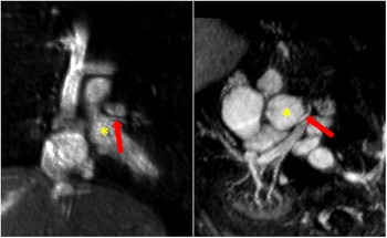
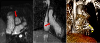
A 36-week gestation newborn male presented with cyanosis at 24 hours of life. A postnatal diagnosis of pulmonary atresia with intact ventricular septum with severe right ventricle hypoplasia was made by echocardiogram. He was intubated and started on intravenous prostaglandin E to maintain patency of the ductus arteriosus and thus provide adequate pulmonary blood flow. The ductus arteriosus was large and tortuous on echocardiogram. The tricuspid valve was diminutive measuring 2.5 mm (z-score −4.7) with no demonstrable antegrade flow or regurgitation. There was a network of vessels in the right ventricular myocardium suggestive of sinusoids. The coronary artery anatomy could not be completely visualised. Right ventricle to coronary artery fistulae was suspected. Invasive cardiac catheterisation with angiography was deferred in this high-risk patient, as ductal anatomy did not appear amenable to stent placement and the medical team wished to avoid an invasive procedure that would therefore be purely diagnostic. Thus, non-invasive ferumoxytol-enhanced 4D cardiac magnetic resonance angiography was obtained to elucidate coronary artery anatomy to inform the choice of surgical single ventricle palliation versus primary orthotopic cardiac transplantation. Under general anaesthesia, ferumoxytol-enhanced 4D cardiac magnetic resonance angiography performed on day of life two showed normal origin of the left main coronary artery but an absent right coronary artery ostium (coronary ostial atresia) with a branch of the right coronary arising directly from the right ventricular apex, consistent with right ventricle-dependent coronary circulation (Figs 1 and 2). Given this finding, he was listed for orthotopic heart transplantation and received a graft at two months of age, rather than undergo the riskier first stage of single ventricle surgical palliation. Pathologic evaluation of the explanted heart confirmed the ferumoxytol-enhanced 4D cardiac magnetic resonance angiography findings.
Coronal and axial 4D MRA MIP images showing the normal origin of the left coronary artery (red arrows) from the aortic root (yellow asterisks). MRA= magnetic resonance angiogram, MIP = maximum intensity projection.

3D volume rendered 4D MRA taken in systole showing a right coronary artery branch (yellow stars) arising from the apex of the hypoplastic RV. The red arrows indicate the area of the expected location of the right coronary ostium, which in this patient is atretic. MRA= magnetic resonance angiogram.

Discussion
Pulmonary atresia with intact ventricular septum is a complex condition with significant anatomic variability leading to multiple potential diagnostic and management algorithms. Treatment strategies range from catheter perforation of the pulmonary valve to primary heart transplant. Reference Chikkabyrappa, Loomba and Tretter1,Reference Gleich, Latham, Joffe and Ross3 As technology and procedural skills advance, there is a continual need to reassess established guidelines and practices for more effective means of diagnosis and treatment. Reference Loomba and Pelech4 When ventriculo-coronary connections are present, there is high likelihood of coronary anomalies such as stenosis or ostial atresia. The gold standard for assessment is invasive coronary angiography to confirm the presence and extent of myocardial dependence on coronary flow from the right ventricle. Reference Gleich, Latham, Joffe and Ross3 Cardiac catheterisation remains the preferred diagnostic tool due to the ability to simultaneously preform therapeutic interventions, but it is not without significant risk. This case illustrates the utility of non-invasive cross-sectional imaging as an alternative to catheterisation. There are increasing reports of the use of CT angiography as an alternative diagnostic modality in the detection of coronary artery fistulae. Reference Gleich, Latham, Joffe and Ross3,Reference Saltik, Bayrak, Guneysu, Sevinc, Oztunc and Degertekin5 In addition to assessment of the coronary arteries, ferumoxytol-enhanced 4D cardiac magnetic resonance angiography allows for further assessment of the right ventricle structure, tricuspid valve competence, as well as the ductus arteriosus flow characteristics (Figs 1 and 2).
In our patient, ferumoxytol-enhanced 4D cardiac magnetic resonance angiography provided critical missing information needed to select the safest management strategy. It should be noted that the sensitivity of the magnetic resonance angiography may be decreased for more distal coronary abnormalities and that the additional information gained from the cardiac magnetic resonance angiography must be weighed against the risk of general anaesthesia required in a neonate. However, MRI has the additional benefits of avoiding ionising radiation exposure, iodinated contrast, and procedural risks of cardiac catheterisation. These are important considerations in the diagnostic algorithm of patients with pulmonary atresia with intact ventricular septum, especially in those with coexisting kidney injury.
Conclusion
The diagnostic use of ferumoxytol-enhanced 4D cardiac magnetic resonance angiography to assess for coronary ostial atresia or proximal stenosis in pulmonary atresia with intact ventricular septum has the potential of avoiding the risks of CT angiography and cardiac catheterisation and may inform the choice between single ventricle palliation and primary heart transplantation in these patients.
Acknowledgements
None.
Financial support
This research received no specific grant from any funding agency, commercial or not-for-profit sectors.
Conflicts of interest
None.