Introduction
Passive exposure to cigarette smoke has been implicated in a number of childhood disorders, ranging from colds and middle-ear diseases to respiratory illness and even cot death.Reference Cook and Strachan1, 2 In addition, smoking has been found by many authors to be associated with poorer outcomes following functional endoscopic sinus surgery (FESS), both in adult populations (i.e. direct smokers)Reference Smith, Mendolia-Loffredo, Lochr, Sparpani, Laud and Nattinger3, Reference Briggs, Wright, Cardes and Calhoun4 and in paediatric populations (i.e. passive smokers).Reference Kim, Dhog, Chung, Chung and Min5 However, all these studies have been based on either subjective outcome measures (i.e. patient or parent questionnaires) or quality of life measures; none have been based on objective outcome measurements. In this study, we used an objective parameter – number of cilia – to study the effect of passive smoking on ciliary regeneration after FESS in a paediatric population.
Material and methods
Thirty-eight children aged between four and 17 years were included prospectively between March 2003 and August 2006. Twenty patients were male and 18 were female.
All our patients were diagnosed as having chronic sinusitis, according to the definition of Melen et al., Reference Melen, Lindahl, Andreasson and Rundcrantz6 and were scheduled for FESS, following failure of medical treatment for three months or more. Twelve of our patients had a history of previous adenoidectomy undertaken to treat chronic paediatric sinusitis, with poor results.
Our exclusion criteria were: (Reference Cook and Strachan1) primary ciliary disease (i.e. cystic fibrosis), craniofacial anomalies and Down syndrome; (2) age younger than four years; (Reference Smith, Mendolia-Loffredo, Lochr, Sparpani, Laud and Nattinger3) clinical, pathological or radiological evidence of fungal infection; and (Reference Briggs, Wright, Cardes and Calhoun4) previous exposure to radiotherapy for head and neck malignancies, previous significant maxillofacial trauma, or nasal fractures.
Two groups were identified within this patient cohort. The first group consisted of 24 children with a history of routine exposure to cigarette smoke within their families; these children were considered as passive smokers. The second group consisted of 14 children with no history of routine exposure to cigarette smoke within their families; they were considered as ‘nonsmokers’.
Exposure to passive smoking was determined subjectively using a parental questionnaire including the question ‘does any member of your household smoke?’, with the possible answers of yes or no. A validated, objective measure of smoke exposure in the preceding 72 hours was determined by measuring the urinary cotininel:creatinine ratio in all children whose parents had responded ‘yes’ to the above question.
Functional endoscopic sinus surgery was performed for our entire study group (i.e. 38/38), under general anaesthesia, according to the technique described by Stammberger and Hawke.Reference Stammberger and Hawke7 In 14 cases, middle meatal antrostomy alone was performed; in another 16 cases, anterior ethmoidectomy was added to middle meatal antrostomy. In the remaining eight cases, total sphenoethmoidectomy with middle meatal antrostomy was performed. In seven cases, adenoidectomy was added to the sinus procedure, and in one case (an eight-year-old boy) minimal septoplasty was performed. In 24 patients, the procedures were bilateral but only one side was tested.
Written consent was obtained from all patients’ parents for the surgical intervention and for the dissemination of research findings, and the study design was approved by the university institutional review board.
Scanning electron microscopy
Biopsies for electron microscopy were taken from the maxillary sinus ostium. Biopsies were taken at the time of surgery, in order to study the condition of the mucosa and the number of cilia present before surgical intervention, and then again after a five to 10 month follow-up period (mean 6.4 months), in order to study cilia regeneration. All follow-up biopsies were taken in the operating room under intravenous sedation, at least two weeks after control of any acute upper respiratory tract infection. Biopsies were taken using fine-tipped cutting forceps, handled with extreme care so as not to crush the cilia, and washed immediately with saline to remove surface mucus. Preparation for scanning electron microscopy included: prefixation with 1 per cent glutraldehyde; postfixation with 1 per cent osmium tetraoxide; sequential dehydration in graded ethanol; substitution with isoamyl acetate; drying with CO2; and sputter-coating with gold. All specimens were observed using a scanning electron microscope (JSM T200; Joel, Tokyou, Japan).
Measurement of ciliary area and image analysis
Cilia growth and regeneration were measured objectively using the concept of ‘ciliary area’. This term designated the area occupied by cilia on a piece of epithelium, i.e. the ratio of ciliated epithelium to total epithelial surface, within the studied picture. Thus, a ciliary area of 30 per cent would indicate that 30 per cent of the total epithelial surface was ciliated while 70 per cent was bare, consisting of cell types other than ciliated columnar cells. The term ciliary area was previously used by Guo et al. (1997), Bassiouny et al. (2003) and Atef and Ayad (2004) in their quantitative studies of cilia of the nose and ear.Reference Guo, Majima, Hattori, Seki and Sakakura8–Reference Atef and Ayad10 In our opinion, this is the most accurate, current method of calculating the number of cilia.
Three scanning electron photomicrographs were taken from random areas of each specimen. Image analysis software (Aquitus-1) was used to sweep the pictures and to calculate automatically the ciliary area depicted. The ciliary area documented for a sample constituted the mean of the ciliary areas from the three photomicrographs.
Statistical analysis
The Statistical Package for the Social Sciences version 12 software was used for data analysis. Data were expressed as means and standard deviations. The Mann–Whitney test was used to compare independent groups, and the paired t-test for dependent groups. A p value of 0.05 or less was considered significant.
Results
This nonrandomised cohort study was conducted on a total of 38 patients, divided into two groups in order to demonstrate the effect of exposure to parental cigarette smoke on the regeneration of cilia after FESS for chronic sinusitis in children. The children were selected from the otolaryngology out-patient clinic of Cairo university hospital between March 2003 and August 2006. All surgical procedures were performed in the same hospital. Ciliary number and regeneration were assessed using the ciliary area concept, measured via image analysis software applied to scanning electron photomicrographs. This method was previously used by many authors as an objective, quantitative technique of counting cilia.
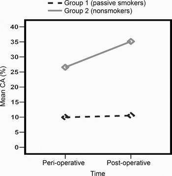
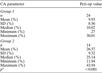
Our results were as follows. In group one (passive smokers, n = 24), the mean peri-operative ciliary area at the maxillary sinus ostium was 9.93 ± 8.56 per cent; this value increased after FESS to a mean of 10.52 ± 10.14 per cent. In group two (nonsmokers; n = 14), the mean peri-operative ciliary area at the maxillary sinus ostium was 26.57 ± 9.31 per cent, increasing after FESS to a mean of 35.14 ± 9.28 per cent.
Statistical analysis of results
In group one (passive smokers), the ciliary area and number of cilia at the maxillary sinus ostium increased slightly following FESS; however, this increase was not statistically significant (p = 0.55, comparing peri- and post-operative ciliary area) (Table I) (Figure 1).
Peri- and post-operative ciliary area: group 1 (passive smokers)

Note that the slight post-operative (post-op) ciliary area (CA) increase was not statistically significant. SD = standard deviation; peri-op = peri-operative
Mean ciliary area (CA) during and after functional endoscopic sinus surgery, for the two groups.

In group two (nonsmokers), the ciliary area and number of cilia showed a marked increase, which was highly statistically significant (p < 0.001, comparing peri- and post-operative ciliary area) (Table II).
Peri- and post-operative ciliary area: group 2 (nonsmokers)

Note the highly statistically significant post-operative (post-op) increase in ciliary area (CA). SD = standard deviation; peri-op = peri-operative
Comparison of ciliary areas between the two groups showed that ciliary area and number were statistically significantly reduced in passive smokers, compared with nonsmokers, before FESS (p < 0.001, comparing the two values) (Table III).
Comparison of peri-operative ciliary areas: groups 1 and 2

* Comparing groups 1 and 2. Note that the peri-operative (peri-op) ciliary area (CA) was statistically significantly less for passive smokers compared with nonsmokers. SD = standard deviation
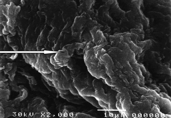
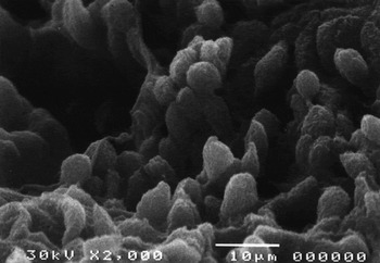
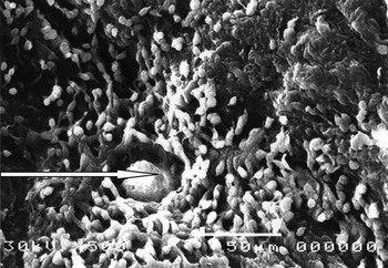
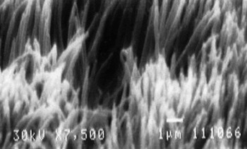
Figures 2 to 5 show examples of scanning electron photomicrographs from both groups, both peri- and post-operatively.
Peri-operative scanning electron photomicrograph from a group 1 patient (i.e. passive smoker), showing that ciliary structures have disappeared and the epithelial surface is covered by thick, matted collagen bundles (arrow) (×2000).

Post-operative scanning electron photomicrograph from same patient as Figure 2, showing regenerating, short, scattered cilia beating in different directions (×2000).

Peri-operative scanning electron photomicrograph from a group 2 patient (i.e. nonsmoker), showing short, sparse cilia beating in different directions, and a submucosal gland opening (arrow) (×1500).

Post-operative scanning electron photomicrograph from same patient as Figure 4, showing regenerating, tall cilia almost covering the entire epithelial surface and beating in one direction (×7500).

Discussion
Paediatric rhinosinusitis is a frequent problem in children, affecting nearly 8 per cent of the paediatric population.Reference Aitken and Taylor11 This disease limits children's physical activities more than other chronic conditions such as asthma and juvenile rheumatoid arthritis.Reference Cunningham, Chiu, Langraf and Gliklich12
Children with chronic rhinosinusitis and symptoms persisting for more than three months are candidates for surgical intervention.Reference Lusk, Bothwell and Piccirillo13 Adenoidectomy and endoscopic sinus surgery have been suggested as viable treatment options for paediatric rhinosinusitis. Adenoidectomy has a 47–58 per cent chance of curing or significantly improving paediatric rhinosinusitis;Reference Vandenberg and Heatley14 however, endoscopic sinus surgery has been shown to be more effective than adenoidectomy, particularly regarding short term results.Reference Ramadan15
Passive exposure to cigarette smoking has been implicated in a number of childhood disorders, ranging from colds and middle-ear diseases to respiratory illness.Reference Cook and Strachan1, 2
Passive exposure to cigarette smoke affects nasal physiology. Ciliary beat frequency is significantly reduced in subjects exposed to passive smoking, compared with controls.Reference Agius, Smallman and Pahor16 Immunoglobulin E producing cells and eosinophils are also present in significantly higher numbers in the nasal mucosa of children exposed to passive smoking.Reference Vinke, Denjan, Severijnen and Fottens17 These children also have a poorer sense of smell than controls.Reference Nageris, Hadar and Hansen18
Only a few studies have assessed the effect of smoking on the results of FESS in patients with chronic sinusitis. Most of these trials have involved adult patients, and all have used subjective outcome measures, e.g. endoscopy scores, quality of life outcomes and computed tomography scores. No previously published studies have used objective outcome measures to study the effect of smoking on FESS results. Briggs et al., in 2004,Reference Briggs, Wright, Cardes and Calhoun4 and Smith et al., in 2005,Reference Smith, Mendolia-Loffredo, Lochr, Sparpani, Laud and Nattinger3 showed that adult smokers had poorer results after FESS compared with nonsmokers, albeit using subjective outcome measures (endoscopy scores or rhinosinusitis-specific quality of life outcome tests).
In 2005, Kim et al. showed that endoscopic surgery in passive-smoking children (n = 97 paediatric patients) was associated with poorer outcomes, compared with children living in a nonsmoking environment. Patients were classified into two groups based on outcome, either good or poor.Reference Kim, Dhog, Chung, Chung and Min5
In 1990, Halma et al. studied the distribution of ciliated cells in normal human nose and paranasal sinuses. They reported that the density of ciliated epithelium inside the maxillary sinus was very great, ranging from 91.35 to 97.75 per cent, except in the ostial region, where the density dropped to 47 per cent.Reference Halma, Decrelon, Bijloos and Clement19
Our study of a paediatric population with chronic sinusitis showed clearly that the ciliary area dropped significantly at the maxillary sinus ostium, compared with normal adult individuals. This drop has previously been recorded in adults, by Bassiouny et al. in 2003, and was attributed to inflammatory processes and mediators which destroy the delicate ciliated epithelium, replacing it with more resistant cuboidal epithelium.Reference Bassiouny, Atef, Abd El Raouf, Nasr, Nasr and Ayad9 We compared our results with findings for normal adults, as no previous study has assessed ciliary distribution in the normal paediatric nose and sinuses. In our study, the reduction in ciliary area was significantly greater in children with chronic exposure to cigarette smoke (i.e. passive smokers), compared with those living in a nonsmoking environment – the mean ciliary area was 9.93 per cent in group one (passive smokers) vs 26.57 per cent in group two (nonsmokers). This finding highlights the role of chronic exposure to cigarette smoke in the destruction of delicate nasal cilia, in addition to the effect of inflammatory mediators active in chronic sinusitis.
• Passive smoking in the paediatric age group is associated with an increased frequency of childhood respiratory disorders
• The effect of passive smoking on ciliary regeneration after functional endoscopic sinus surgery (FESS) for chronic sinusitis has not previously been studied
• This prospective, nonrandomised cohort study of 38 paediatric patients with chronic sinusitis examined the effect of passive smoking on ciliary regeneration after FESS
• Passive smoking appeared to have a negative impact on sinus cilia regeneration after FESS
In our study, passive smoking was associated with a poorer ciliary regeneration capacity of maxillary sinus mucous membrane. Although both groups showed a post-operative increase in cilia number after FESS, this increase was statistically significant in group two but statistically insignificant in group one (the mean ciliary area only increased to 10.52 per cent in group one, but increased to 35.14 per cent in group two). This negative effect of passive smoking on ciliary regeneration after FESS may explain the poorer subjective FESS outcomes observed by most authors as a result of smoking.
Conclusion
In the paediatric population, passive smoking has a negative impact on the regenerative power of sinus cilia following FESS. This negative impact may be the cause of the poor outcomes observed in this patient group.
Acknowledgements
The authors would like to acknowledge the efforts of Dr Hossam El Bosraty, in assisting patient selection, and Dr Esam Ayad, in image processing of the electron microscopy images.