Why diamond?
Single crystals of silicon with an almost perfect crystal lattice are commercially available in large quantities and are commonly used in x-ray optics. However, many relevant applications require materials with better transparency to x-rays, resilience to radiation damage, mechanical stiffness, x-ray Bragg reflectivity, and thermal conductivity. Diamond is the material of choice in such cases; it is superior to silicon in its unrivaled radiation hardness, shows an order of magnitude higher transparency to x-rays, an order of magnitude higher stiffness, Reference Field1,Reference Prelas, Popovici and Bigelow2 orders of magnitude higher thermal conductivity, Reference Prelas, Popovici and Bigelow2,Reference Wei, Kuo, Thomas, Anthony and Banholzer3 superior thermal-expansion properties, Reference Giles, Adriano, Lubambo, Cusatis, Irineu and Hönnicke4–Reference Stoupin and Shvyd’ko6 and nearly 100% reflectivity in Bragg diffraction, even in backscattering. Reference Shvyd’ko, Stoupin, Cunsolo, Said and Huang7,Reference Shvyd’ko, Stoupin, Blank and Terentyev8 This unique combination of outstanding properties makes diamond the most promising material for transparent, resilient, high-resolution, wavefront-preserving x-ray optics components essential for applications in third-generation light sources. These characteristics are especially important in next-generation (fourth-generation) high-brilliance, high-coherence light sources, such as diffraction-limited storage rings (DLSRs) and x-ray free-electron lasers (XFELs).
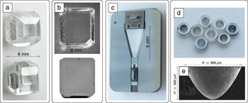
Third-generation storage-ring-based x-ray sources, which emerged in the early 1990s, triggered the quest for new technologies and materials capable of withstanding unparalleled heat and radiation loads on crystal optics elements. Diamond crystals were recognized as the ultimate material for primary monochromators that are resilient to the heat loads of the powerful direct beams. Reference Als-Nielsen and Freund9–Reference Freund14 However, their implementation was hampered by the low quality of the diamond crystals available at that time. Since then, progress in fabricating and characterizing synthetic high-quality diamond, and in developing x-ray optics applications, has been substantial. Reference Pal’yanov, Malinovsky, Borzdov and Khokryakov15–Reference Stoupin, Antipov, Butler, Kolyadin and Katrusha24 Crystals with ≳4 × 4 mm2 defect-free areas Reference Burns, Chumakov, Connell, Dube, Godfried, Hansen, Härtwig, Hoszowska, Masiello, Mkhonza, Rebak, Rommevaux, Setshedi and Van Vaerenbergh20,Reference Polyakov, Denisov, Kuzmin, Kuznetsov, Martyushov, Nosukhin, Terentyev and Blank22–Reference Stoupin, Antipov, Butler, Kolyadin and Katrusha24 became state of the art and enabled numerous diverse applications, far beyond the conventional use of diamond for high-heat-load monochromators ( Figure 1 ). Reference Shvyd’ko, Stoupin, Blank and Terentyev8,Reference Polyakov, Denisov, Kuzmin, Kuznetsov, Martyushov, Nosukhin, Terentyev and Blank22,Reference Terentyev, Blank, Kolodziej and Shvyd’ko25,Reference Terentyev, Blank, Polyakov, Zholudev, Snigirev, Polikarpov, Kolodziej, Qian, Zhou and Shvyd’ko26 We will review advances in the fabrication of defect-free diamond crystals and applications of the diamond optics components at existing and next-generation x-ray sources.
Examples of synthetic diamond crystals and diamond optics components. (a) Raw cuboctahedral high-pressure high-temperature diamond Type IIa crystals. (b) Photograph and x-ray transmission topograph of a 1-mm-thick, nearly flawless diamond plate sliced from the top of a cuboctahedral crystal. Reference Shvyd’ko, Stoupin, Blank and Terentyev8,Reference Polyakov, Denisov, Kuzmin, Kuznetsov, Martyushov, Nosukhin, Terentyev and Blank22 (c) All-diamond assembly of a curved-crystal spectrograph, featuring a curved 20-µm-thin triangular single-crystal diamond in a bender structure; all 10 components of the structure are manufactured from single-crystal or chemical vapor deposited diamond. Reference Terentyev, Blank, Kolodziej and Shvyd’ko25 (d) Concave parabolic diamond x-ray lenses and (e) scanning electron microscope image of a lens revealing its parabolic shape. Reference Terentyev, Blank, Polyakov, Zholudev, Snigirev, Polikarpov, Kolodziej, Qian, Zhou and Shvyd’ko26

Crystal growth, machining, and characterization
Natural diamonds have insufficient crystal lattice perfection for most x-ray optics applications. Man-made diamond crystals feature much higher crystal quality. There are two major approaches currently in use to produce synthetic diamonds—chemical vapor deposition (CVD) and a temperature gradient method under high pressure and high temperature (HPHT). CVD diamonds are less expensive and large crystals can be grown by this process, but the crystal lattice quality of the HPHT-grown crystals is presently the best. Reference Burns, Chumakov, Connell, Dube, Godfried, Hansen, Härtwig, Hoszowska, Masiello, Mkhonza, Rebak, Rommevaux, Setshedi and Van Vaerenbergh20
The HPHT technique was put forward by Leipunski in 1939 Reference Leipunskii27 based on a proposed pressure-temperature phase diagram for carbon, which has been further detailed by many researchers. Reference Berman and Simon28,Reference Bundy29 The modern HPHT process uses a carbon source, a solvent metal and a seed crystal in a high-temperature gradient oven at ≃1650 K under a high pressure of ≃5.5 GPa. The first successful experiments were carried out by researchers at General Electric (USA) and ASEA (Sweden) in the 1950s, and synthesis of diamond microcrystals was reported. Reference Bundy, Hall, Strong and Wentorf30 The first large diamond crystals up to 5-mm dia. were synthesized in 1971 by Wentorf at General Electric. Reference Wentorf31 By the end of 20th century, progress was so significant in advancing synthetic diamond technologies that researchers from General Electric, De Beers, and Sumitomo were able to grow diamond crystals as large as 10-mm dia. (with a mass of approximately 2 g or 10 carats). Reference Sumiya and Satoh16,Reference Sumiya, Toda and Satoh18,Reference Vagarali, Lee and DeVries32–Reference Kanda34
At the beginning of the 1990s, interest in diamond crystals for x-ray optics applications was triggered by the advent of third-generation synchrotron radiation sources. Diamond single crystals were considered the ultimate material for resilient x-ray monochromators, Reference Als-Nielsen and Freund9–Reference Freund14 capable of withstanding the extreme heat and radiation loads of high-power x-ray beams from third-generation synchrotron sources. Because the underlying principle of monochromatization is x-ray Bragg diffraction, this application set a new requirement—along with large crystal size, crystal lattice perfection became important. Additionally, as researchers utilized the coherence properties of the third-generation sources, the ability to preserve the x-ray wavefront as it passes through optical components became critical.
A perfect crystal lattice means an absence of stacking faults, dislocations, precipitates, and inclusions. The importance of diamond crystals with low concentrations of impurities was recognized. Only such crystals, referred to as Type IIa (with nitrogen impurities below 1 ppm), could meet the requirements for wavefront preservation. Reference Burns, Chumakov, Connell, Dube, Godfried, Hansen, Härtwig, Hoszowska, Masiello, Mkhonza, Rebak, Rommevaux, Setshedi and Van Vaerenbergh20 In this regard, the availability of crystal characterization techniques became essential. White-beam and monochromatic beam x-ray diffraction topographies, sequential rocking curve mapping, and high-resolution x-ray diffraction measurements are presently in use for characterizing crystals. Reference Shvyd’ko, Stoupin, Blank and Terentyev8,Reference Sellschop, Connell, Nilen, Detlefs, Freund, Hoszowska, Hustache, Burns, Rebak, Hansen, Welch and Hall17,Reference Burns, Chumakov, Connell, Dube, Godfried, Hansen, Härtwig, Hoszowska, Masiello, Mkhonza, Rebak, Rommevaux, Setshedi and Van Vaerenbergh20,Reference Polyakov, Denisov, Kuzmin, Kuznetsov, Martyushov, Nosukhin, Terentyev and Blank22–Reference Stoupin, Antipov, Butler, Kolyadin and Katrusha24,Reference Macrander, Krasnicki, Zhong, Maj and Chu35,Reference Zhong, Macrander, Krasnicki, Chu, Maj, Assoufid and Qian36
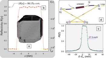
In the past two decades, advances in HPHT crystal growth techniques and crystal characterization have been enormous and rapid. Large HPHT Type IIa diamond crystals with defect-free areas of more than 5 × 5 mm2 became possible with different crystal orientations. Reference Polyakov, Denisov, Kuzmin, Kuznetsov, Martyushov, Nosukhin, Terentyev and Blank22–Reference Stoupin, Antipov, Butler, Kolyadin and Katrusha24 Figure 2 Reference Shvyd’ko, Stoupin, Blank and Terentyev8,Reference Kim, Shvyd’ko and Reiche37,Reference Kim and Shvyd’ko38 shows an example of a 1-mm-thick diamond crystal plate with (001) orientation with a 4 × 4 mm2 flawless area, featuring close to 100% reflectivity in backscattering.
Diamond “x-ray mirrors” with near-100% reflectivity in backscattering. Reference Shvyd’ko, Stoupin, Blank and Terentyev8 (a) X-ray transmission topograph of a 1-mm-thick (001) diamond crystal with almost defect-free 4 × 4 mm2 area. (b) The crystal reflects ≃99% of 13.9-keV x-ray photons backwards in Bragg diffraction, with a small variation in magnitude across the sample and (c) features almost perfect Darwin-shape spectral dependence (close to rectangular dependence predicted by the dynamical theory of x-ray Bragg diffraction) of the reflectivity. (d) High-reflectance backscattering diamond x-ray mirrors (C*) are essential for the realization of x-ray free-electron laser oscillators. Reference Kim, Shvyd’ko and Reiche37,Reference Kim and Shvyd’ko38

The requirements for diamond x-ray optics components are diverse in terms of the desired form—thin or thick; flat, curved, or parabolic; rectangular or trapezoidal. Harsh radiation damage conditions of intense x-ray beams require all-diamond multi-element assemblies. These diverse needs call for the development of sophisticated machining techniques, including diamond crystal cutting, Reference Polikarpov, Snigireva, Morse, Yunkin, Kuznetsov and Snigirev39 three-dimensional machining by reactive-ion etching, Reference Nöhammer, Hoszowska, Freund and David40 or laser milling Reference Terentyev, Blank, Kolodziej and Shvyd’ko25,Reference Terentyev, Blank, Polyakov, Zholudev, Snigirev, Polikarpov, Kolodziej, Qian, Zhou and Shvyd’ko26,Reference Kolodziej, Vodnala, Terentyev, Blank and Shvyd’ko41 and polishing. Reference Burns, Chumakov, Connell, Dube, Godfried, Hansen, Härtwig, Hoszowska, Masiello, Mkhonza, Rebak, Rommevaux, Setshedi and Van Vaerenbergh20 It is essential that the final optical components that users obtain are defect-free, strain-free, and have a well-polished surface.
Diamond x-ray optics applications
Heat-load and radiation-damage management with monochromators
The advent of third-generation synchrotron radiation sources has triggered interest in diamond crystals as the ultimate material for high-heat-load x-ray monochromators (HHLM) capable of withstanding harsh heat-load and radiation-damage conditions due to high-power x-ray beams. Reference Als-Nielsen and Freund9–Reference Freund14 The first crystal of the monochromators is exposed to x-rays with power and power density of up to 1 kW and 400 W/mm2, respectively. Double-crystal water-cooled diamond HHLMs were developed and installed at third-generation synchrotron radiation facilities in the late 1990s at the European Synchrotron Radiation Facility (ESRF, France), Reference Als-Nielsen, Freund, Wulff, Hanfland and Häusermann13,Reference Als-Nielsen, Freund, Grübel, Linderholm, Nielsen, del Rio and Sellschop42–Reference van Vaerenbergh, Detlefs, Härtwig, Lafford, Masiello, Roth, Schmid, Wattecamps, Zhang, Garrett, Gentle, Nugent and Wilkinset44 the Advanced Photon Source (USA), Reference Fernandez, Graber, Lee, Mills, Rogers and Assoufid45,Reference Fernandez, Lee, Mills, Tajiri and Assoufid46 and the Super Photon Ring (SPring-8, Japan). Reference Yabashi, Goto, Shimizu, Tamasaku, Yamazaki, Yoda, Suzuki, Ohishi, Yamamoto and Ishikawa19,Reference Goto, Yamazaki, Shimizu, Suzuki, Kawamura, Mizumaki, Yabashi, Tamasaku and Ishikawa47 The superior thermal and mechanical properties of diamond and higher x-ray transparency resulted in excellent thermal performance at room temperature, comparable to the performance of liquid-nitrogen-cooled silicon monochromators. In contrast to cryo-cooled silicon, water-cooled diamond makes HHLMs vibration-free, which is essential for wavefront preservation. Altogether, the viability of using water-cooled diamonds for high-intensity synchrotron radiation beamlines has been demonstrated.
Despite the initial success, diamond HHLMs did not become the mainstream approach at synchrotron radiation facilities. There are two limitations that make them less attractive in several cases, compared to silicon HHLMs. First, the largest spectral bandwidth and the angular acceptance of x-rays achievable with the (111) Bragg reflection from diamond are one-half of what is possible with the (111) Bragg reflection from silicon. Applications that require high flux therefore favor silicon monochromators. Second, high-heat-load diamond x-ray optics are not yet wavefront preserving, causing problems with focusing x-rays and applying imaging techniques.
To become wavefront-preserving, flawless (111) diamond crystal plates are required that are sufficiently large in size, typically more than 2 × 5 mm2. Because of this, diamond crystal plates in the (111) orientation are in the greatest demand. However, manufacturing flawless diamond plates, particularly in the (111) orientation, is the most challenging task. There is a continuous effort toward ongoing advances. In particular, recent fabrication of crystal plates in the (111) orientation has been reported with a region as large as 3 × 7 mm2 suitable for wavefront-preserving high-heat-load applications. Reference Stoupin, Antipov, Butler, Kolyadin and Katrusha24
Beam multiplexing
Beam multiplexing monochromators
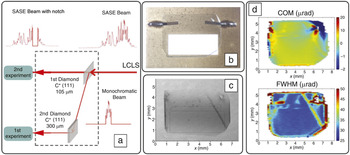
X-ray beams generated in present-day accelerator-based x-ray sources are brilliant, collimated, pulsed, and polarized. Experiment time at these sources is precious and in high demand. The possibility of multiplexing the beams for performing simultaneous experiments using one source has been explored. This was first accomplished at the ESRF using a series of x-ray transparent monochromator crystals. Reference Als-Nielsen, Freund, Grübel, Linderholm, Nielsen, del Rio and Sellschop42,Reference Mattenet, Schneider and Grübel43 A “transparent” crystal is made thick enough to efficiently extract a small part of the x-ray spectrum for the first experiment, but not too thick to let the rest pass through to the next experiments without significant absorption ( Figure 3 ). Diamond is an excellent choice for multiplexing monochromators because of its 10× larger x-ray transparency compared to that of silicon, combined with excellent thermal and mechanical properties. However, beam-multiplexing monochromators have not yet become a popular approach at storage-ring facilities, due to the previously mentioned reasons for diamond HHLMs.
Simultaneous x-ray experiments at the Linac Coherent Light Source (LCLS) x-ray free-electron laser facility (a) made possible by a diamond double-crystal multiplexing monochromator. Reference Zhu, Feng, Stoupin, Terentyev, Lemke, Fritz, Chollet, Glownia, Alonso-Mori, Sikorski, Song, van Driel, Williams, Messerschmidt, Boutet, Blank, Shvyd’ko and Robert48–Reference Feng, Alonso-Mori, Barends, Blank, Botha, Chollet, Damiani, Doak, Glownia, Koglin, Lemke, Messerschmidt, Nass, Nelson, Schlichting, Shoeman, Shvyd’ko, Sikorski, Song, Stoupin, Terentyev, Williams, Zhu, Robert and Boutet50 (b) All-diamond crystal assembly for use as a beam splitter. It consists of a high-quality 105-µm-thick high-pressure high-temperature Type IIa diamond crystal (transparent) in the (111) orientation mounted strain-free on a chemical vapor deposited (CVD) diamond (opaque) substrate with a rectangular window by microperforated CVD diamond springs. (c) X-ray topography and (d) color maps of the Bragg reflection rocking-curve widths full width at half maximum (FWHM) and the center of mass (COM) for the same crystal, showing its good quality in a working area of ≃2 × 4 mm2. Note: SASE, self-amplified spontaneous emission.

X-ray beams generated by XFELs are even more precious. The costs of XFELs and storage-ring facilities are comparable; however, XFELs are linac-based (linear accelerator-based) single-source facilities. Because of this, the Linac Coherent Light Source (LCLS, USA) XFEL initially performed one experiment at a time. X-ray beam splitting with thin diamond crystals now allows two experiments to be performed simultaneously, Reference Zhu, Feng, Stoupin, Terentyev, Lemke, Fritz, Chollet, Glownia, Alonso-Mori, Sikorski, Song, van Driel, Williams, Messerschmidt, Boutet, Blank, Shvyd’ko and Robert48–Reference Feng, Alonso-Mori, Barends, Blank, Botha, Chollet, Damiani, Doak, Glownia, Koglin, Lemke, Messerschmidt, Nass, Nelson, Schlichting, Shoeman, Shvyd’ko, Sikorski, Song, Stoupin, Terentyev, Williams, Zhu, Robert and Boutet50 as illustrated in Figure 3. Implementation of beam multiplexing at XFELs is more challenging than at synchrotrons, since the use of wavefront-preserving optics is a must because of the transversely coherent nature of the source. Two new solutions have been implemented in the realization of the LCLS beam-multiplexing monochromator. First, the diamond crystal plates in the (111) orientation have been cut from the upper part of the (001) growth sector of HPHT diamond crystals of Type IIa. A region approximately 2 × 4 mm2 farthest from the seed was selected as the working area (indicated by the rectangle in Figure 3c) with wavefront-preserving characteristics, as certified by the double-crystal rocking-curve maps in Figure 3d. Second, all-diamond optical assemblies holding high-quality (111) crystal plates were designed, as shown Figure 3b, to provide stable mounting on a rigid CVD diamond substrate. This setup minimizes mounting-induced crystal strain in the ≃100-µm-thin (111) diamond plates and enables them to be resilient to the XFEL x-ray pulses of extreme peak power.
Ultrathin crystal beam splitters and mixers
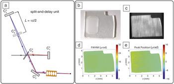
Efficient x-ray beam splitters and mixers are essential for the realization of an x-ray split-and-delay unit (SDU), which is a key optical component of x-ray photon-correlation spectrometers. Reference Roseker, Franz, Schulte-Schrepping, Ehnes, Leupold, Zontone, Lee, Robert and Grübel51–Reference Stetsko, Shvyd’ko and Stephenson53 An SDU splits x-ray pulses, delays them in time, and then recombines them. Because of its high x-ray transparency, diamond is a preferred material for the splitter/mixer crystal. Figure 4 details a particular design of the SDU with a challenging ultrathin (≲50-µm) diamond crystal C0 functioning as a wavefront-preserving beam splitter and beam mixer. Reference Stetsko, Shvyd’ko and Stephenson53
Realization of x-ray photon correlation spectroscopy in the femtosecond to nanosecond regime relies on the feasibility of an x-ray split-and-delay unit. One of its designs is based on a four-crystal scheme (a) with a challenging ultrathin (≲50-µm) diamond crystal C0 functioning as a wavefront-preserving beam splitter and beam mixer. Reference Stetsko, Shvyd’ko and Stephenson53 (b) A diamond drumhead crystal, composed of a 50-µm-thick 1.5 × 2.5 mm2 rectangular membrane machined by laser milling in a 210-µm-thick crystal plate, is a solution that ensures mechanically stable strain-free mounting of the thin membrane. Reference Kolodziej, Vodnala, Terentyev, Blank and Shvyd’ko41 (c) Bragg reflection image and (d–e) rocking-curve maps of a drumhead crystal reveal a flawless and strain-free crystal membrane. Note: FWHM, full width at half maximum.

The fabrication and handling of such thin components without introducing crystal damage and strain are challenging. Drumhead crystals, which are monolithic crystal structures composed of a thin membrane furnished with a surrounding solid collar, are a solution ensuring mechanically stable strain-free mounting of the membranes with efficient thermal transport. Diamond, being one of the hardest and most chemically inert materials, poses significant difficulties in fabrication. Successful fabrication of a diamond drumhead crystal with a 50-µm-thick 1.5 × 2.5 mm2 rectangular defect-free and strain-free membrane, compatible with the requirements for the splitter/mixer crystal of the SDU, has recently been reported. Reference Kolodziej, Vodnala, Terentyev, Blank and Shvyd’ko41 The drumhead crystal (shown in Figure 4b) has been machined by laser milling. Drumhead crystals are anticipated to be a solution for many other diamond thin-crystal wavefront-preserving applications.
Fine spectral control with high-resolution monochromators
Silicon was believed to be unrivaled in terms of crystal lattice perfection. Only silicon crystals of the best available quality have been trusted so far for use in high-resolution x-ray monochromators, devices that filter x-rays within a narrow spectral bandwidth. Silicon monochromators for hard x-rays with resolutions as high as ≃108 and spectral bandwidths as small as ≃100 µeV have been reported. Reference Toellner, Hu, Sturhahn, Bortel, Alp and Zhao54–Reference Shvyd’ko, Stoupin, Mundboth and Kim56 It was almost impossible to believe that the crystal quality of diamond could compete with silicon as a material for ultrahigh-resolution monochromators. However, this was found to be true.
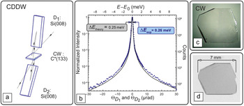
First, it was demonstrated that one single-crystal diamond is capable of monochromatizing 23.7-keV x-rays to a few-meV bandwidth. Reference Shvyd’ko, Stoupin, Cunsolo, Said and Huang7,Reference Shvyd’ko, Stoupin, Blank and Terentyev8 Second, a three-crystal hybrid diamond-silicon x-ray monochromator was demonstrated, which featured close to theoretical performance with a spectral bandwidth of 0.26-meV and a high 68% spectral efficiency Reference Stoupin, Shvyd’ko, Shu, Blank, Terentyev, Polyakov, Kuznetsov, Lemesh, Mundboth, Collins, Sutter and Tolkiehn57 ( Figure 5 ). Both the high resolution and the high efficiency are due to the application of a 100-µm-thick, practically flawless diamond crystal (CW) in the strongly asymmetric (133) Bragg reflection. The CW diamond crystal interacts with the beam sequentially three times. However, because of the very high x-ray transparency and Bragg reflectivity of diamond, the efficiency of the monochromator is very high. It should be compared with ≃5−15% efficiency of all-silicon ultrahigh-resolution monochromators. Reference Toellner, Hu, Sturhahn, Bortel, Alp and Zhao54–Reference Shvyd’ko, Stoupin, Mundboth and Kim56
(a) Three-crystal hybrid diamond-silicon monochromator CDDW (comprising a collimator/wavelength-selector crystal CW and two dispersing crystals D1, D2) for an ultrahigh resolution inelastic x-ray scattering instrument at the Advanced Photon Source featuring (b) a spectral function with an extremely narrow 0.26-meV bandwidth and a very high 68% spectral efficiency. Reference Stoupin, Shvyd’ko, Shu, Blank, Terentyev, Polyakov, Kuznetsov, Lemesh, Mundboth, Collins, Sutter and Tolkiehn57 Both the high resolution and the high efficiency are due to the application of a 100-µm-thick flawless diamond crystal (CW in [a]) in the strongly asymmetric (133) Bragg reflection. Photograph (c) and Lang x-ray topograph (d) of the CW diamond crystal are shown.

Enabling next-generation coherent x-ray sources
Monochromators for self-seeded XFELs
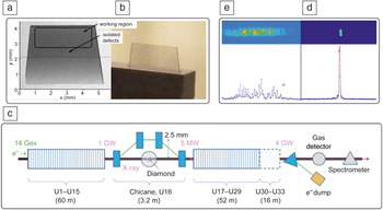
Hard x-rays can be generated with a high degree of transverse coherence by DLSR and self-amplified spontaneous emission (SASE) XFEL sources. However, their performance produces poor temporal coherence and a broad, noisy spectrum. Self-seeded XFELs have been considered as fully coherent sources with Fourier-transform limited x-ray pulses and with a well-defined narrow spectrum. Reference Geloni, Kocharyan and Saldin58,Reference Saldin, Schneidmiller, Shvyd’ko and Yurkov59 Self-seeded XFELs use x-rays from the first half of the undulator to seed the second half through an x-ray monochromator; see Figure 6c. Diamond four-crystal Bragg diffraction Reference Saldin, Schneidmiller, Shvyd’ko and Yurkov59 or diamond single-crystal forward Bragg diffraction (FBD) Reference Geloni, Kocharyan and Saldin58 monochromators have been proposed. Diamond was chosen because of its resilience to the high peak and average powers of the XFEL pulses and because x-ray transparency is crucial for the FBD monochromator. Self-seeding with the FBD diamond monochromator was recently demonstrated. Reference Amann, Berg, Blank, Decker, Ding, Emma, Feng, Frisch, Fritz, Hastings, Huang, Krzywinski, Lindberg, Loos, Lutman, Nuhn, Ratner, Rzepiela, Shu, Shvyd’ko, Spampinati, Stoupin, Terentyev, Trakhtenberg, Walz, Welch, Wu, Zholents and Zhu60 A tiny 100-µm-thin almost flawless diamond crystal (Figure 6b) Reference Stoupin, Blank, Terentyev, Polyakov, Denisov, Kuznetsov, Shvyd’ko, Shu, Emma, Maj and McNulty61 converted a SASE XFEL with a broad noisy spectrum into a fully coherent source of hard x-ray photons with a well-defined sharp spectrum (Figure 6d).
Self-seeding in an x-ray free-electron laser (XFEL) with a diamond monochromator. Reference Geloni, Kocharyan and Saldin58,Reference Amann, Berg, Blank, Decker, Ding, Emma, Feng, Frisch, Fritz, Hastings, Huang, Krzywinski, Lindberg, Loos, Lutman, Nuhn, Ratner, Rzepiela, Shu, Shvyd’ko, Spampinati, Stoupin, Terentyev, Trakhtenberg, Walz, Welch, Wu, Zholents and Zhu60 (a) X-ray white-beam Bragg topograph Reference Stoupin, Blank, Terentyev, Polyakov, Denisov, Kuznetsov, Shvyd’ko, Shu, Emma, Maj and McNulty61 of a diamond crystal flawless in the working region (b) in a graphite holder used as a forward Bragg diffraction monochromator in a hard-x-ray self-seeding system of the (c) Linac Coherent Light Source XFEL. The diamond monochromator enables generation of fully coherent x-rays with a (d) 0.4-eV-sharp well-defined single-line spectrum. Without the monochromator, the XFEL generates x-rays with (e) a 20-eV-broad spiky stochastic spectrum.

X-ray cavities with high-reflectance mirrors
Fully coherent XFEL sources of hard x-rays can be constructed in the oscillator configuration. In an XFEL oscillator (XFELO), x-ray pulses are trapped in a high-quality optical cavity employing diamond crystals as high-reflectance x-ray mirrors (Figure 2d) for repeated low-gain amplification. Reference Kim, Shvyd’ko and Reiche37,Reference Kim and Shvyd’ko38 XFELOs have the potential to generate highly stable, ultrahigh-spectral-purity x-ray pulses with meV bandwidth. X-ray backscattering mirrors with close to 100% reflectivity are critical for the realization of the XFELO. Theory predicts that even at normal incidence, the reflection of x-rays from diamond under the Bragg condition should approach the required 100%—substantially higher than from any other crystal. Experiments Reference Shvyd’ko, Stoupin, Blank and Terentyev8 demonstrated that synthetic flawless diamond crystals feature an unprecedented reflecting power at normal incidence and meV-narrow reflection bandwidths for hard x-rays. Such properties greatly assist the realization of XFELOs.
Permeable mirrors and drumhead crystals
One of the XFELO cavity mirrors has to be permeable to outcouple (let escape) about 5% of the intracavity radiation; see crystal
 $C_1^ *$
in Figure 2d. This is possible by reducing the diamond crystal thickness to a characteristic length of Bragg diffraction (extinction length). It can be as small as a few tenths of a micron, depending on the photon energy. Drumhead crystals, which were previously discussed, are a possible solution. Successful fabrication of diamond drumhead crystals with up to 26-µm-thick, defect-free and strain-free membranes has been reported;
Reference Kolodziej, Vodnala, Terentyev, Blank and Shvyd’ko41
see also Figure 4b.
Focusing with compound refractive lenses
Compound refractive lenses (CRLs) for hard-x-ray focusing were brought into practice two decades ago. Reference Snigirev, Kohn, Snigireva and Lengeler62 Beryllium is the most popular material for CRLs. It was realized that diamond CRLs are also very attractive, especially as primary (front-end) optical components because of their resilience to powerful x-ray beams and their high x-ray transparency.
The fabrication and testing of nanofocusing diamond planar CRLs made from bulk polycrystalline CVD diamond substrates were reported by several groups. Reference Nöhammer, Hoszowska, Freund and David40,Reference Fox, Alianelli, Malik, Pape, May and Sawhney63 The lens structures were generated by electron-beam lithography and transferred to the diamond by reactive-ion etching.
Single-crystal diamond CRLs are considered to be more advantageous, as they can focus x-rays without compromising the brilliance of the beams. Recently, planar parabolic CRLs were fabricated from single-crystal diamond by laser cutting Reference Polikarpov, Snigireva, Morse, Yunkin, Kuznetsov and Snigirev39 and laser milling, Reference Terentyev, Polikarpov, Snigireva, Di Michiel, Zholudev, Yunkin, Kuznetsov, Blank and Snigirev64 featuring large-acceptance, speckle-free, wavefront-preserving front-end optics. Finally, two-dimensional refractive lenses with rotational parabolic profiles focusing x-rays in two dimensions were manufactured by laser milling of single-crystal diamond and tested; Reference Terentyev, Blank, Polyakov, Zholudev, Snigirev, Polikarpov, Kolodziej, Qian, Zhou and Shvyd’ko26,Reference Antipov, Baryshev, Butler, Antipova, Liu and Stoupin65 see also Figure 1d.
Polarization control with phase plates
Producing a circularly polarized beam for magnetic circular dichroism experiments in the hard-x-ray range with quarter-wave (phase) plates was one of the first applications of diamond in x-ray optics. Reference Giles, Malgrange, Goulon, de Bergevin, Vettier, Dartyge, Fontaine, Giorgetti and Pizzini66,Reference Suzuki, Inubushi, Yabashi and Ishikawa67 The x-ray transparency of diamond is a clear advantage over silicon. This application favors, although does not require, extremely high crystal quality, as it works in forward Bragg diffraction with a substantial angular offset from the Bragg peak. It is a popular technique at storage-ring and XFEL sources. It was recently shown that the wavefront-preserving properties of diamond phase plates are good enough to enable Bragg coherent diffractive imaging with circularly polarized x-rays. Reference Logan, Harder, Li, Haskel, Chen, Winarski, Fuesz, Schlagel, Vine, Benson and McNalty68
Noninvasive spectral monitoring
Diffraction gratings
CVD diamond has been used to fabricate transparent x-ray diffraction gratings that outcouple a small portion of the incident radiation into a first-order diffraction peak for noninvasive XFEL spectral monitoring. Reference Karvinen, Rutishauser, Mozzanica, Greiffenberg, Juranic, Menzel, Lutman, Krzywinski, Fritz, Lemke, Cammarata and David69,Reference Makita, Karvinen, Zhu, Juranic, Grnert, Cartier, Jungmann-Smith, Lemke, Mozzanica, Nelson, Patthey, Sikorski, Song, Feng and David70
Bent crystal spectrographs
In an alternative approach, a curved diamond single-crystal spectrograph was designed, fabricated, and tested. Reference Terentyev, Blank, Kolodziej and Shvyd’ko25 The key component is a curved, 20-µm-thin, single crystalline diamond triangular plate in the (110) orientation (Figure 1c), transparent to x-rays. The radius of curvature can be varied between R = 0.6 m and R = 0.1 m in a controlled fashion, ensuring imaging in a spectral window of up to 100 eV for ≃8 keV x-rays. All 10 components of the bending mechanism are manufactured from diamond, thus ensuring safe operation in the intense XFEL beams.
Other Applications
There are several other current or potential applications of diamond that must be mentioned, if only briefly. Commercially available thin CVD diamond has been used for transparent and resilient front-end x-ray windows or screens for photon-beam imaging. Reference Degenhardt, Aprigliano, Schulte-Schrepping, Hahn, Grabosch and Worner71 About 20-µm-thin diamond single crystals are considered to be a coherent bremsstrahlung source of polarized 9-GeV photons for the GlueX experiment, devoted to the search for mesons with gluonic excitations. Reference Shepherd72 The drumhead crystal approach Reference Kolodziej, Vodnala, Terentyev, Blank and Shvyd’ko41 is a viable solution for this application. Because of the nearly 100% reflectivity in Bragg backscattering and high transparency, diamond is the best material for the mirrors of x-ray Fabry–Pérot interferometers and cavities with the largest finesse achievable in the hard-x-ray regime. Reference Shvyd’ko73,Reference Huang, Siddons, Macrander, Peng and Wu74
Outlook
Over the past two decades, there has been impressive ongoing development of advanced technologies for synthesizing high-quality diamond crystals, machining diamond crystal components having sophisticated shapes, and utilizing these components in optical devices at storage-ring and free-electron laser-based x-ray sources. The new emerging x-ray sources are becoming more brilliant and more powerful with a high degree of coherence. This sets more stringent requirements for the quality of the diamond x-ray optics components. To be compatible with the coherent x-rays sources, the optical components must first be wavefront-preserving, which requires the absence of linear defects, low concentration of impurities, and strain-free crystal lattice. Next, they must be high-heat-load compatible. Wavefront-preserving diamond optics have been demonstrated in particular cases. High heat-load diamond optics are in use. Finally, diamond crystal optics must be made both wavefront-preserving and heat-load resilient for optics that will preserve the transverse coherence of the powerful coherent x-ray beams.
The progress in the field has been impressive, however, the technologies need to be further developed to enable synthesis of larger and more cost-efficient diamond crystals having the highest quality. Machining and polishing technologies should be further improved to enable fabrication of the optical components with higher precision and smaller amounts of crystal damage. Diamond x-ray crystal optics is a dynamic field, and we are expecting many new exciting advances in the near future.
Acknowledgments
Work at the Advanced Photon Source was supported by the US Department of Energy, Office of Science, Office of Basic Energy Sciences, under Contract No. DE-AC02-06CH11357. Work at TISCNM was supported by the Ministry of Education and Science of the Russian Federation: Scientific Project RFMEF1586114X0001 Grant No. 14.586.21.0001.

Yuri Shvyd’ko is a senior staff scientist at the Advanced Photon Source, Argonne National Laboratory. He graduated from the Moscow Engineering and Physics Institute, Russia, in 1978, received his PhD degree in physics from Kurchatov Institute, Russia, in 1984, and Habilitation in physics from Hamburg University, Germany, in 2002. He has authored and co-authored more than 130 papers and one book on x-ray optics. His research interests include condensed-matter physics, x-ray optics, Mossbauer spectroscopy, inelastic x-ray scattering, synchrotron radiation instrumentation, and x-ray free-electron lasers. Shvyd’ko can be reached by phone at 630-252-2901 or by email at shvydko@aps.anl.gov.

Vladimir Blank is a professor and the founding Director of the Technological Institute for Superhard and Novel Carbon Materials, Russia. The Institute leads in the development and fabrication of modern superhard materials and has been awarded golden and silver medals for achievements in the field of synthesis and studies of nanocluster materials under high-temperature high-pressure conditions at international exhibitions. He has a doctorate of science in physics and mathematics. He is an author and co-author of more than 250 scientific articles, three monographs, and 22 patents. Blank can be reached by email at vblank@tisnum.ru.

Sergey Terentyev is head of the Department of Single Crystal Growth at the Technological Institute for Superhard and Novel Carbon Materials, Russian Federation, since 1995. He received his degree from the Moscow Steel and Alloy Institute, Russia. The activities of this department include the creation of methods to produce high purity and semiconductor diamonds for optical and electronic applications. Terentyev can be reached by email at s.ter@bk.ru.